

Activation of extracellular signal-regulated protein kinase 5 is essential for cystitis- and nerve growth factor-induced calcitonin gene-related peptide expression in sensory neurons
- PMID: 22742729
- PMCID: PMC3502118
- DOI: 10.1186/1744-8069-8-48
Activation of extracellular signal-regulated protein kinase 5 is essential for cystitis- and nerve growth factor-induced calcitonin gene-related peptide expression in sensory neurons
Abstract
Background: Cystitis causes considerable neuronal plasticity in the primary afferent pathways. The molecular mechanism and signal transduction underlying cross talk between the inflamed urinary bladder and sensory sensitization has not been investigated.
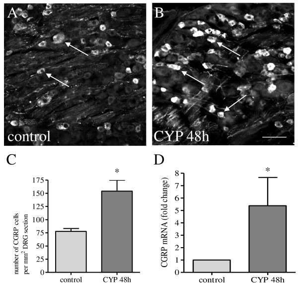
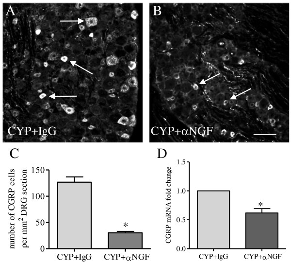
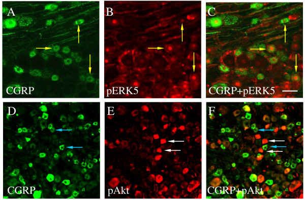
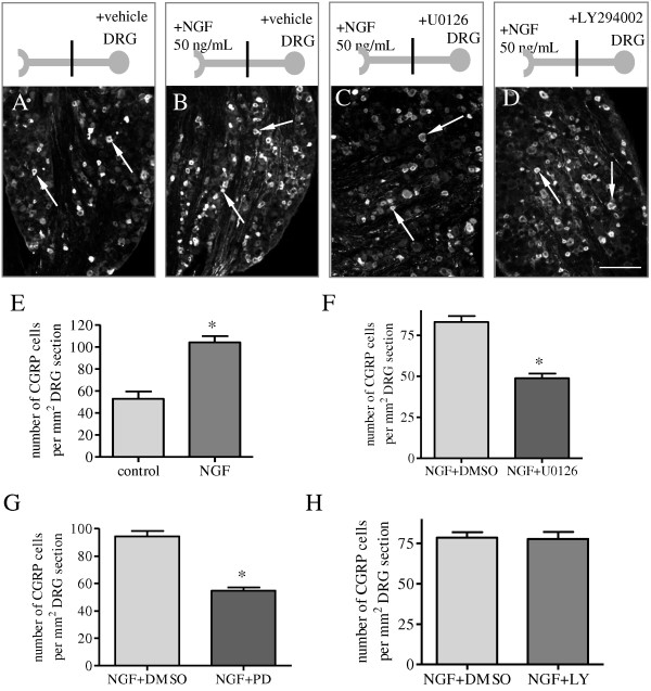
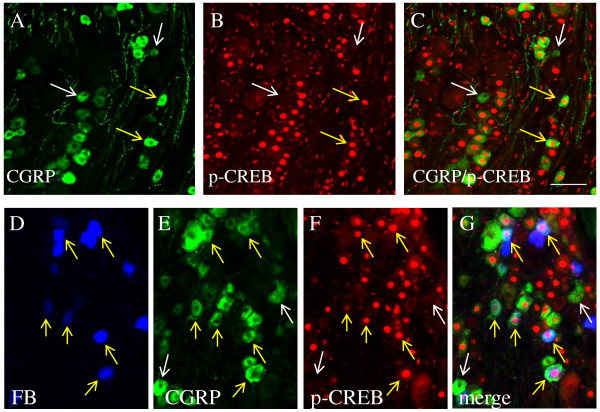
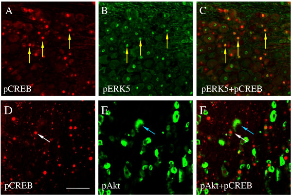
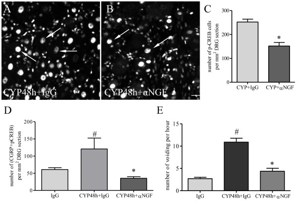
Results: In a rat cystitis model induced by cyclophosphamide (CYP) for 48 h, the mRNA and protein levels of the excitatory neurotransmitter calcitonin gene-related peptide (CGRP) are increased in the L6 dorsal root ganglia (DRG) in response to bladder inflammation. Cystitis-induced CGRP expression in L6 DRG is triggered by endogenous nerve growth factor (NGF) because neutralization of NGF with a specific NGF antibody reverses CGRP up-regulation during cystitis. CGRP expression in the L6 DRG neurons is also enhanced by retrograde NGF signaling when NGF is applied to the nerve terminals of the ganglion-nerve two-compartmented preparation. Characterization of the signaling pathways in cystitis- or NGF-induced CGRP expression reveals that the activation (phosphorylation) of extracellular signal-regulated protein kinase (ERK)5 but not Akt is involved. In L6 DRG during cystitis, CGRP is co-localized with phospho-ERK5 but not phospho-Akt. NGF-evoked CGRP up-regulation is also blocked by inhibition of the MEK/ERK pathway with specific MEK inhibitors U0126 and PD98059, but not by inhibition of the PI3K/Akt pathway with inhibitor LY294002. Further examination shows that cystitis-induced cAMP-responsive element binding protein (CREB) activity is expressed in CGRP bladder afferent neurons and is co-localized with phospho-ERK5 but not phospho-Akt. Blockade of NGF action in vivo reduces the number of DRG neurons co-expressing CGRP and phospho-CREB, and reverses cystitis-induced increases in micturition frequency.
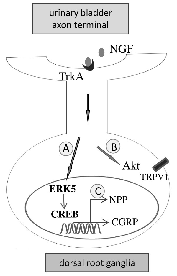
Conclusions: A specific pathway involving NGF-ERK5-CREB axis plays an essential role in cystitis-induced sensory activation.
Figures
Similar articles
-
In vivo regulation of brain-derived neurotrophic factor in dorsal root ganglia is mediated by nerve growth factor-triggered Akt activation during cystitis.PLoS One. 2013 Nov 26;8(11):e81547. doi: 10.1371/journal.pone.0081547. eCollection 2013. PLoS One. 2013. PMID: 24303055 Free PMC article.
-
Chronic morphine exposure increases the phosphorylation of MAP kinases and the transcription factor CREB in dorsal root ganglion neurons: an in vitro and in vivo study.Eur J Neurosci. 2001 Oct;14(7):1091-104. doi: 10.1046/j.0953-816x.2001.01731.x. Eur J Neurosci. 2001. PMID: 11683901
-
Up-regulation of brain-derived neurotrophic factor is regulated by extracellular signal-regulated protein kinase 5 and by nerve growth factor retrograde signaling in colonic afferent neurons in colitis.Exp Neurol. 2012 Dec;238(2):209-17. doi: 10.1016/j.expneurol.2012.08.007. Epub 2012 Aug 19. Exp Neurol. 2012. PMID: 22921460 Free PMC article.
-
Distribution and immunocytochemical characterization of dorsal root ganglion neurons innervating the lumbar intervertebral disc in rats: a review.Life Sci. 2004 Apr 9;74(21):2627-42. doi: 10.1016/j.lfs.2004.01.008. Life Sci. 2004. PMID: 15041445 Review.
-
Molecular mechanisms of sensitization of pain-transducing P2X3 receptors by the migraine mediators CGRP and NGF.Mol Neurobiol. 2008 Feb;37(1):83-90. doi: 10.1007/s12035-008-8020-5. Epub 2008 May 6. Mol Neurobiol. 2008. PMID: 18459072 Review.
Cited by
-
Endogenous PI3K/Akt and NMDAR act independently in the regulation of CREB activity in lumbosacral spinal cord in cystitis.Exp Neurol. 2013 Dec;250:366-75. doi: 10.1016/j.expneurol.2013.10.015. Epub 2013 Oct 30. Exp Neurol. 2013. PMID: 24184018 Free PMC article.
-
Research progress of the role and mechanism of extracellular signal-regulated protein kinase 5 (ERK5) pathway in pathological pain.J Zhejiang Univ Sci B. 2016 Oct.;17(10):733-741. doi: 10.1631/jzus.B1600188. J Zhejiang Univ Sci B. 2016. PMID: 27704743 Free PMC article. Review.
-
Neuroimmune Mechanisms in Signaling of Pain During Acute Kidney Injury (AKI).Front Med (Lausanne). 2020 Aug 7;7:424. doi: 10.3389/fmed.2020.00424. eCollection 2020. Front Med (Lausanne). 2020. PMID: 32850914 Free PMC article. Review.
-
Treatment of painful bladder syndrome/interstitial cystitis with botulinum toxin A: why isn't it effective in all patients?Transl Androl Urol. 2015 Oct;4(5):543-54. doi: 10.3978/j.issn.2223-4683.2015.10.02. Transl Androl Urol. 2015. PMID: 26816853 Free PMC article. Review.
-
Colitis-induced bladder afferent neuronal activation is regulated by BDNF through PLCγ pathway.Exp Neurol. 2016 Nov;285(Pt B):126-135. doi: 10.1016/j.expneurol.2015.12.006. Epub 2015 Dec 11. Exp Neurol. 2016. PMID: 26687970 Free PMC article.
References
Publication types
MeSH terms
Substances
Grants and funding
LinkOut - more resources
Full Text Sources
Research Materials
Miscellaneous
