. 2012 Oct;61(5):721-30.
doi: 10.1016/j.neuint.2012.06.013. Epub 2012 Jun 26.

Ogg1 null mice exhibit age-associated loss of the nigrostriatal pathway and increased sensitivity to MPTP

Affiliations

Ogg1 null mice exhibit age-associated loss of the nigrostriatal pathway and increased sensitivity to MPTP

Fernando Cardozo-Pelaez et al. Neurochem Int. 2012 Oct.

Abstract

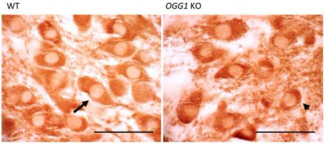
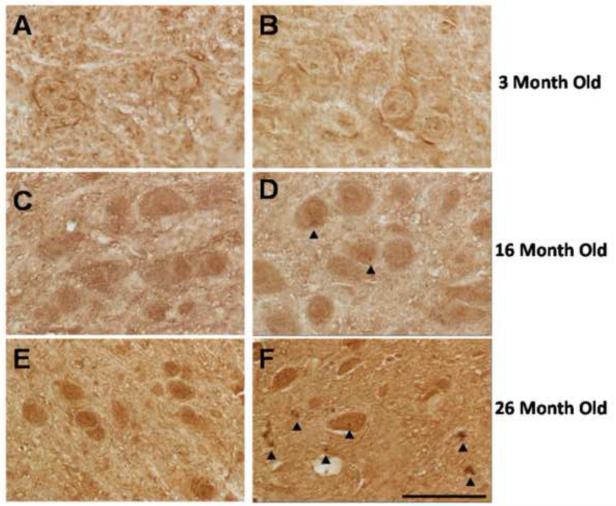
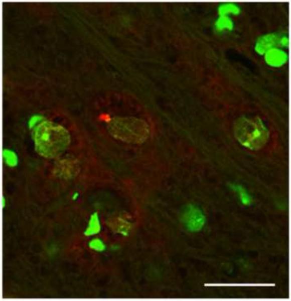
Cumulative damage to cellular macromolecules via oxidative stress is a hallmark of aging and neurodegenerative disease. Whether such damage is a cause or a subsequent effect of neurodegeneration is still unknown. This paper describes the development of an age-associated mild parkinsonian model in mice that lack the DNA repair enzyme 8-oxoguanine glycosylase 1 (Ogg1). Aged OGG1 knock-out (OGG1 KO) mice show a decreased spontaneous locomotor behavior and evidence a decrease in striatal dopamine levels, a loss of tyrosine hydroxylase (TH)-positive neurons in the substantia nigra (SN), and an increase in ubiquitin-positive inclusions in their remaining SN neurons. In addition, young OGG1 KO mice are more susceptible to the dopaminergic toxin 1-methyl-4-phenyl-1,2,3,6-tetrahydropyridine (MPTP) than their wild-type (WT) counterparts. Age-associated increases in 7,8-dihydro-2'-deoxyguanine (oxo(8)dG) have been reported in brain regions and neuronal populations affected in Parkinson's disease (PD), toxin-induced parkinsonian models, and mice harboring genetic abnormalities associated with PD. Because of these increased oxo(8)dG levels, the OGG1 KO mouse strain could shed light on molecular events leading to neuronal loss as a consequence of cumulative oxidative damage to DNA during aging and after toxicological challenge.


Figures

Fig. 1
Fig. 1
(A) oxo8dG levels in DNA from caudate putamen (CP) of aged (26 month old) WT (white bar) and OGG1 KO (black bar) mice. Data expressed as mean ± SEM. *Significant difference as compared to age-matched control values (p<0.05; n=8-9). (B) Microphotograph of oxo8dG immunostaining in the substantia nigra (SN) of aged (26 months old) WT and OGG1 KO mice.
Fig. 2
Fig. 2
Open field test in aged WT (white bars) and OGG1 KO (black bars) mice. A) Line crossings; B) Mean speed; C) Number of rearings; D) Time immobile. * denotes significant difference from WT mice (p<0.05; n=10). Insets are data for same analysis in young WT (white bars) and OGG1 KO (black bars) mice.
Fig. 3
Fig. 3
A) Striatal DA levels in young WT (white bar) and OGG1 KO (black bar); B) Striatal DA levels in aged WT (white bar) and OGG1 KO (black bar); C) number of TH-positive neurons in SN aged WT (white bar) and OGG1 KO (black bar) mice. D) Low magnification of TH-positive neurons in SN of WT (left) and OGG1 KO (right) aged mice. Data expressed as mean ± SEM. *Significant difference as compared to age-matched control values (p<0.05; n=6).
Fig. 4
Fig. 4
High magnification of TH-positive neurons in the substantia nigra (SN) of WT (left) and OGG1 KO (right) aged mice. Neurons in the aged OGG1 KO mice exhibit increased levels of atrophy, vacuolization, and cytosolic condensation (arrow head). Long arrow shows a typical healthy TH-positive neuron in the SN of the aged WT mice. Scale Bar 50 μm.
Fig. 5
Fig. 5
Representative pictures of hematoxylin and eosin staining in the substantia nigra (A, B), caudate putamen (C, D) and hippocampus (E, F) of aged WT (A, C, E) and OGG1 KO (B, D, F) mice. Long arrows show nuclear location, indicating nuclear condensation in the SN of the OGG1 KO. Open arrowhead shows a chromatolytic neuron with a displaced nucleus. Scale Bar 75 μm.
Fig. 6
Fig. 6
Glutamate levels in the caudate putamen of WT (white bars) and OGG1 KO (black bars) mice at 3 and 26 months of age. Data expressed as mean ± SEM (n=5-7).
Fig. 7
Fig. 7
Representative microphotographs of the substantia nigra (A, B) and ventral tegmental area (C, D) of aged WT (A,C) and OGG1 KO (B,D) stained for ubiquitin. Long arrows denote neuronal localization; arrowheads identify intraneuronal ubiquitin aggregates. Scale Bar 50 μm.
Fig. 8
Fig. 8
Microscopic assessment of age-associated accumulation of ubiquitin-positive intraneuronal inclusions in the WT and OGG1 KO mice. Representative photographs of the substantia nigra of WT (A,C,E) and OGG1 KO (B,D,F) mice. Arrow heads identify intraneuronal ubiquitin aggregates. Scale Bar 50 m.
Fig. 9
Fig. 9
Representative confocal micrograph of green Nissl NeurTrace staining (green) and ubiquitin positive immunostaining (red) in the SN of aged OGG1 KO mouse.
Fig. 10
Fig. 10
Assessment of MPTP susceptibility in 3 month old wild-type (A) Striatal DA levels in young WT treated with saline (white bar) or with MPTP (black bar) (B) Striatal DA levels in young OGG1 KO treated with saline (white bar) or with MPTP (black bar). Data expressed as mean ± SEM, *Significant differences from saline treated control. (p<0.05; n=6-8) (C) TH-positive staining in young WT after MPTP treatment (D) TH-positive staining in young OGG1 KO after MPTP treatment

References

    1. Abe T, Isobe C, Murata T, Sato C, Tohgi H. Alteration of 8-hydroxyguanosine concentrations in the cerebrospinal fluid and serum from patients with Parkinson’s disease. Neurosci Lett. 2003;336:105–108. - PubMed
    1. Alam ZI, Jenner A, Daniel SE, Lees AJ, Cairns N, Marsden CD, Jenner P, Halliwell B. Oxidative DNA damage in the parkinsonian brain: an apparent selective increase in 8-hydroxyguanine levels in substantia nigra. J Neurochem. 1997;69:1196–1203. - PubMed
    1. Alves-Rodrigues A, Gregori L, Figueiredo-Pereira ME. Ubiquitin, cellular inclusions and their role in neurodegeneration. Trends Neurosci. 1998;21:516–520. - PubMed
    1. Beretta S, Wood JP, Derham B, Sala G, Tremolizzo L, Ferrarese C, Osborne NN. Partial mitochondrial complex I inhibition induces oxidative damage and perturbs glutamate transport in primary retinal cultures. Relevance to Leber Hereditary Optic Neuropathy (LHON) Neurobiol Dis. 2006;24:308–317. - PubMed
    1. Betarbet R, Sherer TB, MacKenzie G, Garcia-Osuna M, Panov AV, Greenamyre JT. Chronic systemic pesticide exposure reproduces features of Parkinson’s disease. Nat Neurosci. 2000;3:1301–1306. - PubMed

Publication types

MeSH terms