

doi: 10.1038/nmeth.2084.
Biological imaging software tools
Affiliations
- PMID: 22743775
- PMCID: PMC3659807
- DOI: 10.1038/nmeth.2084
Item in Clipboard
Biological imaging software tools
Nat Methods.
.
Erratum in
- Nat Methods. 2012 Oct;9(10):1031. Stuurmann, Nico [corrected to Stuurman, Nico]
Abstract
Few technologies are more widespread in modern biological laboratories than imaging. Recent advances in optical technologies and instrumentation are providing hitherto unimagined capabilities. Almost all these advances have required the development of software to enable the acquisition, management, analysis and visualization of the imaging data. We review each computational step that biologists encounter when dealing with digital images, the inherent challenges and the overall status of available software for bioimage informatics, focusing on open-source options.
Figures
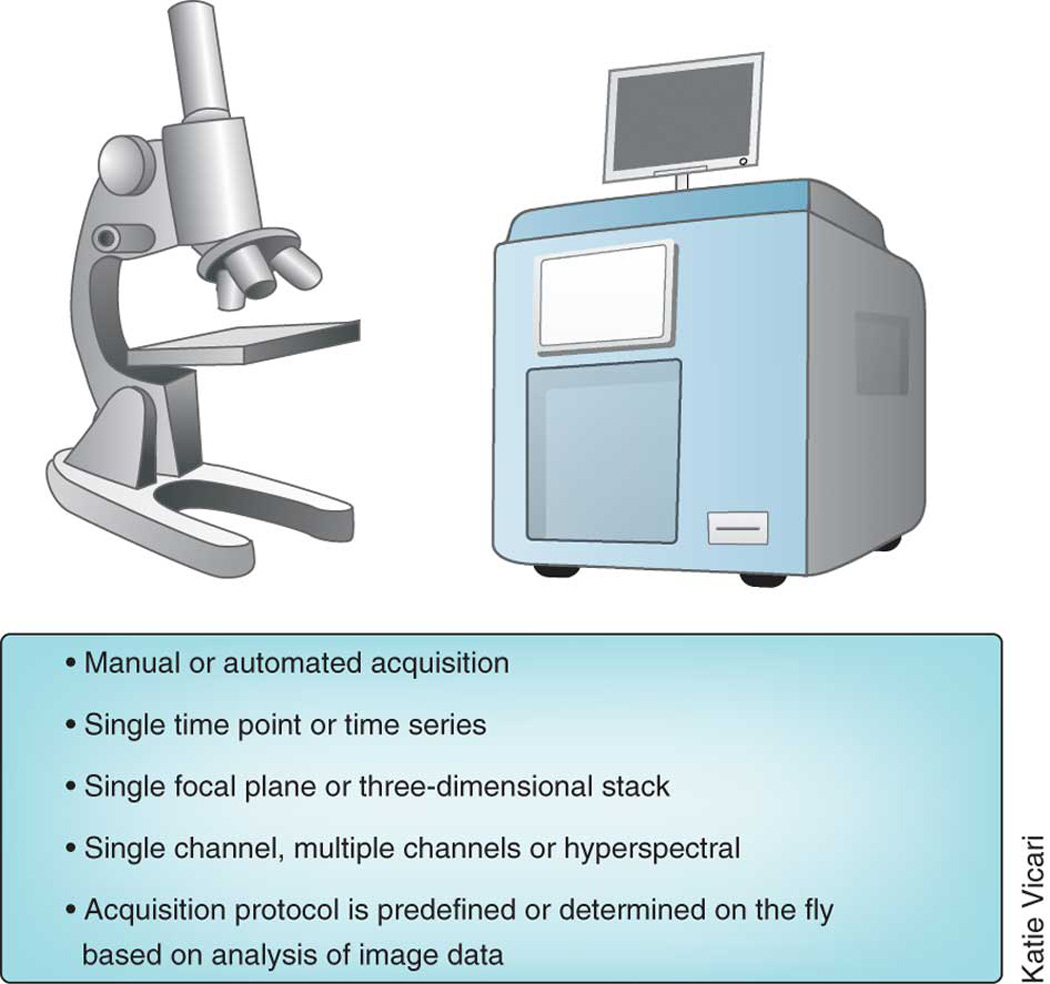
Legend: Image acquisition spans a range of complexity and variation.
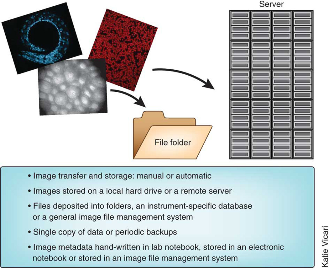
Legend: Options for image storage span a range of complexity and variation.
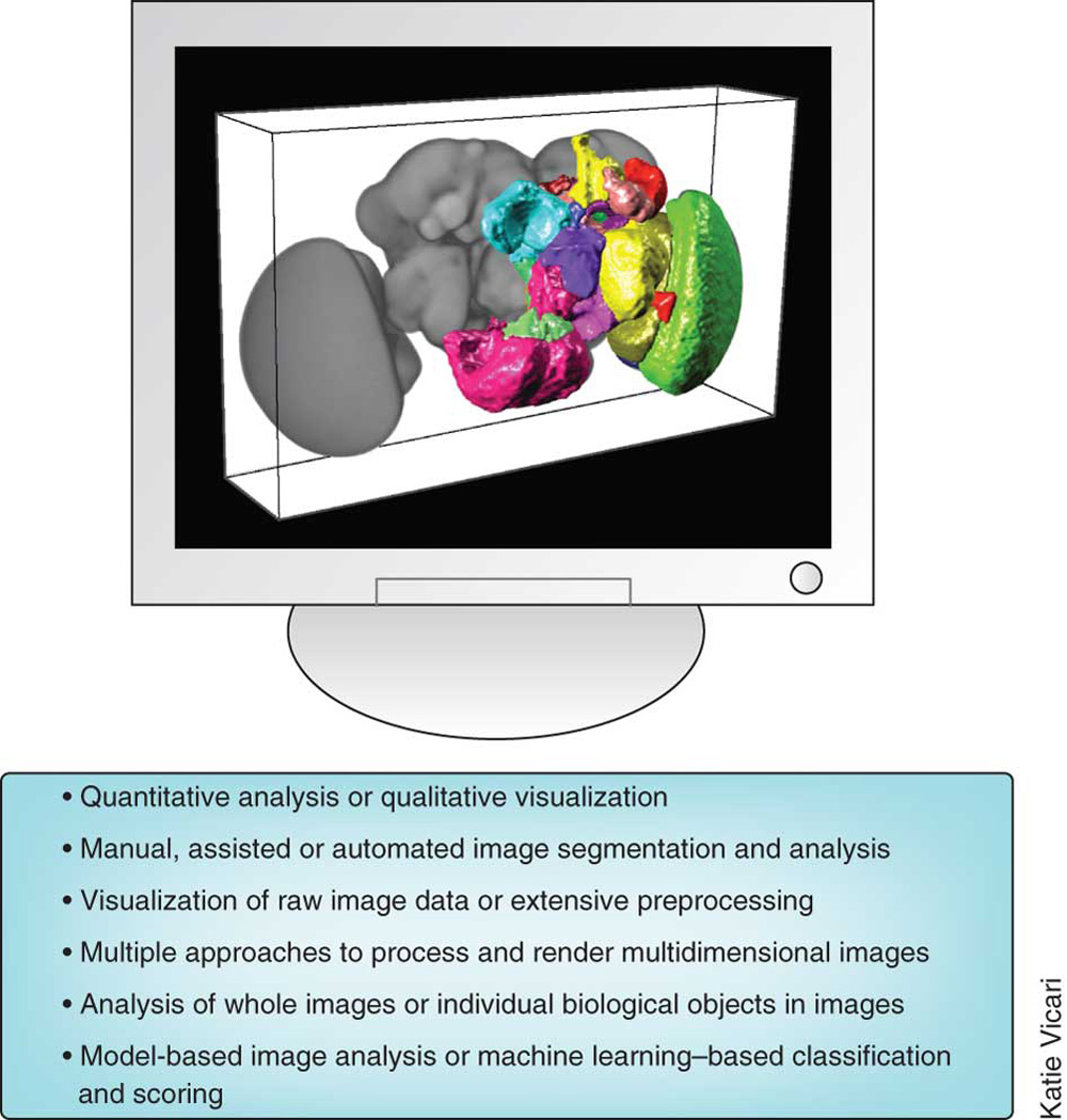
Legend: Image analysis and visualization span a range of complexity and variation.
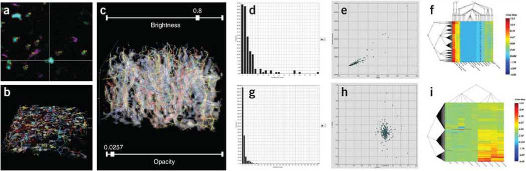
Actual screen view illustrating the series of image analysis steps starting from a multi-channel multi-photon time-lapse movie culminating in a bio-informatic profiling of the extracted spatio-temporal data, using the FARSIGHT toolkit. This movie (Courtesy Dr. Ellen Robey, UC Berkeley) recorded the three-dimensional (3-D) movements of thymocytes in an ex-vivo preparation of a live developing mouse thymus at two-minute intervals, with wild-type thymocytes displayed in cyan, F5 thymocytes in green, and dendritic cells in violet. (a) The first step is cell segmentation, shown as an orthogonal (x, y, z, t) view. Cells are delineated, and identified with numbers that correspond to rows of a table of cell measurements (not shown). The cell tracking results are displayed in multiple ways in panels B and C. (b) "Beads on strings" view showing the 3-D movement paths of cells for detecting anomalies. (c) "3-D kymograph view" showing the same movement paths overlaid on a spatio-temporal (x, y, t) projection for convenience of assessing cell tracking accuracy. (d) Histogram of cell morphological measurements (size). (e) Scatter plots provide a visual cytometric summary of pairs of measurements. (f) Coifman bi-cluster plots organize the cell data into groups based on the cytometric data. (g) Histogram of cell tracking measurements (track tortuosity).(h) Scatter plot view of pairs of cell-track measurements. (i) Coifman bi-cluster plot organizing the cell tracks into groups based on the track-based measurements. The bi-cluster modules are courtesy of Drs. Ronald Coifman (Yale University), and Lawrence Carin (Duke University).
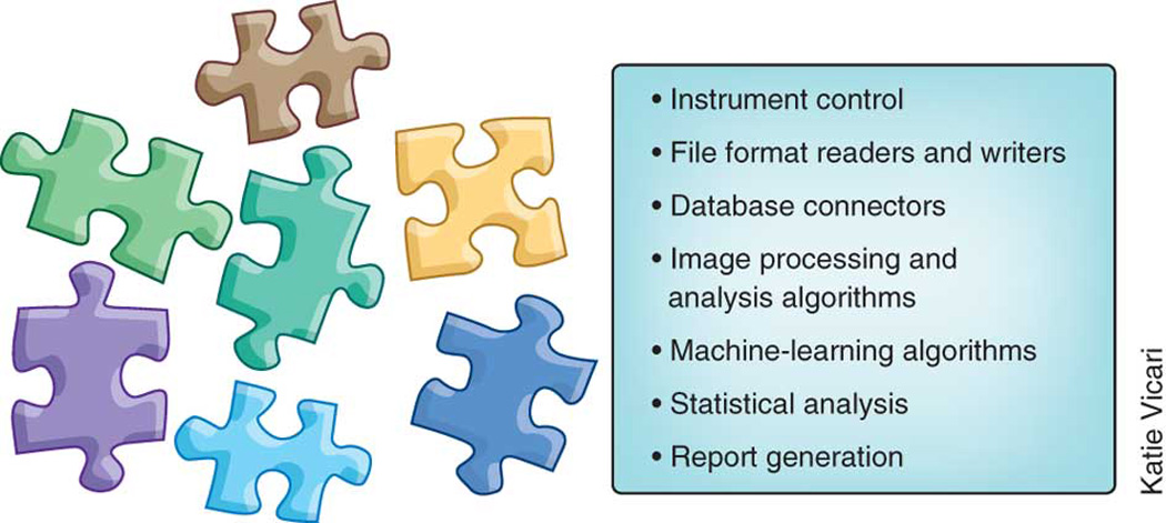
Legend: Bioimaging libraries and toolkits are available to cover a range of functionalities.
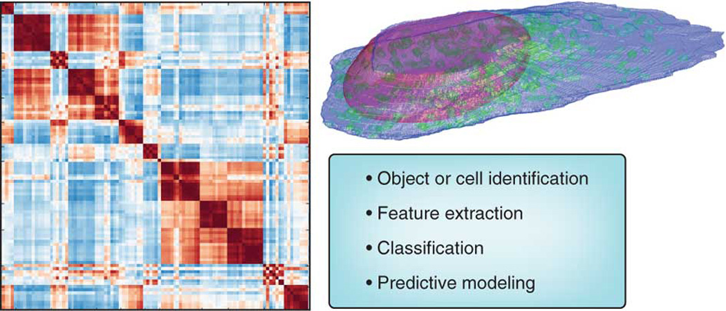
Legend: Application areas where machine learning is used in bioimaging.
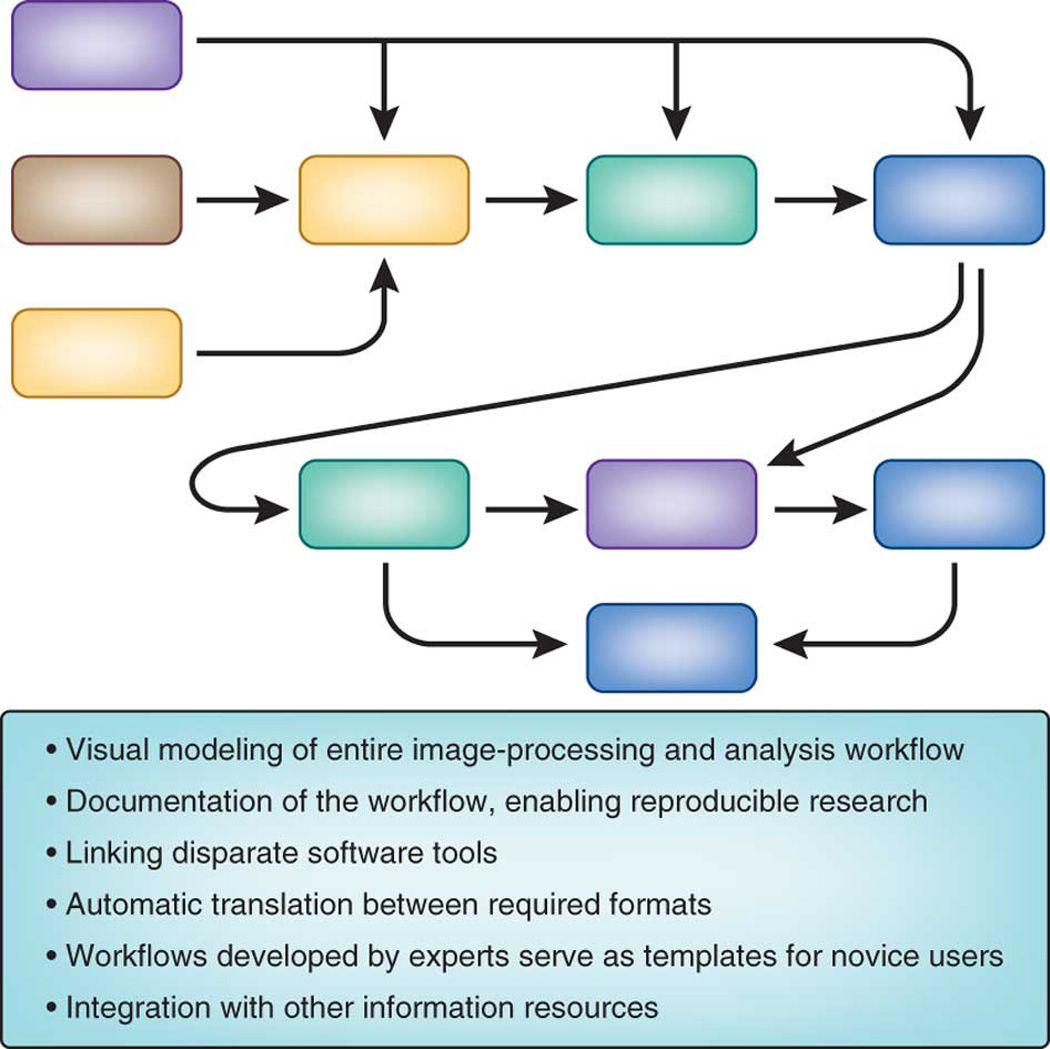
Legend: Benefits of using a workflow system.
References
Publication types
MeSH terms
Grants and funding
LinkOut - more resources
Full Text Sources
Other Literature Sources
