. 2012 Jun 28;7(7):1397-409.
doi: 10.1038/nprot.2012.067.

Protein misfolding cyclic amplification of infectious prions

Affiliations

Protein misfolding cyclic amplification of infectious prions

Rodrigo Morales et al. Nat Protoc. .

Abstract

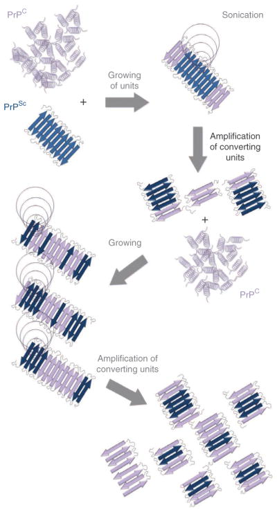
Prions are proteinaceous infectious agents responsible for the transmission of prion diseases. The lack of a procedure for cultivating prions in the laboratory has been a major limitation to the study of the unorthodox nature of this infectious agent and the molecular mechanism by which the normal prion protein (PrP(C)) is converted into the abnormal isoform (PrP(Sc)). Protein misfolding cyclic amplification (PMCA), described in detail in this protocol, is a simple, fast and efficient methodology to mimic prion replication in the test tube. PMCA involves incubating materials containing minute amounts of infectious prions with an excess of PrP(C) and boosting the conversion by cycles of sonication to fragment the converting units, thereby leading to accelerated prion replication. PMCA is able to detect the equivalent of a single molecule of infectious PrP(Sc) and propagate prions that maintain high infectivity, strain properties and species specificity. A single PMCA assay takes little more than 3 d to replicate a large amount of prions, which could take years in an in vivo situation. Since its invention 10 years ago, PMCA has helped to answer fundamental questions about this intriguing infectious agent and has been broadly applied in research areas that include the food industry, blood bank safety and human and veterinary disease diagnosis.


Conflict of interest statement

COMPETING FINANCIAL INTERESTS

The authors declare competing financial interests: details are available in the online version of the paper.

Figures

Figure 1
Figure 1
Schematic diagram for protein misfolding cyclic amplification. PMCA is based on the assumption that prion replication occurs by a seeding/nucleation model, in which PrPSc seeds bind and convert PrPC by incorporating the protein into the polymer. In PMCA, PrPSc and PrPC are mixed and incubated, allowing the misfolding of PrPC, which permits the incorporation and growth of PrPSc aggregates. After incubation, samples are submitted to sonication in order to fragment PrPSc polymers, thereby generating new free ends suitable for continued prion replication. This process is cyclically repeated in order to produce an exponential amplification of PrPC conversion.
Figure 2
Figure 2
PMCA flowchart. A conventional PMCA assay is usually composed of four main stages: substrate preparation, inoculum preparation, PMCA procedure and visualization of the extent of prion formation. After mixing PMCA substrate and inoculum, samples are submitted to incubation/sonication cycles that will allow an exponential generation of infectious prions. After the PMCA reaction is completed, samples are digested with PK and visualized by western blotting. Modifications to this technique have been used to replicate prions from different strains/species. PMCA has been adapted for many different applications, including PrPSc detection in body fluids and tissues and interspecies prion replication. PROCEDURE Steps are indicated in purple boxes with the step number(s) in parentheses. Critical steps are labeled in blue and Troubleshooting steps in green.
Figure 3
Figure 3
Schematic representation of the serial PMCA (sPMCA) assay. In a typical sPMCA assay, the resulting product of a PMCA round is used as an inoculum in a new PMCA reaction (usually by diluting the PMCA product tenfold in fresh brain homogenate substrate). (a,b) By using this procedure, it is possible to increase the detection levels in order to detect up to a single infectious prion. In the left side of the figure, schematic representations of the dilution setup is shown both for the experimental samples (a) and negative controls (b). (a) Western blots show how the detection of PrP27–30 increases among PMCA rounds (right), detecting the equivalent of a 10−10 263K brain dilution in a second PMCA round. Numbers on top of each lane correspond to the dilution of the brain homogenate used to trigger amplification. Gaps in PrP27–30 detection as shown for the second PMCA round (10−9 dilution) are infrequently seen and are usually normalized in the following PMCA rounds. (b) Negative controls consisting only of NBH and submitted to the same procedures do not show any PrPSc signal if all proper precautions are followed (for details, please refer to the TROUBLESHOOTING section and references). In this panel, the numbers on top of the gel refer to multiple replicates. Results showed in this figure are obtained from PMCA assays performed as explained in the protocol using four silica beads per tube. MW, molecular weight ladder.
Figure 4
Figure 4
Distinguishing complete and incomplete proteolytic digestion of PrPSc. A frequent problem associated with the detection of PrPSc after PMCA amplification is ensuring that the product being detected is bona fide PrPSc and not some kind of amorphous aggregate that prevents access to the protease used to eliminate unconverted PrPC. Fortunately, gel electrophoresis and western blotting enable these possibilities to be discriminated. Incomplete digestion (lane 1) leads to a signal with the molecular weight similar to PrPC not treated with PK (lane 4), whereas the typical signal for PrP27–30 has a clearly lower molecular weight (around 29 kDa; lane 3). Lanes 1–3 show the results of experiments containing a similar amount of hamster PrPSc, in which PK digestion did not digest (lane 1), digested partially (lane 2) and digested completely (lane 3) the N-terminal fragment of the protein. To depict the migration of full-length PrPC, lane 4 corresponds to a hamster NBH not treated with PK.

References

    1. Prusiner SB. Prions. Proc Natl Acad Sci USA. 1998;95:13363–13383. - PMC - PubMed
    1. Collinge J. Prion diseases of humans and animals: their causes and molecular basis. Annu Rev Neurosci. 2001;24:519–550. - PubMed
    1. Collee JG, Bradley R. BSE: a decade on—Part I. Lancet. 1997;349:636–641. - PubMed
    1. Collee JG, Bradley R. BSE: a decade on—Part 2. Lancet. 1997;349:715–721. - PubMed
    1. Llewelyn CA, et al. Possible transmission of variant Creutzfeldt-Jakob disease by blood transfusion. Lancet. 2004;363:417–421. - PubMed

Publication types