

Evaluation of CT angiography for visualisation of the lenticulostriate artery: difference between normotensive and hypertensive patients
- PMID: 22744324
- PMCID: PMC3500797
- DOI: 10.1259/bjr/67294268
Evaluation of CT angiography for visualisation of the lenticulostriate artery: difference between normotensive and hypertensive patients
Abstract
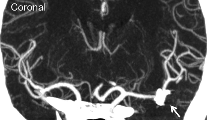
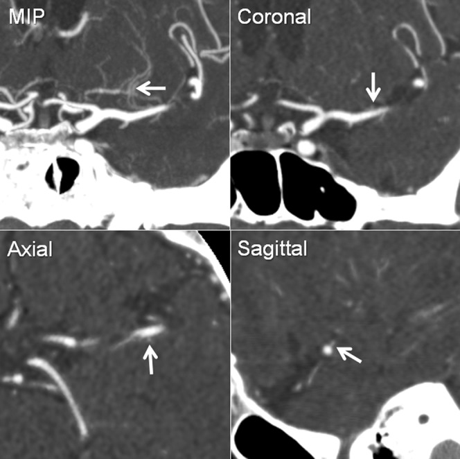
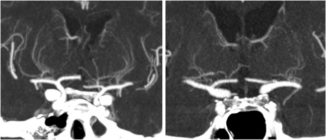
Objective: High-resolution CT angiography (CTA) is currently available using multidetector row CT (MDCT); however, its use for small artery visualisation has been limited. To evaluate its capability, we investigated CTA visualisation for difference in number of the lenticulostriate artery (LSA) branches between normotensive and hypertensive patients, because hypertension is a major cause of LSA damage.
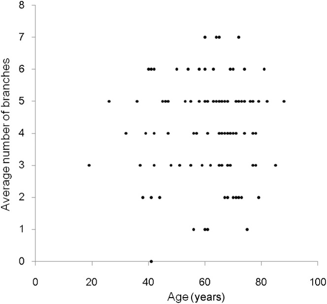
Methods: This was a retrospective study evaluating cerebrovascular CTA at our hospital conducted from February 2008 to June 2009 under approval of the institutional review board. 117 patients (39 males and 78 females, 19-88 years old) were included. CTA was conducted using a 64 channel MDCT. Total numbers of LSA branches were examined for differences by age with regression analysis and the presence or absence of hypertension and/or aneurysm using two-sample t-tests. A p-value <0.016 was considered statistically significant after correction for multiple comparisons. A multiple variable analysis of three factors was also conducted.
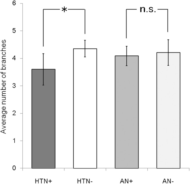
Results: The average number of LSA branches was 3.6 [95% confidence interval (CI) 3.0-4.1] and 4.4 (95% CI 4.1-4.7), respectively, for a patient with and without history of hypertension, and the difference was statistically significant (p=0.013). The difference was approximately one branch in the multiple variable analysis. No significant correlation was observed for age and no significant difference was observed for the presence or absence of aneurysms.
Conclusions: Contrast-enhanced CTA can visualise significant differences in the number of LSA branches among patients with and without hypertension. Advances in knowledge Current high-resolution CTA can visualise LSA well, which enables finding a difference in the LSA between normotensive subjects and hypertensive patients.
Figures
References
-
- Marinkovic SV, Gibo H. The surgical anatomy of the perforating branches of the basilar artery. Neurosurgery 1993;33:80–7 - PubMed
-
- Greenberg SM. Small vessels, big problems. N Engl J Med 2006;354:1451–3 - PubMed
-
- Wardlaw JM, Dennis MS, Warlow CP, Sandercock PA. Imaging appearance of the symptomatic perforating artery in patients with lacunar infarction: occlusion or other vascular pathology? Ann Neurol 2001;50:208–15 - PubMed
-
- Feekes JA, Cassell MD. The vascular supply of the functional compartments of the human striatum. Brain 2006;129:2189–201 - PubMed
-
- Caplan LR. Intracranial branch atheromatous disease: a neglected, understudied, and underused concept. Neurology 1989;39:1246–50 - PubMed
