. 2012 Sep;26(9):1157-72.
doi: 10.1038/eye.2012.126. Epub 2012 Jun 29.

Progress in the management of patients with uveal melanoma. The 2012 Ashton Lecture

Affiliations

Progress in the management of patients with uveal melanoma. The 2012 Ashton Lecture

B Damato. Eye (Lond). 2012 Sep.

Abstract

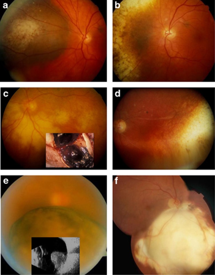
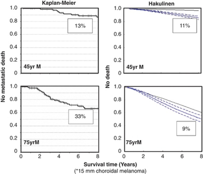
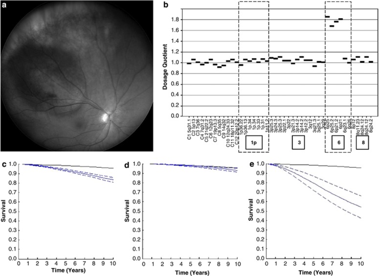
Uveal melanomas are diverse in their clinical features and behaviour. More than 90% involve the choroid, the remainder being confined to the ciliary body and iris. Most patients experience visual loss and more than a third require enucleation, in some cases because of pain. Diagnosis is based on slit-lamp biomicroscopy and/or ophthalmoscopy, with ultrasonography, autofluorescence photography, and/or biopsy in selected cases. Conservation of the eye with useful vision has improved with advances in brachytherapy, proton beam radiotherapy, endoresection, exoresection, transpupillary thermotherapy, and photodynamic therapy. Despite ocular treatment, almost 50% of patients develop metastatic disease, which occurs almost exclusively in patients whose tumour shows chromosome 3 loss and/or class 2 gene expression profile. When the tumour shows such lethal genetic changes, the survival time depends on the anatomical stage and the histological grade of malignancy. Prognostication has improved as a result of progress in multivariate analysis including all the major risk factors. Screening for metastases is more sensitive as a consequence of advances in liver scanning with magnetic resonance imaging and other methods. More patients with metastases are living longer, benefiting from therapies such as: partial hepatectomy; radiofrequency ablation; ipilumumab immunotherapy; selective internal radiotherapy; intra-hepatic chemotherapy, possibly with isolated liver perfusion; and systemic chemotherapy. There is scope for improvement in the detection of uveal melanoma so as to maximise any opportunities for conserving the eye and vision, as well as preventing metastatic spread. Patient management has been enhanced by the formation of multidisciplinary teams in specialised ocular oncology centres.


Figures

Figure 1
Figure 1
(a) Choroidal melanoma in the left eye of a 40-year-old woman treated with an eccentrically positioned, ruthenium plaque in 1986; (b) Fundus appearance 3 years later. Ten years post-operatively, the visual acuity was 6/6 and 26 years after treatment the patient was still alive. (c) Exudative retinopathy after ruthenium plaque radiotherapy of a ciliary body melanoma in the left eye of a 51-year-old man with diabetes. The ‘toxic tumour' was excised in 1989. (d) Fundus appearances 3 years later, showing resolution of the exudation. (e) Inferior, juxtapapillary, choroidal melanoma with a basal diameter of 10.4 mm and a thickness of 9.6 mm, in the right eye of a 65-year-old man. The patient was treated by endoresection in 1999. (f) Post-operative fundus appearance. Thirteen years after surgery, the visual acuity with the operated eye was 6/5. All procedures performed by the author.
Figure 2
Figure 2
Risk of metastatic death in 45-year-old and 75-year-old men having a 15-mm choroidal melanoma, estimated by the Kaplan–Meier analysis and using the Hakulinen method. According to the Kaplan–Meier analysis, the risk of metastatic death increases with age from 13 to 33% whereas with the Hakulinen method these risks diminish from 11% in young males to 9% in older men. Adapted from Damato et al.
Figure 3
Figure 3
(a) Right eye of a 51-year-old woman with a 14.1-mm diameter choroidal melanoma having a thickness of 2.6 mm. The patient was treated with proton beam radiotherapy and TTT with a good ocular result. (b) MLPA showed the melanoma to be of disomy 3 type, with gains in chromosome 6p and no gains in 8q. (c) According to LUMPO analysis based on clinical features alone, the 10-year risk of metastatic death would have been 13%. (d) Biopsy showed no chromosome 3 loss, indicating this risk of metastatic death to be only 3%. (e) If the patient had undergone local resection or enucleation and if the tumour had been found to be of monosomy 3 type with epithlioid cells, closed loops and a high-mitotic rate, the 10-year risk of metastatic death would have been as high as 42%.

References

    1. Damato BE, Coupland SE. Differences in uveal melanomas between men and women from the British Isles. Eye. 2012;26:292–299. - PMC - PubMed
    1. Damato EM, Damato BE.Detection and time to treatment of uveal melanoma in the United Kingdom: an evaluation of 2384 patients Ophthalmology 2012. e-pub ahead of print 11 April 2012doi:10.1016/j.ophtha.2012.01.048 - DOI - PubMed
    1. Damato BE, Foulds WS. Tumour-associated retinal pigment epitheliopathy. Eye. 1990;4:382–387. - PubMed
    1. Coupland SE, Campbell I, Damato B. Routes of extraocular extension of uveal melanoma: risk factors and influence on survival probability. Ophthalmology. 2008;115:1778–1785. - PubMed
    1. Eskelin S, Pyrhönen S, Hahka-Kemppinen M, Tuomaala S, Kivelä T. A prognostic model and staging for metastatic uveal melanoma. Cancer. 2003;97:465–475. - PubMed