. 2012 Aug 24;287(35):29599-609.
doi: 10.1074/jbc.M112.379313. Epub 2012 Jun 28.

The Drosophila neurally altered carbohydrate mutant has a defective Golgi GDP-fucose transporter

Affiliations

The Drosophila neurally altered carbohydrate mutant has a defective Golgi GDP-fucose transporter

Christoph Geisler et al. J Biol Chem. .

Abstract

Studying genetic disorders in model organisms can provide insights into heritable human diseases. The Drosophila neurally altered carbohydrate (nac) mutant is deficient for neural expression of the HRP epitope, which consists of N-glycans with core α1,3-linked fucose residues. Here, we show that a conserved serine residue in the Golgi GDP-fucose transporter (GFR) is substituted by leucine in nac(1) flies, which abolishes GDP-fucose transport in vivo and in vitro. This loss of function is due to a biochemical defect, not to destabilization or mistargeting of the mutant GFR protein. Mass spectrometry and HPLC analysis showed that nac(1) mutants lack not only core α1,3-linked, but also core α1,6-linked fucose residues on their N-glycans. Thus, the nac(1) Gfr mutation produces a previously unrecognized general defect in N-glycan core fucosylation. Transgenic expression of a wild-type Gfr gene restored the HRP epitope in neural tissues, directly demonstrating that the Gfr mutation is solely responsible for the neural HRP epitope deficiency in the nac(1) mutant. These results validate the Drosophila nac(1) mutant as a model for the human congenital disorder of glycosylation, CDG-IIc (also known as LAD-II), which is also the result of a GFR deficiency.


Figures
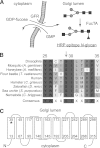
FIGURE 1.
FIGURE 1.
Production of the HRP epitope and the Drosophila nac1 GFR mutation. A, GDP-fucose is produced in the cytoplasm and transported into the Golgi lumen by the GFR transporter in exchange for GMP. FucTA uses GDP-fucose in the Golgi lumen to produce the HRP epitope consisting of core α1,3-fucosylated N-glycans. Squares, N-acetylglucosamine; circles, mannose; triangles, fucose. B, amino acid sequence comparison of known and predicted GDP-fucose transporters. The arrow indicates the conserved serine residue that is mutated to a leucine in Drosophila nac1 GFR. Amino acid residue numbering is according to the Drosophila gene product. GenBankTM accession numbers are as follows: Drosophila, NP_649782.1; mosquito, XP_312562.2; honeybee, XP_623632.1; flour beetle, XP_967192.1; human, NP_001138737.1; Chinese hamster, BAE16173.1; zebrafish: NP_001008590.1; sea urchin, XP_798515.1; nematode, XP_002637574.1. C, Ser-29 is located in the middle of the first predicted transmembrane domain of Drosophila GFR.
FIGURE 2.
FIGURE 2.
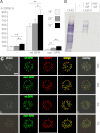
WT, but not nac1 GFR can transport GDP-fucose in vitro and in vivo, and both are Golgi-localized. A, [3H]GDP-fucose import activity of Golgi-enriched microsomes from Sf9 cells infected with baculovirus vectors encoding WT GFR, nac1 GFR, or no exogenous transporter (−) at 18, 25, or 32 °C. Background import at 18 °C (1.5 fmol of GDP-fucose/μg of total protein/min) is set at 100% (−). Error bars, 95% confidence interval. p values for different samples at the same temperatures were all <0.01. *, p < 0.05; **, p < 0.01. B, A. aurantia lectin (AAL) blot of CHO cells or CDG-IIc (LAD-II) cells transfected with expression plasmids encoding WT GFR, nac1 GFR, or nothing (−). C, subcellular distribution of WT and nac1 GFR in Sf9 and Drosophila S2 cells. Columns, phase-contrast, GFP-tagged GFR, RFP-tagged MGAT1 (insect Golgi marker), GFP and RFP merge, and overlay. Scale bar, 10 μm.
FIGURE 3.
FIGURE 3.
Core di-, α1,3-, and α1,6-fucosylated N-glycan levels are strongly reduced in nac1 flies. Peptide-N-glycosidase A-released N-glycans from WT Canton S (A, C, E, and F) and nac1 adults (B, D, E, and G) were subjected to analysis by ESI-MS (A–D), RP-HPLC (E), and MALDI-TOF MS (F and G). F, fucose; G, glucose; M, mannose; N, N-acetylhexosamine. Red triangles, fucose; green circles, mannose; blue squares, N-acetylglucosamine. The late elution time of M3N2F (E) indicates that it is a core α1,6-fucosylated glycan, and its reduced relative intensity in nac1 flies is shown by all three methods, MALDI-TOF MS analysis (not shown) of individual RP-HPLC fractions indicated trace levels of difucosylated glycans co-eluting with M3N2 in nac1 flies. The exploded views of the ESI-MS spectra (C and D; m/z 1265–1335) are set to the same ion count (y axis; 1.5 × 10⁵) and show the almost complete absence in nac1 flies of the difucosylated HRP epitope MMF3F6 glycan in its [M + H]+ and [M + Na]+ forms.
FIGURE 4.
FIGURE 4.
Reduced HRP epitope expression in nac1 homozygous embryos is rescued by transgenic expression of WT Gfr. A, C, and E, lateral view. B, D, and F, ventral view. All embryos are late stage 12 to early stage 13. In nac1/nac1 embryos (A and B), HRP epitope expression is reduced in comparison with WT embryos (E and F). A WT Gfr transgene driven by the neuron-specific elav promoter rescues neural HRP epitope expression (C and D). Scale bar, 70 μm.
FIGURE 5.
FIGURE 5.
Drosophila GFR Ser-29 is conserved in other GDP-sugar transporters. Shown is an alignment of Drosophila GFR with other characterized GDP-sugar transporters from Leishmania donovani (GDP-SugarT) (64), Saccharomyces cerevisiae (yeast GDP-ManT) (59), Pichia pastoris (Pichia GDP-ManT) (57), Candida albicans (C. albicans GDP-ManT) (62), Candida glabrata (C. glabrata GDP-ManT) (61), Cryptococcus neoformans (Cryptococcus GDP-ManT-1 and -2) (58), Aspergillus nidulans (Aspergillus GMT-1 and -2) (60), Volvox carteri (Volvox GDP-ManT) (67), and Arabidopsis thaliana (Arabidopsis GONST-1 and -2) (65, 66) and similar NSTs of Caenorhabditis elegans (nematode SQV-7) (71), Drosophila melanogaster (Drosophila FRC) (69), and humans (human FRC1 and hUGTrel7) (68, 70).

References

    1. Freeze H. H., Eklund E. A., Ng B. G., Patterson M. C. (2012) Neurology of inherited glycosylation disorders. Lancet Neurol. 11, 453–466 - PMC - PubMed
    1. Morava E., Lefeber D. J., Wevers R. A. (2011) Protein glysosylation and congenital disorders of glycosylation. in Post-translational Modifications in Health and Disease (Vidal C. J., ed) pp. 97–117, Springer, New York
    1. Jaeken J. (2010) Congenital disorders of glycosylation. Ann. N.Y. Acad. Sci. 1214, 190–198 - PubMed
    1. Fukuda M. N., Papayannopoulou T., Gordon-Smith E. C., Rochant H., Testa U. (1984) Defect in glycosylation of erythrocyte membrane proteins in congenital dyserythropoietic anemia type II (HEMPAS). Br. J. Haematol. 56, 55–68 - PubMed
    1. Jaeken J., van Eijk H. G., van der Heul C., Corbeel L., Eeckels R., Eggermont E. (1984) Sialic acid-deficient serum and cerebrospinal fluid transferrin in a newly recognized genetic syndrome. Clin. Chim. Acta 144, 245–247 - PubMed

Publication types