. 2012 Aug 17;287(34):28646-55.
doi: 10.1074/jbc.M112.362608. Epub 2012 Jun 28.

TRIM32 protein modulates type I interferon induction and cellular antiviral response by targeting MITA/STING protein for K63-linked ubiquitination

Affiliations

TRIM32 protein modulates type I interferon induction and cellular antiviral response by targeting MITA/STING protein for K63-linked ubiquitination

Jing Zhang et al. J Biol Chem. .

Abstract

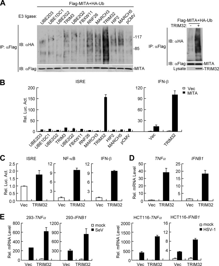
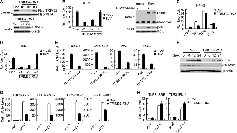
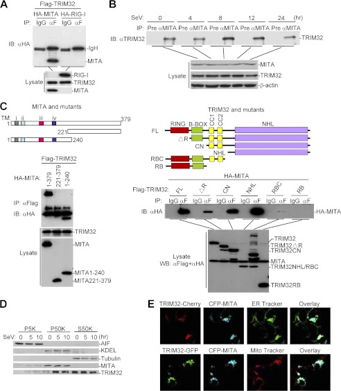
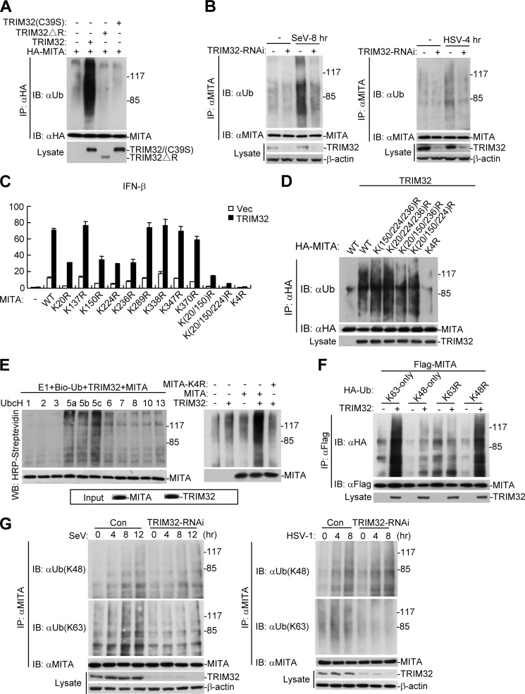
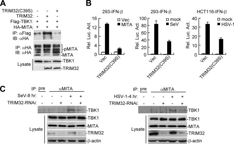
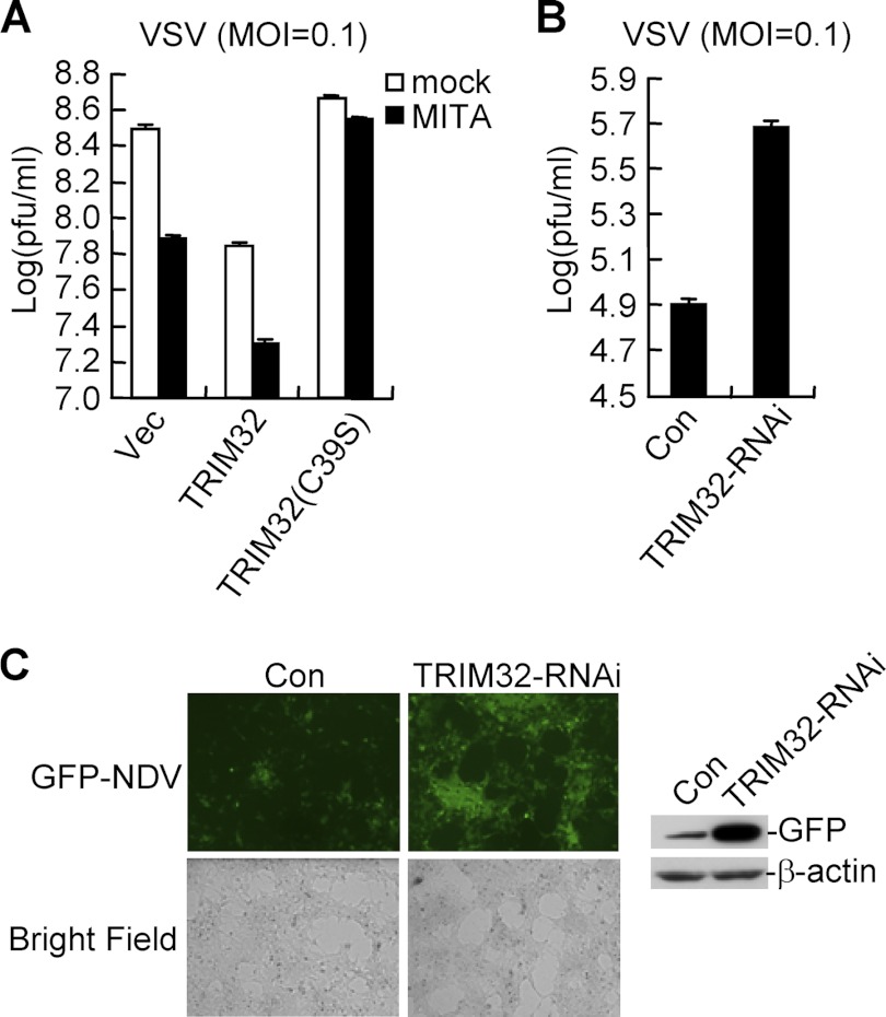
Viral infection activates several transcription factors including NF-κB and IRF3, which collaborate to induce type I interferons (IFNs) and innate antiviral response. MITA (also called STING) is a critical adaptor protein that links virus-sensing receptors to IRF3 activation upon infection by both RNA and DNA pathogens. Here we show that the E3 ubiquitin ligase tripartite motif protein 32 (TRIM32) ubiquitinated MITA and dramatically enhanced MITA-mediated induction of IFN-β. Overexpression of TRIM32 potentiated virus-triggered IFNB1 expression and cellular antiviral response. Consistently, knockdown of TRIM32 had opposite effects. TRIM32 interacted with MITA, and was located at the mitochondria and endoplasmic reticulum. TRIM32 targeted MITA for K63-linked ubiquitination at K20/150/224/236 through its E3 ubiquitin ligase activity, which promoted the interaction of MITA with TBK1. These findings suggest that TRIM32 is an important regulatory protein for innate immunity against both RNA and DNA viruses by targeting MITA for K63-linked ubiquitination and downstream activation.


Figures

FIGURE 1.
FIGURE 1.
TRIM32 plays a role in virus-triggered induction of IFN-β and TNFα. A, screens for E3 ubiquitin ligases that cause MITA ubiquitination. Left panels: the 293 cells (4 × 10⁵) were transfected with Flag-MITA (2 μg) along with HA-ubiquitin (0.2 μg) and the indicated E3 ligases (0.2 μg each). Cell lysates were immunoprecipitated with anti-Flag and the immunoprecipitates were analyzed by immunoblots with anti-HA or anti-Flag. Right panels: the 293 cells were transfected with the indicated plasmids. Cell lysates were denatured by adding 1% SDS and heating for 10 min. The denatured lysates were diluted for immunoprecipitation and immunoblot analysis as described above. B, effects of the indicated E3 ligases on MITA-mediated activation of ISRE and the IFN-β promoter. The 293 cells (1 × 10⁵) were transfected with MITA (0.1 μg) and the indicated E3 ligases (0.1 μg) along with ISRE or the IFN-β promoter reporter plasmid (0.1 μg). Luciferase assays were performed 22 h after transfection. Graphs show mean ± S.D., n = 3. C, overexpression of TRIM32 results in activation of NF-κB and the IFN-β promoter. The 293 cells (1 × 10⁵) were transfected with the indicated reporter (0.1 μg) and TRIM32 (0.1 μg) plasmids. Luciferase assays were performed 22 h after transfection. Graphs show mean ± S.D., n = 3. D, overexpression of TRIM32 leads to transcription of TNFα and IFNB1 genes. The 293 cells (2 × 10⁵) were transfected with the indicated expression plasmids (0.1 μg each). Real-time PCR was performed 20 h after transfection. E, TRIM32 potentiates SeV- and HSV-1-induced transcription of TNFα and IFNB1 genes. For the left histographs, the 293 cells (2 × 10⁵) were transfected with the indicated expression plasmids (0.1 μg each). 18 h after transfection, cells were left untreated or infected with SeV for 12 h before real-time PCR were performed. For the right histograph, HCT116 cells (2 × 10⁵) were transfected with the indicated expression plasmids (0.1 μg each). 18 h after transfection, cells were left untreated or infected with HSV-1 for 24 h before real-time PCR was performed.
FIGURE 2.
FIGURE 2.
Effects of TRIM32 knockdown on virus-triggered induction of downstream genes. A, effects of TRIM32-RNAi plasmids on the expression of transfected and endogenous TRIM32. In the upper panels, the 293 cells (2 × 10⁵) were transfected with expression plasmids for Flag-TRIM32 and Flag-MITA (0.1 μg each), and the indicated RNAi plasmids (1 μg each). At 24 h after transfection, cell lysates were analyzed by immunoblot with anti-Flag. In the lower panels, the 293 cells (1 × 10⁷) were transfected with control or the indicated TRIM32-RNAi plasmids (8 μg each) for 24 h. Cell lysates were immunoprecipitated with anti-TRIM32, and the immunoprecipitates were analyzed by immunoblots with anti-TRIM32. The lysates were also analyzed by immunoblot with anti-β-actin as control. B, effects of TRIM32-RNAi on SeV-induced ISRE activation in 293 cells. In the left panel, the 293 cells (1 × 10⁵) were transfected with the indicated RNAi plasmids (0.5 μg each) and ISRE reporter plasmid (0.1 μg). Twenty-four hours after transfection, cells were left untreated or infected with SeV for 12 h before luciferase assays were performed. Graphs show mean ± S.D., n = 3. In the right panels, the 293 cells (2 × 10⁵) were transfected with the indicated RNAi plasmids (2 μg each). Twelve hours after transfection, cells were selected with puromycin (1 μg/ml) for 24 h, then infected with SeV or left uninfected for 8 h. Cell lysates were separated by native (upper panel) and SDS (bottom two panels) PAGE and analyzed with the indicated antibodies. C, effects of TRIM32-RNAi on SeV-, TNFα-, and IL-1β-induced NF-κB activation in 293 cells. Reporter assays were performed similarly as in B except that TNFα (10 ng/ml) and IL-1β (10 ng/ml) were used as stimuli. D, effects of TRIM32-RNAi on SeV-induced activation of the IFN-β promoter in 293 cells. Reporter assays were similarly performed as in B. E, effects of TRIM32-RNAi on SeV-induced transcription of IFNB1, RANTES, RIG-I, and TNFα genes. 293 cells (2 × 10⁵) were transfected with the indicated RNAi plasmids (2 μg each). 12 h after transfection, cells were selected with puromycin (1 μg/ml) for 24 h, then infected with SeV or left uninfected for 12 h before real-time PCR was performed. F, knockdown of TRIM32 inhibited SeV-induced expression of RIG-I. The 293 cells (2 × 10⁵) were transfected with TRIM32-RNAi plasmid for 12 h, selected with puromycin (1 μg/ml) for 24 h, then infected with SeV or left uninfected for the indicated time points. Cell lysates were analyzed by immunoblots with the indicated antibodies. G, effects of TRIM32-RNAi on HSV-1-induced transcription of downstream genes in THP-1 cells. THP-1 cells were transduced with GFP-RNAi or TRIM32-RNAi by retrovirus-mediated gene transfer. The cells were then either untreated or infected with HSV-1 for 24 h before real-time PCR experiments were performed. H, effects of TRIM32-RNAi on poly(I:C)-induced activation of ISRE and the IFN-β promoter in TLR3 cell lines. TLR3-stable cells (2 × 10⁵) were transfected with TRIM32-RNAi plasmid (0.5 μg each) along with the indicated reporter plasmids (0.1 μg). 24 h after transfection, cells were left untreated or treated with poly(I:C) (20 μg/ml) in the medium for 12 h before luciferase assays were performed. Graphs show mean ± S.D., n = 3.
FIGURE 3.
FIGURE 3.
TRIM32 interacts and colocalizes with MITA. A, TRIM32 interacts with MITA in mammalian overexpression system. 293 cells (1 × 10⁷) were transfected with the indicated plasmids (5 μg each). Coimmunoprecipitation and immunoblot analysis were performed with the indicated antibodies. B, endogenous association between TRIM32 and MITA. The 293 cells (3 × 10⁷) were left uninfected or infected with SeV for the indicated time points before coimmunoprecipitation and immunoblot analysis were performed with the indicated antibodies. C, domain mapping of the interaction between TRIM32 and MITA. Upper panels: a schematic presentation of MITA, TRIM32 and their mutants. Lower panels: the 293 cells (1 × 10⁷) were transfected with the indicated plasmids (5 μg each), followed by coimmunoprecipitation and immunoblot analysis with the indicated antibodies. D, subcellular distribution of TRIM32. The 293 cells (5 × 10⁷) were left untreated or infected with SeV for the indicated times. The cellular fractions were analyzed by immunoblots with the indicated antibodies. E, TRIM32 and MITA colocalize to the mitochondria and ER in 293 cells. The 293 cells were transfected with the indicated plasmids. 20 h after transfection, cells were stained with Mito-Tracker Red or ER-Tracker Green for 30 min. The cells were fixed with 4% paraformaldehyde and subjected for confocal microscopy.
FIGURE 4.
FIGURE 4.
TRIM32 targets MITA for K63-linked ubiquitination at K20/150/224/236. A, overexpression of wild-type but not the mutant TRIM32 promotes ubiquitination of MITA. The 293 cells (1 × 10⁷) were transfected with MITA (5 μg) together with TRIM32 or its mutants (2 μg). 24 h after transfection, coimmunoprecipitation was performed with anti-HA. The immunoprecipitates were denatured and reimmunoprecitated with anti-HA and then analyzed by immunoblot with anti-ubiquitin (upper panel). The expression levels of the proteins were examined by immunoblots with the indicated antibodies (lower panels). B, knockdown of TRIM32 attenuated virus-induced ubiquitination of endogenous MITA. The 293 (left panel) or HeLa (right panel) cells were transduced with GFP-RNAi or TRIM32-RNAi by retrovirus-mediated gene transfer and selected with puromycin to establish stable cell lines. The cells (3 × 10⁷) were then left uninfected or infected with SeV or HSV-1 respectively for the indicated times before coimmunoprecipitation and immunoblot analysis were performed with the indicated antibodies. C, effects of MITA mutation on activation of the IFN-β promoter. The 293 cells (1 × 10⁷) were transfected with TRIM32 and the indicated MITA mutants along with the IFN-β promoter reporter plasmid. Luciferase assays were performed 24 h after transfection. Graphs show mean ± S.D., n = 3. D, TRIM32 targets MITA for ubiquitination at K20/150/224/236. 293 cell (1 × 10⁷) were transfected with TRIM32 (3 μg) and the indicated MITA mutant plasmids (6 μg). Ubiquitination experiments were carried out similarly as in A. E, TRIM32 ubiquitinates MITA but not MITA-K4R in vitro. TRIM32 and MITA were translated in vitro, and biotin-ubiquitin, E1 and the indicated E2s were added for ubiquitination assays. Ubiquitin-conjugated proteins were detected by immunoblot with HRP-streptavidin. The input levels of the translated proteins were detected by immunoblots. F, TRIM32 targetes MITA for K63-linked ubiquitination. The 293 cells (2 × 10⁶) were transfected with MITA (2 μg) along with the indicated plasmids (0.2 μg). 24 h after transfection, immunoprecipitation, and immunoblot analysis were performed with the indicated antibodies. G, knockdown of TRIM32 inhibits virus-induced K63-linked but not K48-linked ubiquitination of endogenous MITA. Experiments were carried out similar to B except that different antibodies were used for the immunoblot.
FIGURE 5.
FIGURE 5.
TRIM32 is required for the interaction between MITA and TBK1. A, effects of TRIM32 and TRIM32(C39S) on interaction between MITA and TBK1. The 293 cells (1 × 10⁷) were transfected with the indicated plasmids for 22 h, and then coimmunoprecipitation and immunoblot analysis were performed with the indicated antibodies. B, effects of TRIM32(C39S) on MITA-, SeV-, and HSV-1-induced activation of the IFN-β promoter. The 293 or HCT116 cells were transfected with the indicated expression and reporter plasmids. The cells were left untreated, or infected with SeV (12 h) or HSV-1 (24 h) before reporter assays were performed. C, effects of knockdown of TRIM32 on the endogenous interaction between MITA and TBK1. The control or TRIM32 knockdown 293 (left panels) or HeLa (right panels) cells were left uninfected with infected with SeV or HSV-1 as indicated. Coimmunoprecipitation and immunoblots were performed with the indicated antibodies.
FIGURE 6.
FIGURE 6.
Roles of TRIM32 in cellular antiviral response. A, effects of TRIM32 and its mutant on VSV replication. The 293 cells (1 × 10⁵) were transfected with the indicated expression plasmids (0.5 μg each). 24 h after transfection, cells were infected with VSV (MOI = 0.1). The supernatants were harvested 24 h after infection for standard plaque assays. Graphs show mean ± S.D., n = 3. B, effects of TRIM32-RNAi on VSV replication. Plaque assays were similarly performed as in A except that a TRIM32-RNAi plasmid (#1) (1 μg) was transfected and cells were infected with VSV (MOI = 0.1) for 16 h. Graphs show mean ± S.D., n = 3. C, effects of TRIM32-RNAi on NDV replication. The 293 cells were transfected with TRIM32-RNAi plasmid (1 μg each). 24 h later, cells were infected with NDV-GFP (MOI = 0.1) for 36 h and imaged by microscopy or analyzed by immunoblots with the indicated antibodies.

References

    1. Takeuchi O., Akira S. (2010) Pattern recognition receptors and inflammation. Cell 140, 805–820 - PubMed
    1. Akira S., Uematsu S., Takeuchi O. (2006) Pathogen recognition and innate immunity. Cell 124, 783–801 - PubMed
    1. Durbin J. E., Fernandez-Sesma A., Lee C. K., Rao T. D., Frey A. B., Moran T. M., Vukmanovic S., García-Sastre A., Levy D. E. (2000) Type I IFN modulates innate and specific antiviral immunity. J. Immunol. 164, 4220–4228 - PubMed
    1. Levy D. E., García-Sastre A. (2001) The virus battles: IFN induction of the antiviral state and mechanisms of viral evasion. Cytokine Growth Factor Rev. 12, 143–156 - PubMed
    1. Levy D. E., Marié I. J. (2004) RIGging an antiviral defense–it's in the CARDs. Nat Immunol 5, 699–701 - PubMed

Publication types

MeSH terms