. 2012 Jul 31;109(31):12794-9.
doi: 10.1073/pnas.1209014109. Epub 2012 Jun 28.

Identification of cytoskeletal elements enclosing the ATP pools that fuel human red blood cell membrane cation pumps

Affiliations

Identification of cytoskeletal elements enclosing the ATP pools that fuel human red blood cell membrane cation pumps

Haiyan Chu et al. Proc Natl Acad Sci U S A. .

Abstract

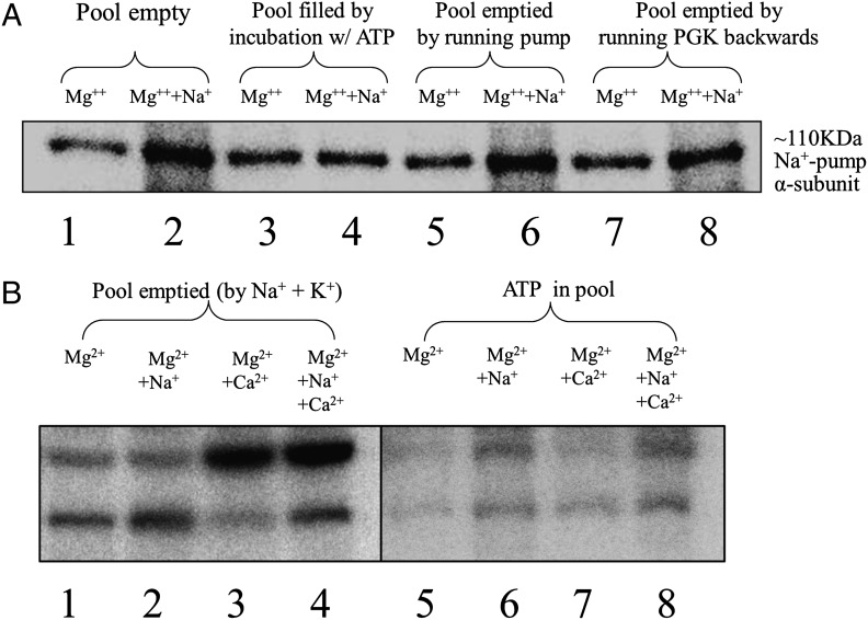
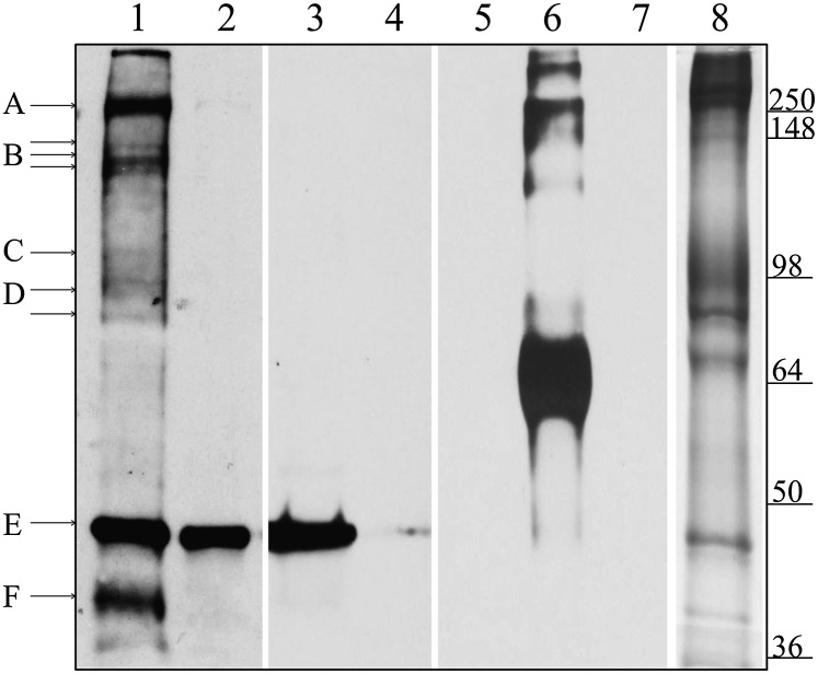
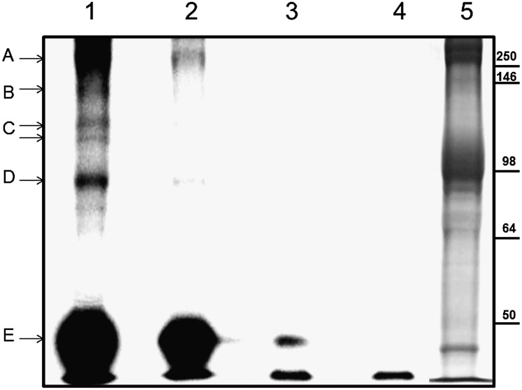
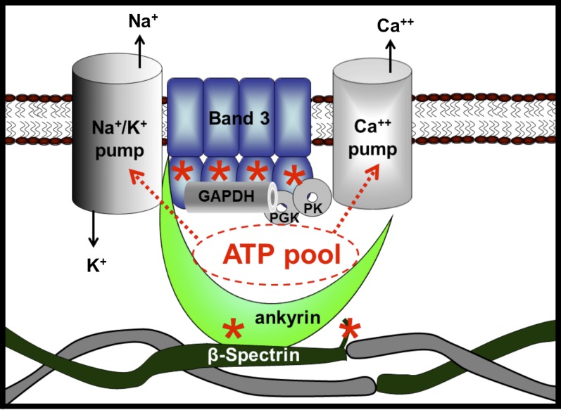
The type of metabolic compartmentalization that occurs in red blood cells differs from the types that exist in most eukaryotic cells, such as intracellular organelles. In red blood cells (ghosts), ATP is sequestered within the cytoskeletal-membrane complex. These pools of ATP are known to directly fuel both the Na(+)/K(+) and Ca(2+) pumps. ATP can be entrapped within these pools either by incubation with bulk ATP or by operation of the phosphoglycerate kinase and pyruvate kinase reactions to enzymatically generate ATP. When the pool is filled with nascent ATP, metabolic labeling of the Na(+)/K(+) or Ca(2+) pump phosphoproteins (E(Na)-P and E(Ca)-P, respectively) from bulk [γ-(32)P]-ATP is prevented until the pool is emptied by various means. Importantly, the pool also can be filled with the fluorescent ATP analog trinitrophenol ATP, as well as with a photoactivatable ATP analog, 8-azido-ATP (N(3)-ATP). Using the fluorescent ATP, we show that ATP accumulates and then disappears from the membrane as the ATP pools are filled and subsequently emptied, respectively. By loading N(3)-ATP into the membrane pool, we demonstrate that membrane proteins that contribute to the pool's architecture can be photolabeled. With the aid of an antibody to N(3)-ATP, we identify these labeled proteins by immunoblotting and characterize their derived peptides by mass spectrometry. These analyses show that the specific peptides that corral the entrapped ATP derive from sequences within β-spectrin, ankyrin, band 3, and GAPDH.


Conflict of interest statement

The authors declare no conflict of interest.

Figures

Fig. 1.
Fig. 1.
Effect of loading the ATP pool in ghosts with nonradioactive ATP on the ability to detect [γ-32P]-labeled ENa-P and ECa-P by phosphoimaging. The ATP pool in ghosts was filled with nonradioactive ATP and then either emptied or left filled, as indicated. After addition of [γ-32P]-ATP and the desired cations, the ENa-P and ECa-P were detected by SDS/PAGE, followed by phosphoimaging. The relative intensities of these bands reflect the ability to label the ENa-P and ECa-P with a 20-s pulse of 2 μM [γ-32P]-ATP. (A) Lanes 1 and 2 show labeling of the EMg-P and ENa-P under conditions with all ATP in the pools removed by previous washing. Lanes 3 and 4 show the same experiment as in lanes 1 and 2, except with the membrane pools filled with unlabeled ATP before the addition of [γ-32P]-ATP. Lanes 5 and 6 and lanes 7 and 8 show the E-P values when the pools of ATP have been depleted either by running the Na+/K+ pump forward with Na+ and K+ (lanes 5 and 6) or by running the PGK reaction backward (lanes 7 and 8). Note that when the ATP pools are filled with unlabeled ATP (lanes 3 and 4), ENa-P and EMg-P are of similar intensity; however, if the ATP pools are emptied, thereby allowing [γ32P]-ATP to label the pumps, then the relative intensity of the band representing the ENa-P is greater than its Mg2+ counterpart. Quantitation ratios of the intensities of the bands for each pair of lanes were 2.9 for lanes 1 and 2, 0.88 for lanes 3 and 4, 2.2 for lanes 5 and 6, and 1.6 for lanes 7 and 8, with an SEM of ±3% for n = 2. (B) These lanes show the same band intensity relationships as in A for both the ENa-P (lower bands) and the ECa-P (upper bands), even when both types of E-Ps are labeled in the same incubation. Note that when the pools are filled with ATP (lanes 5–8), labeling of the E-Ps with [γ-32P]-ATP is inhibited. Moreover, when the ATP pool is emptied by running the Na+/K+ pump forward (lanes 1–4), the calcium pump also can be labeled with [γ-32P]-ATP, indicating that the Na+/K+ and Ca2+ pumps share the same ATP pools. The phosphoimages shown here are representative of multiple experiments.
Fig. 2.
Fig. 2.
Dynamic imaging by confocal microscopy of the loading and emptying of membrane pools of ATP using the fluorescent ATP analog TNP-ATP. ATP pools in porous ghosts were either loaded or loaded and then emptied of TNP-ATP by running the PGK reaction backward. Ghosts were then observed by confocal microscopy. (A) Ghosts with their ATP pools filled with TNP-ATP. Because a large ensemble of ghosts is shown, the plane of focus passes through the center of most ghosts, showing the fluorescent ATP only on the cell periphery where the plane of focus coincides with the membrane. However, in a few ghosts, the plane of focus passes through part of the membrane, revealing pool ATP in the mid region of the cell. The fact that ghosts exhibit only punctate fluorescence in a ring on the cell periphery indicates that the TNP-ATP–loaded pools are localized to the membrane (12). (B) The entrapped TNP-ATP is labile and susceptible to removal by running the PGK reaction backward. (C and D) Controls, with one of the substrates (PGA) needed to run the PGK reaction backward omitted (C) and both substrates (PGA and NADH) omitted (D). In D, the fluorescent images of the loaded ghosts are stable even after continued incubation in the absence of both substrates. These images are similar to ones as studied by Hoffman et al. (12).
Fig. 3.
Fig. 3.
Identification of ghost proteins labeled with pool-associated N3-ATP. Here the ghosts were filled or emptied of N3-ATP, as described in Materials and Methods. After washing, the membranes were photolyzed and separated by SDS/PAGE. Equal amounts of ghost proteins (30 μg) were loaded in all lanes. Lanes 1–7 were transferred to nitrocellulose and immunoblotted with an antibody to N3-ATP, whereas lane 8 was stained with Coomassie blue. Lane 1 shows pools filled with N3-ATP; lanes 2 and 3, pools first filled with N3-ATP and then emptied by either running the Na+ pump forward or running PGK reaction backward, respectively. In lane 4, pools are filled with N3-ATP in the presence of excess unlabeled ATP to block any specific ATP-binding sites. Lanes 5–7 show the specificity of the N3-ATP antibody, demonstrating that it labels only BSA photolyzed with N3-ATP (lane 6), not unlabeled BSA (lane 5) or unlabeled RBC ghosts (lane 7). Lane 8 shows a Coomassie blue stain of ghost proteins after separation by SDS/PAGE. The proteins tentatively identified are indicated with lettered lines. Possible protein candidates for the labeled bands are A, β-spectrin and/or ankyrin; B, Ca2+ pump (Mr ∼140 kDa) or fragments of spectrin/ankyrin; C, α-subunit of the Na+/K+ pump (Mr ∼110 kDa); D, band 3; E, actin; and F, GAPDH.
Fig. 4.
Fig. 4.
Labeling of ATP pool-associated membrane proteins with [α-32P]-N3-ATP. This was accomplished following the same protocol used for N3-ATP. Labeled proteins were separated by SDS/PAGE and analyzed with a Cyclone phosphoimager. Lanes 1–4 show phosphoimages of ghost proteins after separation. Lane 5 is the Coomassie blue-stained counterpart. The proteins tentatively identified are indicated with arrows. Possible protein candidates for the labeled bands are: A, β-spectrin and/or ankyrin; B, Ca2+ pump (Mr ∼140 kDa) or fragments of spectrin/ankyrin; C, α-subunit of the Na+/K+ pump (Mr ∼110 kDa); D, band 3; and E, actin.
Fig. 5.
Fig. 5.
Possible arrangement of membrane components that form the ATP pools that fuel the cation pumps. Membrane components that were photolabeled with N3-ATP and identified by MS are shown in color, and other membrane components implicated in the pool’s architecture but not identified by MS are shown in gray. The locations of the major N3-ATP–labeled peptides in ankyrin (residues 961–974), band 3 (residues 361–378), and β-spectrin (residues 2124–2137) are marked with an asterisk.

References

    1. Alberts B, et al. In: Molecular Biology of the Cell. 4th Ed. Alberts B, et al., editors. New York: Garland Science; 2002. pp. 659–669.
    1. Srere PA. Complexes of sequential metabolic enzymes. Annu Rev Biochem. 1987;56:89–124. - PubMed
    1. Skou JC. The influence of some cations on an adenosine triphosphatase from peripheral nerves. Biochim Biophys Acta. 1957;23:394–401. - PubMed
    1. Schatzmann HJ. ATP-dependent Ca++-extrusion from human red cells. Experientia. 1966;22:364–365. - PubMed
    1. Knauf PA, Proverbio F, Hoffman JF. Electrophoretic separation of different phophosproteins associated with Ca-ATPase and Na, K-ATPase in human red cell ghosts. J Gen Physiol. 1974;63:324–336. - PMC - PubMed

Publication types

MeSH terms