. 2012 Jul 10;109(28):11348-53.
doi: 10.1073/pnas.1201428109. Epub 2012 Jun 27.

Structure of the VirB4 ATPase, alone and bound to the core complex of a type IV secretion system

Affiliations

Structure of the VirB4 ATPase, alone and bound to the core complex of a type IV secretion system

Karin Walldén et al. Proc Natl Acad Sci U S A. .

Abstract

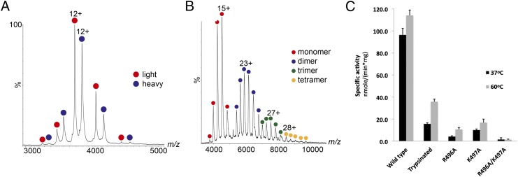
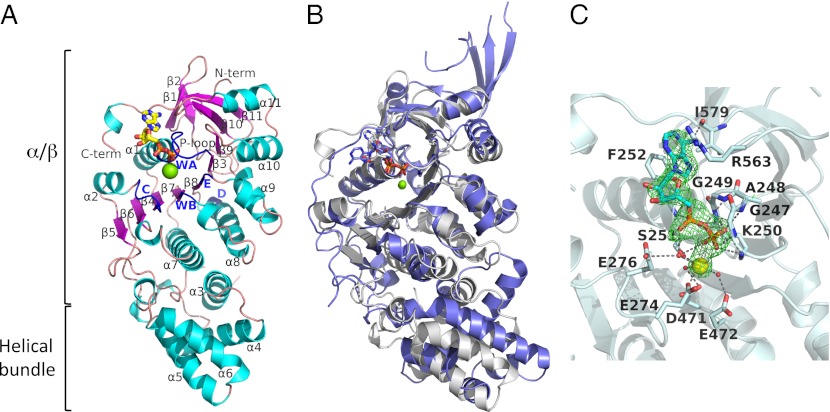
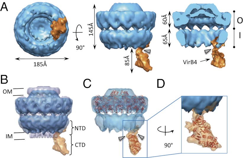
Type IV secretion (T4S) systems mediate the transfer of proteins and DNA across the cell envelope of bacteria. These systems play important roles in bacterial pathogenesis and in horizontal transfer of antibiotic resistance. The VirB4 ATPase of the T4S system is essential for both the assembly of the system and substrate transfer. In this article, we present the crystal structure of the C-terminal domain of Thermoanaerobacter pseudethanolicus VirB4. This structure is strikingly similar to that of another T4S ATPase, VirD4, a protein that shares only 12% sequence identity with VirB4. The VirB4 domain purifies as a monomer, but the full-length protein is observed in a monomer-dimer equilibrium, even in the presence of nucleotides and DNAs. We also report the negative stain electron microscopy structure of the core complex of the T4S system of the Escherichia coli pKM101 plasmid, with VirB4 bound. In this structure, VirB4 is also monomeric and bound through its N-terminal domain to the core's VirB9 protein. Remarkably, VirB4 is observed bound to the side of the complex where it is ideally placed to play its known regulatory role in substrate transfer.


Conflict of interest statement

The authors declare no conflict of interest.

Figures

Fig. 2.
Fig. 2.
Investigation of the oligomeric state of TpsVirB4 and TpsVirB4CTD. Mass spectrometry analysis of TpsVirB4CTD (A) and TpsVirB4 (B). (A) Mass spectrum showing two dominant products of the TpsVirB4CTD protein after limited proteolysis by trypsin. Using electrospray, multiple charged ions are generated for each of the components present in solution. This result is because of the many different protonation sites being available for each protein. The charge states series corresponding to the lighter and heavier TpsVirB4CTD monomers are indicated in red and blue, respectively. (B) Mass spectrum showing the different oligomerization states of the TpsVirB4 protein. Charge state series corresponding to monomer, dimer, trimer, and tetramer of the protein are indicated in red, blue, green, and yellow, respectively. (C) ATPase activities of TpsVirB4 and its motif D mutants at two different temperatures, 37 °C (black) and 60 °C (gray). From left: wild-type TpsVirB4, TpsVirB4CTD, TpsVirB4R496A, TpsVirB4K497A, and TpsVirB4R496AK497A.
Fig. 1.
Fig. 1.
Structure of TpsVirB4CTD. (A) Ribbon representation of the structure of TpsVirB4CTD, covering residues 205–587. Secondary structure elements, β-strands and α-helixes, are marked β1–β11 and α1–11, respectively. Walker A (WA) and B (WB) motifs and motifs C, D, and E are colored in blue. (B) Superposition of TpsVirB4CTD (white) and TrwBCTD (slate; PDB code 1GKI). ADP molecules and Mg2+ from each structure are shown. (C) Nucleotide-binding site of TpsVirB4CTD. An OMIT Fo-Fc map is shown for the product ADP (green) and the catalytic Mg2+ (yellow) contoured at 3σ. Water molecules are shown as red balls. Oxygen is shown in red, nitrogen in blue, and phosphorus in orange. Residues involved in interactions with the nucleotide are in stick representation with oxygen, nitrogen, and carbon atoms color-coded red, blue, and white, respectively.
Fig. 3.
Fig. 3.
Structure of the pkVirB4 bound to the core complex. (A) Surface representation of the core/pkVirB4 complex. Bottom, side, and section views are shown at Left, Center, and Right, respectively. The core is shown in blue and pkVirB4 is in orange. The O and I layers are indicated as O and I, respectively. This structure was solved to a resolution of 20 Å. (B) Superposition of the negative stain core/pkVirB4 complex structure (in blue) and the cryo-EM structure of the core complex alone. The cryo-EM structure of the core complex is shown in half-transparent gray. (C) Fit of the crystal structure of the O layer (in orange; PDB ID 3JQO) into the negative stain EM structure of core-pkVirB4 complex. (D) TpsVirB4CTD atomic structure (in red ribbon representation) docked into the pkVirB4 electron density of the core-pkVirB4 complex. In A, Right, and in C, the gray arrowhead indicates links between the core and pkVirB4.

Similar articles

Cited by

References

    1. Grohmann E, Muth G, Espinosa M. Conjugative plasmid transfer in gram-positive bacteria. Microbiol Mol Biol Rev. 2003;67:277–301. - PMC - PubMed
    1. Wallden K, Rivera-Calzada A, Waksman G. Type IV secretion systems: Versatility and diversity in function. Cell Microbiol. 2010;12:1203–1212. - PMC - PubMed
    1. Kwok T, et al. Helicobacter exploits integrin for type IV secretion and kinase activation. Nature. 2007;449:862–866. - PubMed
    1. Zhao Y, et al. Role of a type IV-like secretion system of Streptococcus suis 2 in the development of streptococcal toxic shock syndrome. J Infect Dis. 2011;204:274–281. - PubMed
    1. Christie PJ, Atmakuri K, Krishnamoorthy V, Jakubowski S, Cascales E. Biogenesis, architecture, and function of bacterial type IV secretion systems. Annu Rev Microbiol. 2005;59:451–485. - PMC - PubMed

Publication types

MeSH terms

LinkOut - more resources