. 2012 Aug;26(8):1417-27.
doi: 10.1210/me.2012-1102. Epub 2012 Jun 28.

Evidence of the importance of the first intracellular loop of prokineticin receptor 2 in receptor function

Affiliations

Evidence of the importance of the first intracellular loop of prokineticin receptor 2 in receptor function

Ana Paula Abreu et al. Mol Endocrinol. 2012 Aug.

Abstract

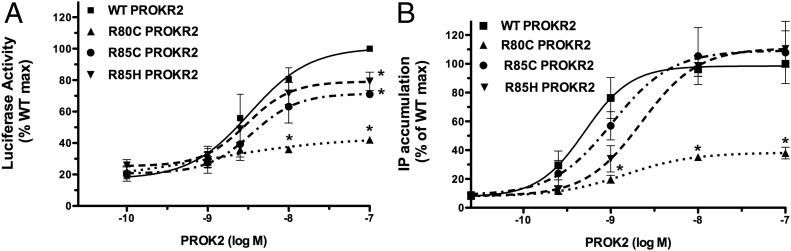
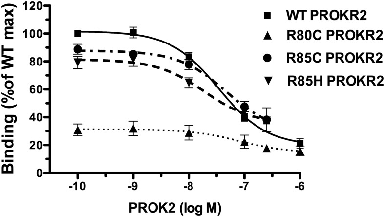
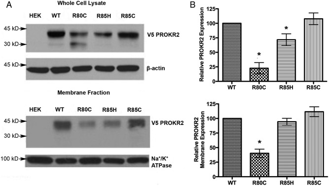
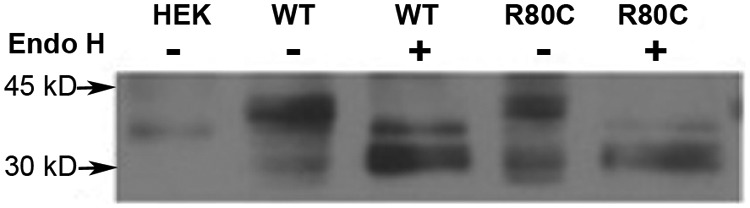
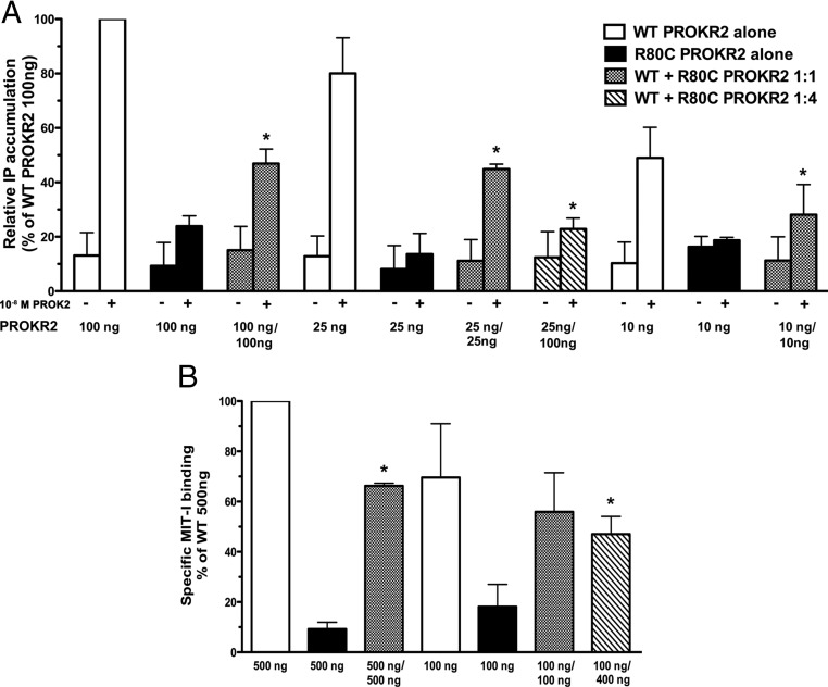
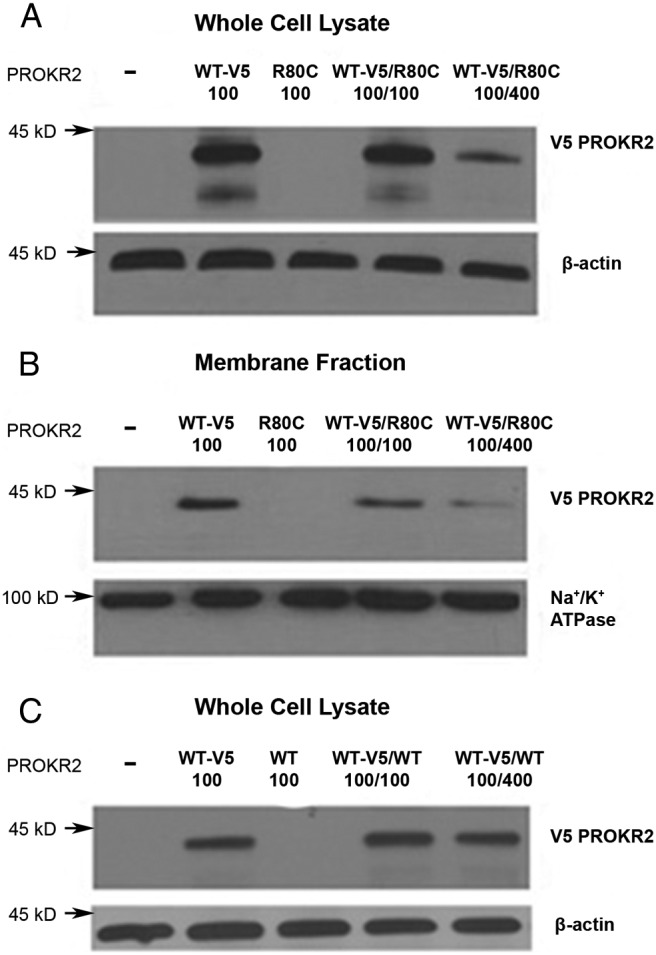
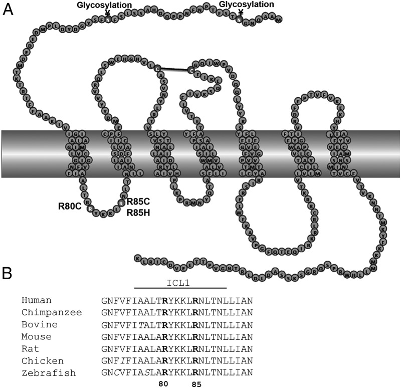
Prokineticin receptors (PROKR) are G protein-coupled receptors (GPCR) that regulate diverse biological processes, including olfactory bulb neurogenesis and GnRH neuronal migration. Mutations in PROKR2 have been described in patients with varying degrees of GnRH deficiency and are located in diverse functional domains of the receptor. Our goal was to determine whether variants in the first intracellular loop (ICL1) of PROKR2 (R80C, R85C, and R85H) identified in patients with hypogonadotropic hypogonadism interfere with receptor function and to elucidate the mechanisms of these effects. Because of structural homology among GPCR, clarification of the role of ICL1 in PROKR2 activity may contribute to a better understanding of this domain across other GPCR. The effects of the ICL1 PROKR2 mutations on activation of signal transduction pathways, ligand binding, and receptor expression were evaluated. Our results indicated that the R85C and R85H PROKR2 mutations interfere only modestly with receptor function, whereas the R80C PROKR2 mutation leads to a marked reduction in receptor activity. Cotransfection of wild-type (WT) and R80C PROKR2 showed that the R80C mutant could exert a dominant negative effect on WT PROKR2 in vitro by interfering with WT receptor expression. In summary, we have shown the importance of Arg80 in ICL1 for PROKR2 expression and demonstrate that R80C PROKR2 exerts a dominant negative effect on WT PROKR2.


Figures

Fig. 1.
Fig. 1.
Effects of ICL1 PROKR2 mutations on ligand-mediated activation of signal transduction. PROKR2 signaling activity in response to ligand was evaluated by measuring IP accumulation and using an Egr1-Luc reporter assay to measure MAPK activation. The PROK2 dose-response curves of [3H]IP accumulation and Egr1-Luc activity were used to calculate the EC50 for each receptor. Results are expressed as percentage of the maximal stimulation of WT PROKR2 (mean ± se for three independent experiments, each performed in triplicate). A, Activation of Egr1-Luc by WT and ICL1 PROKR2 mutants. HEK293 cells were transiently transfected with WT PROKR2, R80C PROKR2, R85C PROKR2, or R85H PROKR2 together with the Egr1-Luc reporter and PGL3 empty vector. Twenty-four hours after transfection, cells were stimulated with increasing concentrations of PROK2 for 16 h, and cell extracts were assayed for luciferase activity. B, Stimulation of PROK2-mediated [3H]IP production by WT and ICL1 PROKR2 mutants. COS7 cells were transiently transfected with WT PROKR2, R80C PROKR2, R85C PROKR2, or R85H PROKR2. Forty-eight hours after transfection, cells were stimulated with increasing concentrations of PROK2 for 1 h, and [3H]IP were measured. *, P < 0.05 at that specific dose compared with WT.
Fig. 2.
Fig. 2.
Displacement binding assay of WT and ICL1 PROKR2 mutants. To measure PROK2 binding by displacement analysis, COS7 cells transfected with either WT, R80C, R85C, or R85H PROKR2 were incubated with 125I-MIT-1 in the presence of increasing concentrations (10−10 to 10−6 m) of unlabeled PROK2 for 15 min, after which cells were washed and radioactivity assayed in cell lysates. The PROK2 displacement curves were used to calculate Bmax and Kd for each PROKR2 receptor. Results are expressed as percentage of the WT PROKR2 maximal binding. Each point is the mean ± se of three independent experiments.
Fig. 3.
Fig. 3.
Analysis of WT and ICL1 PROKR2 mutant protein expression. A, Representative Western blot analysis showing total cellular (upper panel) and membrane (lower panel) expression of WT and ICL1 mutant V5-tagged PROKR2 in stable transfectants of HEK293 cells. B, Quantification of the relative total cellular (upper panel) and membrane (lower panel) expression of the PROKR2 mutants compared with WT PROKR2, using data pooled from at least three independent experiments. The intensity of the PROKR2 band (detected using anti-V5 antibody) was normalized to β-actin in whole-cell lysates and to Na+/K+ATPase in the membrane fraction. Both the 45- and 30-kDa bands were included in the quantification. HEK, Untransfected HEK293 cells. Bars represent mean ± sem. *, P < 0.05 compared with WT.
Fig. 4.
Fig. 4.
Evaluation of PROKR2 glycosylation. Evaluation of N-glycosylation of WT and R80C PROKR2 by means of Endo H treatment. HEK293 cells stably expressing WT and R80C V5-tagged PROKR2 were used to prepare whole-cell protein extracts. These samples were either left untreated (−) or treated with Endo H (+) and then analyzed by reducing SDS-PAGE and Western blot analysis using an anti-V5 antibody; 45-kDa (glycosylated) and 30-kDa (deglycosylated) bands were visualized as marked. HEK, Untransfected HEK293 cells.
Fig. 5.
Fig. 5.
Evaluation for dominant negative effects of R80C PROKR2 on the WT receptor. A, Measurement of [3H]IP accumulation in COS7 cells after cotransfection with varying amounts of WT and R80C PROKR2 plasmids as indicated and stimulation with 10−8 m PROK2. Results are expressed as percentage of the maximal stimulation of WT PROKR2 (100 ng) alone. Bars represent mean ± sem for three independent experiments, each performed in triplicate. B, Displacement binding assay in COS7 cells transfected with either WT PROKR2 alone or cotransfected with WT and R80C PROKR2 in varying amounts as indicated. Cells were incubated with 125I-MIT-1 with or without 10−7 m of unlabeled PROK2 for 15 min, after which cells were washed and radioactivity assayed in cell lysates. Specific binding activity was calculated and expressed as percentage of binding to WT PROKR2 (500 ng) alone. Bars represent mean ± sem for three independent experiments, each performed in triplicate. *, P < 0.05 compared with the same amount of WT PROKR2 alone; (−) untreated and (+) treated with 10−8 m PROK2; 10, 25, 100, 400, and 500 ng represent the amount of each PROKR2 plasmid transfected.
Fig. 6.
Fig. 6.
Evaluation for potential dominant negative effects of R80C PROKR2 on WT PROKR2 expression. Representative Western blot analysis of whole-cell lysates (A) and membrane fraction (B) from HEK 293 cells transiently transfected with either 100 ng of V5-tagged WT PROKR2 plasmid alone (WT-V5 100), 100 ng of untagged R80C PROKR2 plasmid alone (R80C 100), or 100 ng of V5-tagged WT PROKR2 cotransfected with 100 ng (WT-V5/R80C 100/100) or 400 ng (WT-V5/R80C 100/400) of untagged R80C PROKR2. C, Representative Western blot analysis of whole-cell lysates from HEK 293 cells transiently transfected with either 100 ng of V5-tagged WT PROKR2 plasmid alone (WT-V5 100), 100 ng of untagged WT PROKR2 plasmid alone (WT 100), or 100 ng of V5-tagged WT PROKR2 cotransfected with 100 ng (WT-V5/WT 100/100) or 400 ng (WT-V5/WT 100/400) of untagged WT PROKR2. The intensity of the PROKR2 band (detected using anti-V5 antibody) was normalized to β-actin in whole-cell lysates and to Na+/K+ATPase in the membrane fractions. −, Untransfected HEK 293 cells.
Fig. 7.
Fig. 7.
PROKR2 ICL1 amino acid sequence alignment among species. A, Schematic representation of mutations in the ICL1 of PROKR2. Residues in dark gray represent the ICL1 mutations. Residues involved in disulfide bond formation (black line) and in glycosylation are represented in gray. B, Amino acid sequence alignment among species, showing that arginines in position 80 and 85 are highly conserved among different species. Sequences were taken from the GPCR database (www.gpcr.org), and the alignment was produced in ClustalW.

References

    1. Ngan ES, Tam PK. 2008. Prokineticin-signaling pathway. Int J Biochem Cell Biol 40:1679–1684 - PubMed
    1. Lin DC, Bullock CM, Ehlert FJ, Chen JL, Tian H, Zhou QY. 2002. Identification and molecular characterization of two closely related G protein-coupled receptors activated by prokineticins/endocrine gland vascular endothelial growth factor. J Biol Chem 277:19276–19280 - PubMed
    1. Soga T, Matsumoto S, Oda T, Saito T, Hiyama H, Takasaki J, Kamohara M, Ohishi T, Matsushime H, Furuichi K. 2002. Molecular cloning and characterization of prokineticin receptors. Biochim Biophys Acta 1579:173–179 - PubMed
    1. Masuda Y, Takatsu Y, Terao Y, Kumano S, Ishibashi Y, Suenaga M, Abe M, Fukusumi S, Watanabe T, Shintani Y, Yamada T, Hinuma S, Inatomi N, Ohtaki T, Onda H, Fujino M. 2002. Isolation and identification of EG-VEGF/prokineticins as cognate ligands for two orphan G-protein-coupled receptors. Biochem Biophys Res Commun 293:396–402 - PubMed
    1. Chen J, Kuei C, Sutton S, Wilson S, Yu J, Kamme F, Mazur C, Lovenberg T, Liu C. 2005. Identification and pharmacological characterization of prokineticin 2β as a selective ligand for prokineticin receptor 1. Mol Pharmacol 67:2070–2076 - PubMed

Publication types

MeSH terms

LinkOut - more resources