. 2012 Jul 20;337(6092):358-62.
doi: 10.1126/science.1222381. Epub 2012 Jun 28.

Regional astrocyte allocation regulates CNS synaptogenesis and repair

Affiliations

Regional astrocyte allocation regulates CNS synaptogenesis and repair

Hui-Hsin Tsai et al. Science. .

Abstract

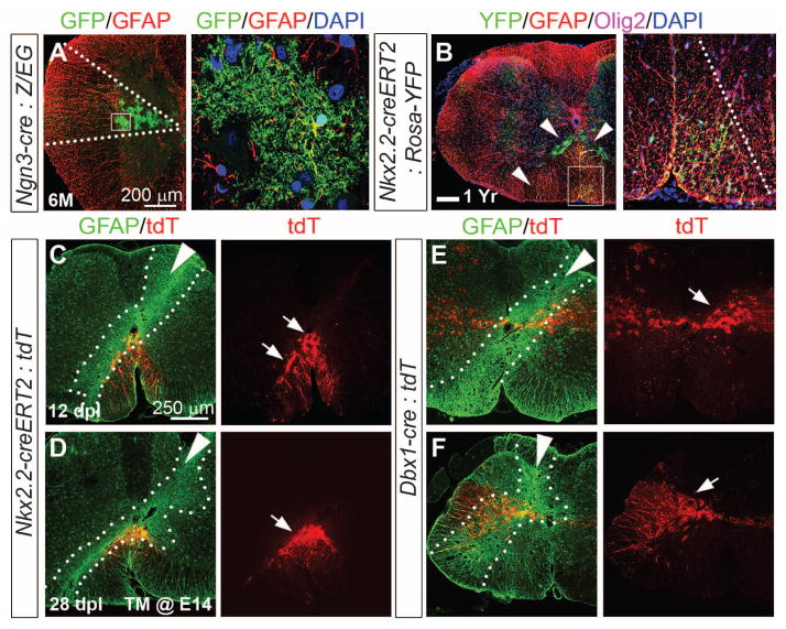
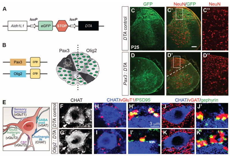
Astrocytes, the most abundant cell population in the central nervous system (CNS), are essential for normal neurological function. We show that astrocytes are allocated to spatial domains in mouse spinal cord and brain in accordance with their embryonic sites of origin in the ventricular zone. These domains remain stable throughout life without evidence of secondary tangential migration, even after acute CNS injury. Domain-specific depletion of astrocytes in ventral spinal cord resulted in abnormal motor neuron synaptogenesis, which was not rescued by immigration of astrocytes from adjoining regions. Our findings demonstrate that region-restricted astrocyte allocation is a general CNS phenomenon and reveal intrinsic limitations of the astroglial response to injury.


Figures

Fig. 1
Fig. 1
Segmental distribution of fibrous and protoplasmic astrocytes in spinal cord. (A) Nkx2.2-creERT2 (tamoxifen induction E10.5 to E12.5):Rosa26-YFP fate map shows YFP+, GFAP+ cells at the ventral mid-line at P0. YFP, yellow fluorescent protein. (B) In P2 Olig2-tva-cre:CAG-GFP mice, astrocytes remain in register with pMN, whereas Olig2+ OPs distribute widely. (C) Ngn3-cre:Z/EG P1 cord shows intermediate wedge of astrocytes. (D, G, G′) At P2, FAs and PAs in Pax3-cre animals remain dorsally restricted. DAPI, 4′,6-diamidino-2-phenylindole. (E and F) Aldh1L1-GFP coexpression with GFAP+ (FAs) and AldoC+ (FAs and PAs) cells. (H to N) Astrocytes from Olig2cre/+ spinal cord have a restricted ventral distribution. In Olig2cre/cre nulls, we observe significantly (*P < 0.0001) increased p2-type (GFAP+, Pax6+, AldoC+) astrocytes (fig. S1I), which fail to migrate from the ventral domain. Scale bars, 200 μm.
Fig. 2
Fig. 2
Absence of tangential astrocyte migration in adult spinal cord even after injury. (A) Bushy GFP+ PAs in Ngn3-cre:Z/EG cord persisted after 6 months of age. (B) Astrocytes in Nkx2.2-creERT2 (induced E10 to E11):Rosa26-YFP cords remain ventrally restricted at 1 year. (C to F) Post-stab gliosis does not recruit astrocytes from adjacent domains. Fate-mapped astrocytes (arrows) in Rosa26-tdTomato on the Nkx2.2-creERT2 [induced E14 (C and D)] or Dbx1-cre (E and F) background remain confined to ventral or intermediate cord, respectively, 12 and 28 days postlesion (dpl). Intense GFAP staining indicates lesion site (dashed lines, white arrowheads indicate needle trajectory).
Fig. 3
Fig. 3
Regional astrocyte depletion results in neuronal abnormalities. (A) Cartoon of Aldh1L1-DTA transgene; cre excises eGFP-Stop cassette allowing DTA transcription. (B) Regions targeted by Pax3-cre or Olig2-cre. (C and D) Pax3-cre:Aldh1L1-DTA mice show absence of GFP, neuropil, and congested appearance of NeuN (red) neurons in dorsal cord. (E) Cartoon of MN soma and synapse subtypes. CST, corticospinal tract. (F and G) We observed no differences in the number of cholinergic CHAT or vGlut2 (fig. S5) synapses. (H and I) Numbers of excitatory vGluT1-PSD95 synapses were significantly decreased, whereas (J and K) inhibitory vGAT-gephyrin synapses were significantly increased in bigenic animals compared with controls. For quantification, see fig. S5.
Fig. 4
Fig. 4
Region-restricted astrocyte investment from forebrain radial glia. (A to D) Emx1-creERT2 (induced E17):Rosa26-tdTomato labeled cells; note (A′) distribution of astrocytes (green) confined to cortical plate and corpus callosum. Red box indicates region of cortex analyzed in (B) to (D). (E to I) Distribution of Dbx1-cre astrocytes in striatum. Red boxes indicate regions of cortex and striatum analyzed in (F) to (I). (J to N) Distribution of Nkx2.1-creERT2 (induced E17) astrocytes in ventromedial forebrain. Red box in cortex indicates fate-mapped interneurons. (O to S) Distribution of astrocytes after radial glial Ad-cre infection of P1 Z/EG reporter mice in the forebrain regions indicated analyzed at P4 or P28. Astrocytes were recognized by morphology and AldoC immunolabeling. We injected dorsal (n = 20), ventral (n = 21), medial (n = 19), and cortical (n = 17) brain regions (red arrows). No tangential astrocyte migration was observed. PALv, ventral pallidum.

Comment in

References

    1. Kettenmann H, Ransom BR. Neuroglia. Oxford Univ. Press; Oxford: 2005.
    1. Wang DD, Bordey A. Prog Neurobiol. 2008;86:342. - PMC - PubMed
    1. Doyle JP, et al. Cell. 2008;135:749. - PMC - PubMed
    1. Houades V, Koulakoff A, Ezan P, Seif I, Giaume C. J Neurosci. 2008;28:5207. - PMC - PubMed
    1. Nimmerjahn A, Mukamel EA, Schnitzer MJ. Neuron. 2009;62:400. - PMC - PubMed

Publication types

MeSH terms