Review
. 2012 Aug 1;4(8):a013680.
doi: 10.1101/cshperspect.a013680.

Synaptic vesicle pools and dynamics

Affiliations
Review

Synaptic vesicle pools and dynamics

AbdulRasheed A Alabi et al. Cold Spring Harb Perspect Biol. .

Abstract

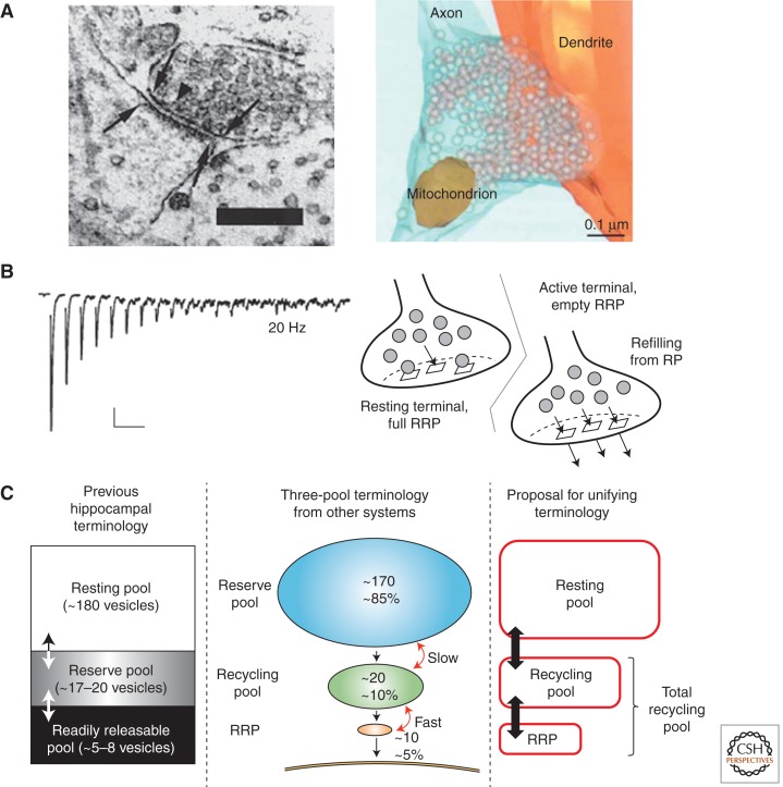
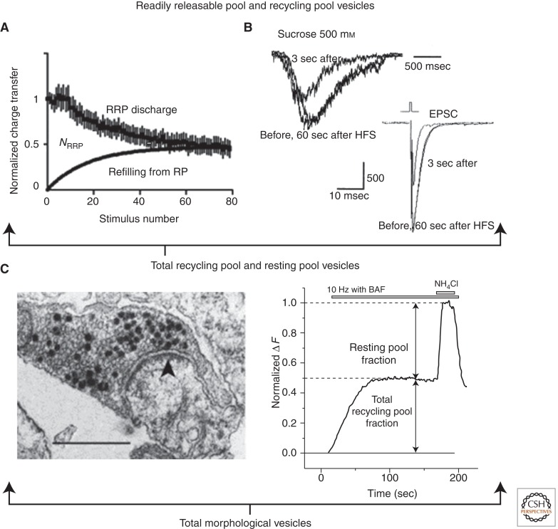
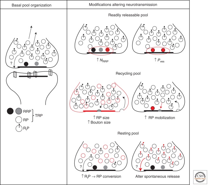
Synaptic vesicles release neurotransmitter at chemical synapses, thus initiating the flow of information in neural networks. To achieve this, vesicles undergo a dynamic cycle of fusion and retrieval to maintain the structural and functional integrity of the presynaptic terminals in which they reside. Moreover, compelling evidence indicates these vesicles differ in their availability for release and mobilization in response to stimuli, prompting classification into at least three different functional pools. Ongoing studies of the molecular and cellular bases for this heterogeneity attempt to link structure to physiology and clarify how regulation of vesicle pools influences synaptic strength and presynaptic plasticity. We discuss prevailing perspectives on vesicle pools, the role they play in shaping synaptic transmission, and the open questions that challenge current understanding.


Figures

Figure 1.
Figure 1.
Release dynamics and vesicle pool terminology. (A) Left panel: Ultrastructural image from mouse hippocampal neurons in culture. Boundaries of the presynaptic active zone and postsynaptic density (arrows) anatomically define a synaptic contact. Few vesicles appear docked to the active zone (example arrowhead), whereas the vast majority are distributed within the greater bouton volume. Scale bar, 0.25 µm (Schikorski and Stevens 1997). Right panel: 3D reconstruction of serial sections like those in (A) displays overall architecture of synapse and vesicles, devoid of any distinguishing morphological features. (B) Left panel: Synaptic depression recorded from postsynaptic responses in hippocampal synapses during 20 Hz stimulation. Note the increase in asynchronous release, occurring between the large-amplitude synchronous peaks, during the stimulus train. Scale bars, 500 ms and 100 pA (Stevens and Williams 2007). Right panel: Simple model suggesting depression results from sequential recruitment of functionally heterogeneous vesicle pools. Depletion of a readily releasable pool (RRP) of vesicles gives way to a rate-limiting refilling process from a general recycling pool (RP). (C) Vesicle pool terminology from hippocampal terminals (left panel) (Südhof 2000) and an alternative three-pool model (middle panel) (Rizzoli and Betz 2005). A proposed unifying scheme (right panel) avoids the conflicting term “reserve pool” and merges the remaining terminology for the final lexicon used in this review. Arrows denote interconversion of vesicles between pools and numerical values represent absolute number or relative percentage of vesicles within each pool (see text for more details). (Left side of Panel A is from Schikorski and Stevens 1997; reprinted, with permission, from J Soc Neurosci © 1997; right side of Panel A is from Rizzoli and Betz 2005; reprinted, with permission, from Macmillan Publishers Ltd., Nat Rev Neurosci © 2005 (originally sourced from Schikorski and Stevens 2001, Nat Neurosci © 2001; left side of Panel B is from Stevens and Williams 2007; reprinted, with permission, from the American Physiological Society © 2007; right side of panel B is from Wesseling and Lo 2002; reprinted, with permission, from J Soc Neurosci © 2002; left side of Panel C is from Südhoff 2000; reprinted, with permission, from Elsevier © 2000; middle of Panel C is from Rizzoli and Betz 2005; reprinted, with permission, from Nature Rev Neurosci © 2005.)
Figure 2.
Figure 2.
Summary of functional characterization of vesicle pools. (A) Analysis of the action potential (AP) responses from Figure 1B (left panel) indicates the normalized charge transferred per 50 msec time window (interstimulus interval) declines, reflecting a discharge and depletion of readily releasable pool (RRP) vesicles. Note that applying the charge integral and not simply peak amplitude captures synchronous and asynchronous release. Refilling of the RRP from the RP is approximated as a first-order exponential process. RRP size (NRRP) is proportional to the total charge integrated over the stimulus duration less that resulting from the refilling process (area between the two curves) (Stevens and Williams 2007). (B) Cross-depletion with high-frequency stimulation (HFS) (100 APs at 20 Hz) reveals that both hypertonic sucrose and AP challenges access the same RRP vesicles (Rosenmund and Stevens 1996). (C) Left panel: Example of photoconverted hippocampal terminal after loading with FM1-43 using 20 Hz, 60 sec stimulation with an additional 60 sec rest for residual endocytic activity poststimulus. The aim was to label the total recycling pool TRP. Approximately 39% of vesicles recycled and thus contained electron-dense puncta (dark vesicle lumen), with some in close proximity to the active zone (arrowhead) and others intermixed within the greater bouton area. Scale bar, 0.5 µm (Harata et al. 2001b). (Right panel) 10 Hz stimulation of hippocampal neurons expressing a pHluorin-tagged vesicular protein in the presence of proton-pump inhibitor bafilomycin reveals the TRP. Ammonium chloride pulse at the end of the stimulus neutralizes the remaining pHluorin constructs in acidic compartments, thereby unmasking nonrecycling vesicles in the resting pool (Kim and Ryan 2010). (Panel A is from Stevens and Williams 2007; reprinted, with permission, from the American Physiological Society © 2007; Panel B is from Rosenmund and Stevens 1996; reprinted, with permission, from Elsevier © 1996; right side of Panel C is from Kim and Ryan 2010; reprinted, with permission, from Elsevier © 2010.)
Figure 3.
Figure 3.
Simplified overview of vesicle pool function and regulation. Left column: Vesicles in RRP are presumed to be in close proximity to active zone for highest Pves at the bouton and rapid fusion. RP and RtP vesicles are interspersed within vesicle cluster. Both contribute to the vesicle superpool (arrows leaving bouton). RtP vesicles are distinguished by molecular labels (Hua et al. 2011; Ramirez et al. 2012) and possibly contribute to spontaneous release at sites outside AZ (Zenisek 2008). Shading of RRP vesicles reflects potential RRP heterogeneity. The number of vesicles depicted within the bouton is scaled down by a factor of 5 for easy visualization. Right column: Modifications to vesicle pool number or properties are marked in red when compared to basal state. Each modification contributes to shaping neurotransmission and short-term plasticity (see text for further details). For RRP, the number of vesicles (NRRP) or their fusion probability (Pves) may change. For RP, increases in bouton size can accommodate more RP (and RtP) vesicles and increase active zone length (which could possibly enhance RRP capacity) (Welzel et al. 2011). Additionally, calcium or protein kinase C activation can accelerate RP mobilization, mitigating RRP depletion. For RtP, inhibition of CDK5 recruits vesicles to the total recycling pool (TRP) (Kim and Ryan 2010) and modification of a proposed molecular label can alter the frequency of spontaneous release events (Ramirez et al. 2012).

References

    1. Abbott LF, Regehr WG 2004. Synaptic computation. Nature 431: 796–803 - PubMed
    1. Abbott LF, Varela JA, Sen K, Nelson SB 1997. Synaptic depression and cortical gain control. Science 275: 220–224 - PubMed
    1. Ahmari SE, Buchanan J, Smith SJ 2000. Assembly of presynaptic active zones from cytoplasmic transport packets. Nat Neurosci 3: 445–451 - PubMed
    1. Allen C, Stevens CF 1994. An evaluation of causes for unreliability of synaptic transmission. Proc Natl Acad Sci 91: 10380–10383 - PMC - PubMed
    1. Armitage BA, Siegelbaum SA 1998. Presynaptic induction and expression of homosynaptic depression at Aplysia sensorimotor neuron synapses. J Neurosci 18: 8770–8779 - PMC - PubMed

Publication types

LinkOut - more resources