. 2012 Aug;139(15):2763-72.
doi: 10.1242/dev.074179. Epub 2012 Jun 28.

Concerted control of gliogenesis by InR/TOR and FGF signalling in the Drosophila post-embryonic brain

Affiliations

Concerted control of gliogenesis by InR/TOR and FGF signalling in the Drosophila post-embryonic brain

Amélie Avet-Rochex et al. Development. 2012 Aug.

Abstract

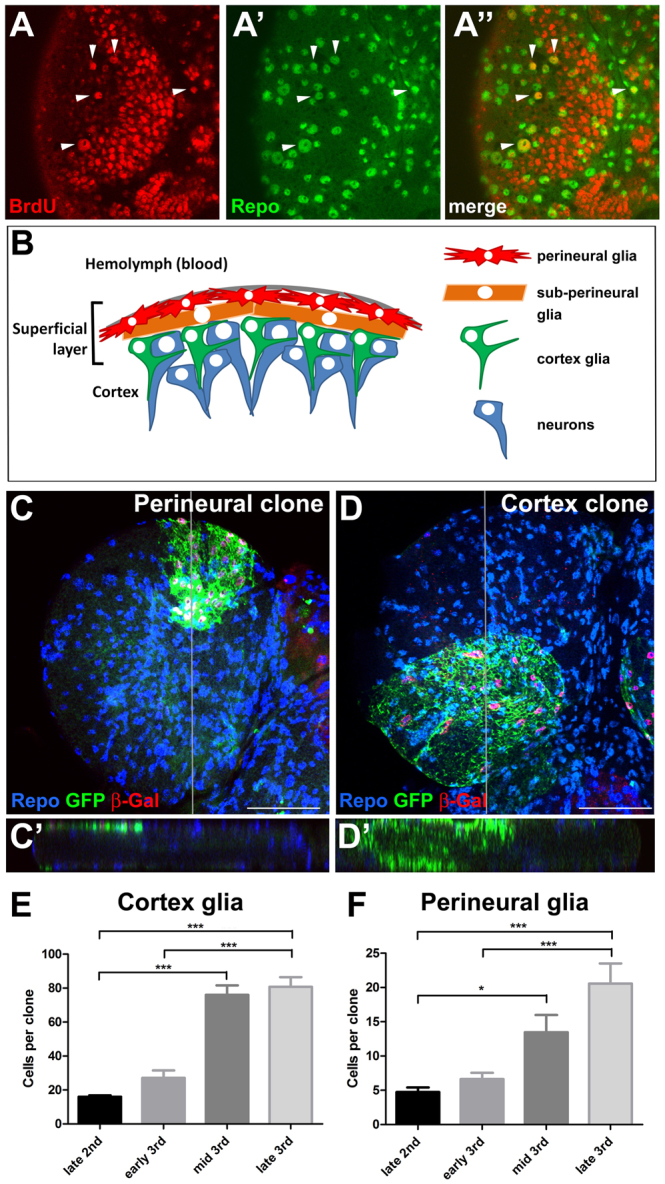
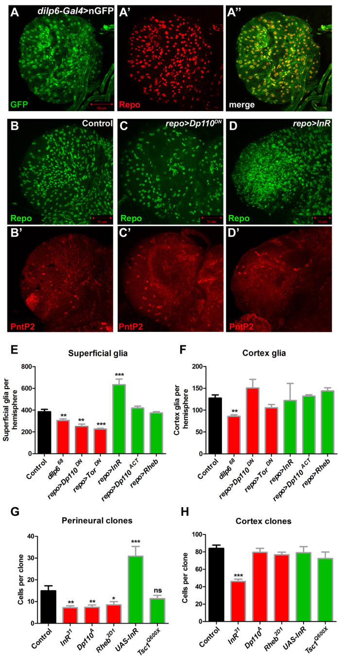
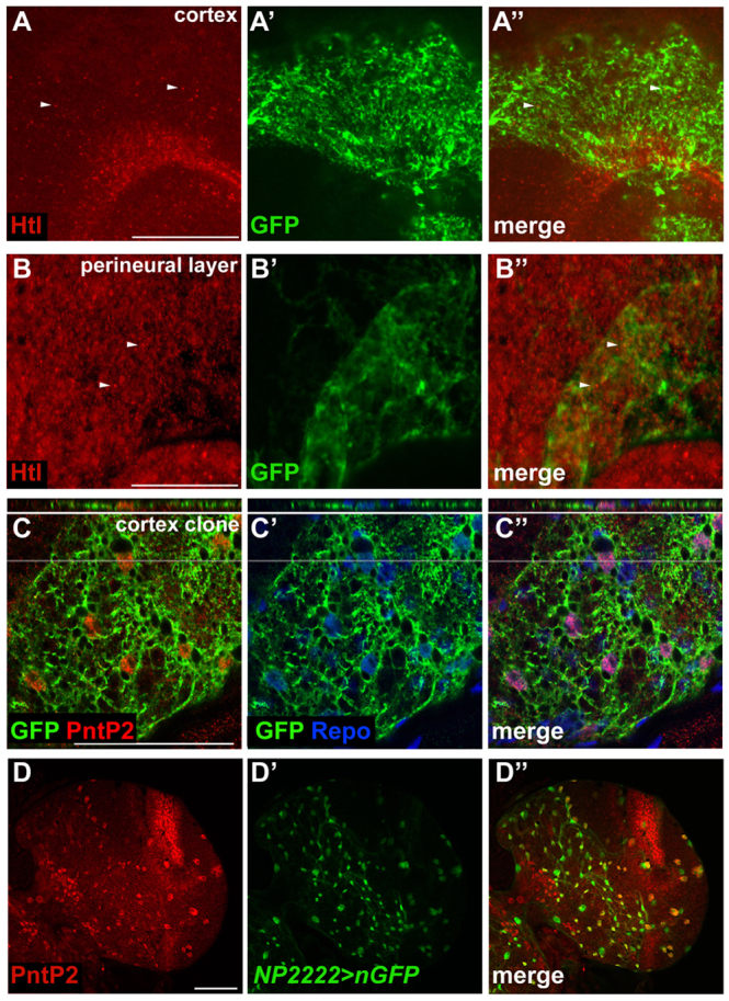
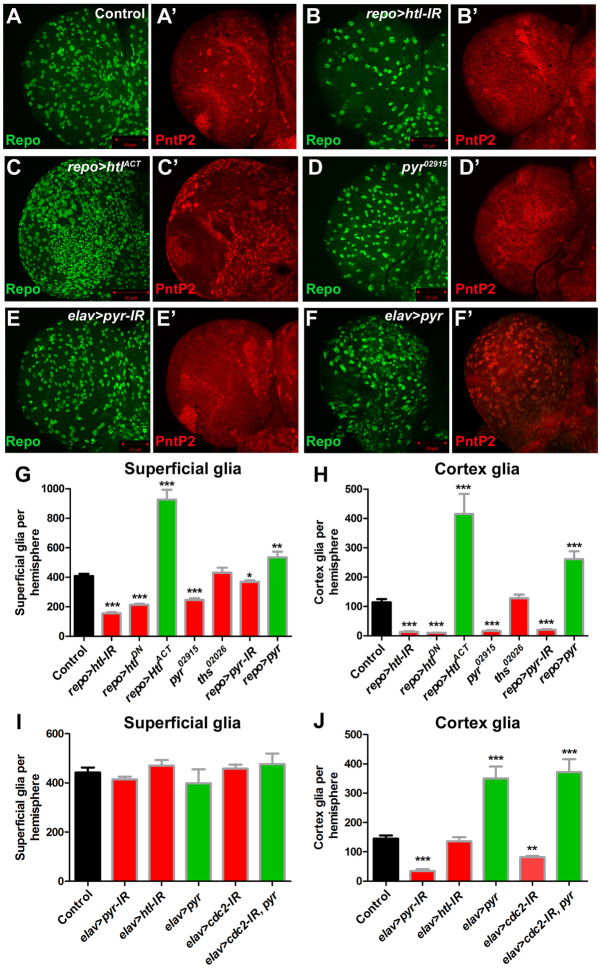
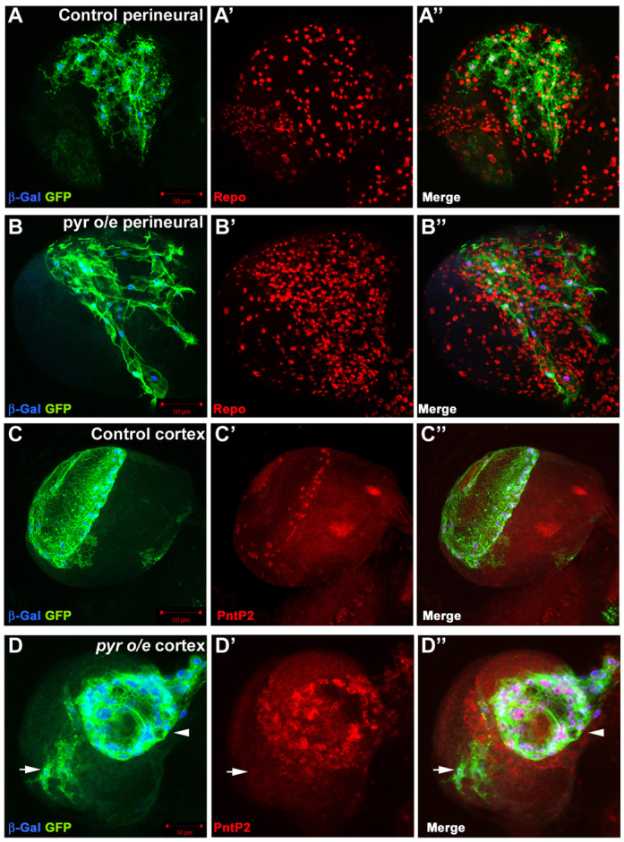
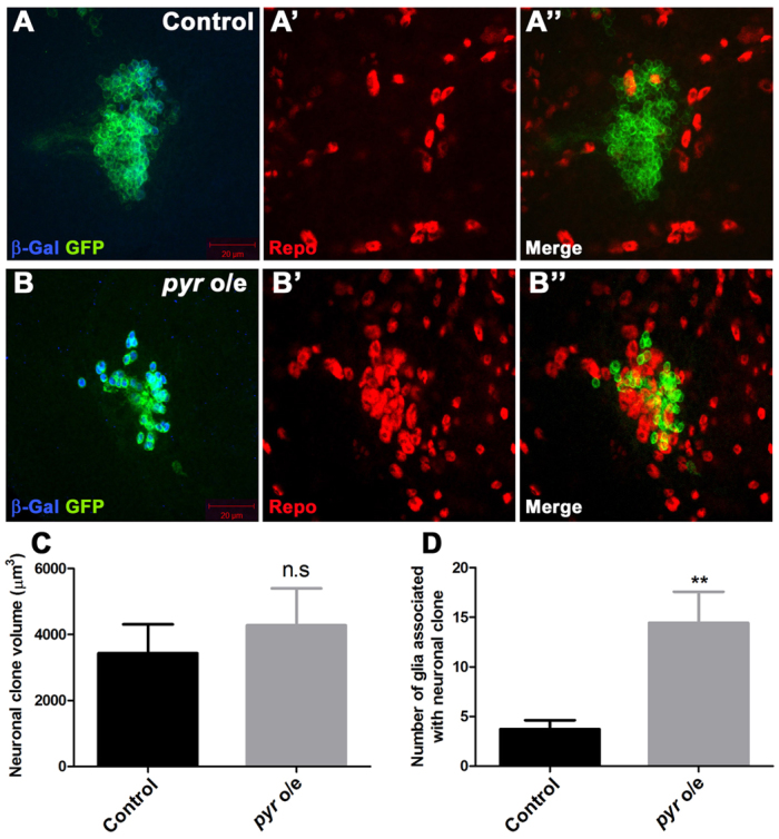
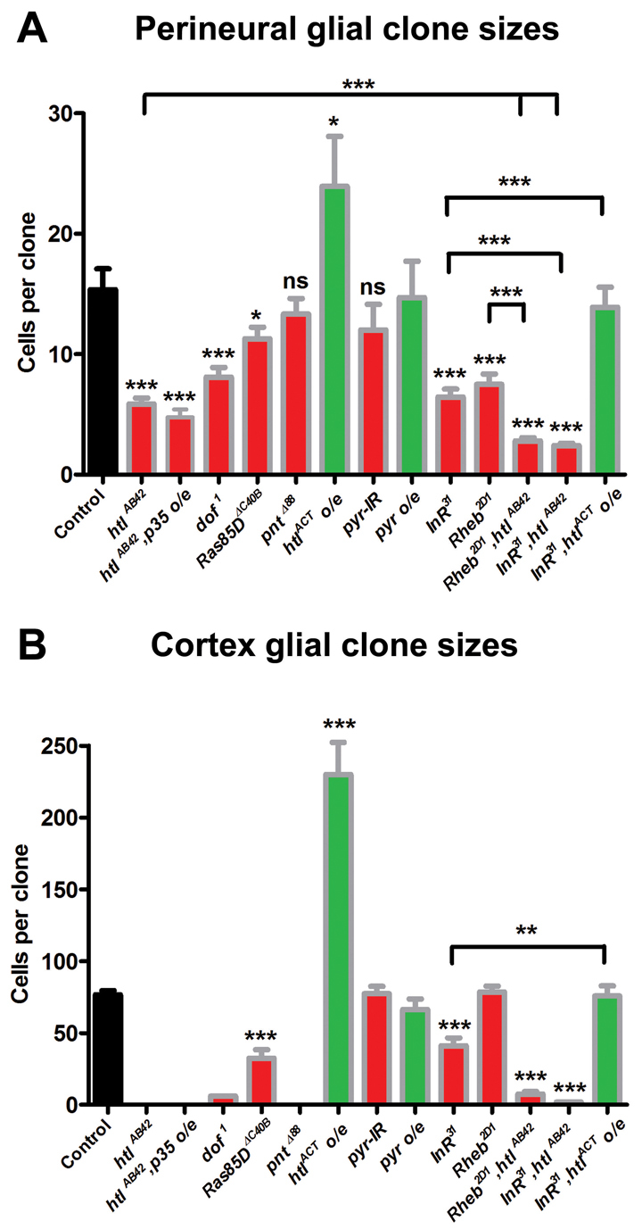
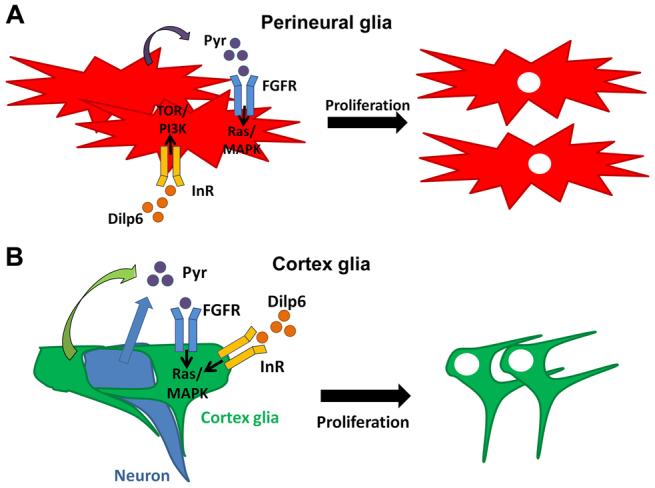
Glial cells are essential for the development and function of the nervous system. In the mammalian brain, vast numbers of glia of several different functional types are generated during late embryonic and early foetal development. However, the molecular cues that instruct gliogenesis and determine glial cell type are poorly understood. During post-embryonic development, the number of glia in the Drosophila larval brain increases dramatically, potentially providing a powerful model for understanding gliogenesis. Using glial-specific clonal analysis we find that perineural glia and cortex glia proliferate extensively through symmetric cell division in the post-embryonic brain. Using pan-glial inhibition and loss-of-function clonal analysis we find that Insulin-like receptor (InR)/Target of rapamycin (TOR) signalling is required for the proliferation of perineural glia. Fibroblast growth factor (FGF) signalling is also required for perineural glia proliferation and acts synergistically with the InR/TOR pathway. Cortex glia require InR in part, but not downstream components of the TOR pathway, for proliferation. Moreover, cortex glia absolutely require FGF signalling, such that inhibition of the FGF pathway almost completely blocks the generation of cortex glia. Neuronal expression of the FGF receptor ligand Pyramus is also required for the generation of cortex glia, suggesting a mechanism whereby neuronal FGF expression coordinates neurogenesis and cortex gliogenesis. In summary, we have identified two major pathways that control perineural and cortex gliogenesis in the post-embryonic brain and have shown that the molecular circuitry required is lineage specific.


Figures

Fig. 1.
Fig. 1.
Glial proliferation in the Drosophila larval brain. (A-A″) Ventral superficial layer of a third instar larval brain stained for BrdU incorporation (red) and Repo expression (green) in glia. Arrowheads indicate glia that have incorporated BrdU. (B) Schematic of the superficial layer of the larval brain. Below the dense extracellular matrix, known as the neural lamella (grey), lie perineural glia (red) that are star-shaped and extend thin protrusions laterally but do not contact neurons (blue). Below the perineural layer are large sub-perineural cells (orange) connected by septate junctions required for the integrity of the blood-brain barrier. Cortex glia (green), which are found below the sub-perineural layer and throughout the brain, send out fine processes that ensheath neuronal cell bodies and neurites as they project between cell bodies towards the neuropil (supplementary material Fig. S2). (C) Perineural repo-MARCM clone in the dorsal surface of the larval brain stained for Repo (blue), GFP (green) and β-Gal (red) expression. (C′) Orthogonal view shows GFP expression in the perineural layer. (D) Cortex repo-MARCM clone in the ventral surface of the larval brain. (D′) Orthogonal view shows cortex glia processes extending into the brain. Note the difference in appearance to the perineural clone in C. Grey lines indicate positions of the orthogonal sections. (E,F) Average clone size of cortex glia (E) and perineural glia (F) during larval development. Error bars indicate s.e.m. *P<0.05; ***P<0.001. Scale bars: 50 μm.
Fig. 2.
Fig. 2.
InR/TOR signalling is required for perineural gliogenesis. (A-A″) dilp6-Gal4 driving nuclear GFP (green) colocalises with Repo expression (red) in glia in the Drosophila larval brain. (B-D′) The ventral superficial layer of larval brain stained for Repo (green) or PntP2 (red) from control (B,B′), or repo-Gal4 driving expression of (C,C′) a dominant-negative form of Dp110 (repo>Dp110DN) or (D,D′) InR (repo>InR). (E,F) Quantification of superficial (E) or cortex (F) glia from homozygous dilp6 mutant larvae (dilp668) or brains expressing inhibitors (red bars) or activators (green bars) of the InR/TOR pathway in glia using repo-Gal4; control is repo-Gal4/+ (G) Quantification of perineural repo-MARCM clone size for FRT82B controls (n=71), InR31 (n=40), Dp110A (n=38), Rheb2D1 (n=53), UAS-InR (n=52) or Tsc1Q600X (n=46). (H) Quantification of cortex repo-MARCM clone size for FRT82B controls (n=15), InR31 (n=19), Dp110A (n=16), Rheb2D1 (n=10), UAS-InR (n=13) or Tsc1Q600X (n=7). Error bars indicate s.e.m. *P<0.05; **P<0.01; ***P<0.001; ns, not significant.
Fig. 3.
Fig. 3.
Expression of FGF pathway components in the larval brain. (A-B″) Htl expression (red) in glia in the cortex (A-A″) or perineural (B-B″) layers of the Drosophila larval brain. repo-MARCM clones expressing GFP (green) were used to identify the respective glial types. (C-C″) Third instar larval brain hemisphere showing a repo-MARCM cortex clone stained for GFP (green), PntP2 (red) and Repo (blue) expression. Note that PntP2-positive cortex glia colocalise with GFP in the orthogonal view (top; grey lines indicate the position of orthogonal section). (D-D″) Cortex glia-specific NP2222-Gal4 driving UAS-nGFP (green) showing colocalisation with PntP2 expression (red). Scale bars: 50 μm.
Fig. 4.
Fig. 4.
FGF signalling is necessary and sufficient for glial proliferation. (A-C′) Ventral superficial layer of Drosophila larval brain stained for Repo (green) and PntP2 (red, to visualise cortex glia) expressing the following transgenes in glia: (A) repo-Gal4/+ control, (B) dsRNA against htl (repo>htl-IR), (C) an activated form of the Htl receptor (repo>htlACT). (D,D′) pyr02915 homozygous larval brain stained as in A. (E-F′) The ventral superficial layer of larval brains expressing either dsRNA against pyr (elav>pyr-IR) (E), or wild-type pyr (elav>pyr) (F) in neurons, stained as in A. (G) Superficial glia numbers from larvae with repo-Gal4 driving the expression of a dsRNA against htl (repo>htl-IR), a dominant-negative form of Htl (repo>htlDN), an activated form of Htl (repo>htlACT), a dsRNA against pyr (repo>pyr-IR), or wild-type pyr (repo>pyr), or in mutants for pyr (pyr02915) or ths (ths02026). (H) Quantification of cortex glia numbers in the same brains as in G. (I) Quantification of superficial glia numbers in larvae with elav-Gal4 driving the expression of a dsRNA against pyr (elav>pyr-IR), a dsRNA against htl (elav>htl-IR), wild-type pyr (elav>pyr), dsRNA against cdc2 (elav>cdc2-IR), or combined cdc2 dsRNA and pyr (elav>cdc2-IR,pyr) in neurons. (J) Quantification of cortex glia numbers in the same brains as in I. Green bars indicate pathway activation, red bars indicate pathway inhibition. Error bars indicate s.e.m. *P<0.05; **P<0.01; ***P<0.001.
Fig. 5.
Fig. 5.
pyr overexpression in glia causes non-cell-autonomous glial proliferation. (A-B″) Control (A-A″) or pyr-overexpressing (B-B″) repo-MARCM perineural clones stained for β-Gal (blue) and Repo (red) expression. Note the extended processes (B) and the increased number of glial cells surrounding the clone (B′,B″). (C-D″) Control (C-C″) or pyr-overexpressing (arrowhead in D,D″) repo-MARCM cortex clones stained for β-Gal (blue) and PntP2 (red) expression. Note the non-cell-autonomous proliferation of PntP2-expressing cortex glia, clustered cell bodies and shortened processes in the pyr-overexpressing cortex clone (arrowhead in D,D″). Note also that the adjacent small perineural clone overexpressing pyr (arrow in D-D″) does not cause cortex glia proliferation.
Fig. 6.
Fig. 6.
pyr overexpression in neurons induces gliogenesis. (A-B″) Control (A-A″) or pyr-overexpressing (B-B″) elav-MARCM clones stained for β-Gal (blue) and Repo (red) expression. Note the cluster of glial cells surrounding the clone in B″. (C) Quantification of neuronal clone volume for control or pyr-overexpressing neuronal clones (pyr o/e) (n=10 clones per genotype). (D) Quantification of glia associated with neuronal clones for control or pyr-overexpressing neuronal clones (pyr o/e) (n=10 clones per genotype). Error bars indicate s.e.m. **P<0.01; n.s, not significant.
Fig. 7.
Fig. 7.
FGF signalling acts together with InR/TOR signalling to control the genesis of perineural and cortex glia. (A) Quantification of perineural repo-MARCM clone size. Average clone size of FRT82B control clones (n=71), htlAB42 (n=28), htlAB42,p35 overexpression (htlAB42,p35 o/e, n=28), dof1 (n=63), Ras85DΔC40B (n=88), pntΔ88 (n=67), htlACT overexpression (htlACT o/e, n=23), pyr RNAi (pyr-IR, n=31), pyr overexpression (pyr o/e, n=20), InR31 (n=37), Rheb2D1 (n=66), Rheb2D1, htlAB42 (n=74), InR31, htlAB42 (n=54), and InR31,htlACT overexpression (InR31,htlACT o/e, n=41). (B) Quantification of cortex repo-MARCM clone size. FRT82B control clones (n=13), dof1 (n=1), Ras85DΔC40B (n=10), htlACT overexpression (htlACT o/e, n=5), pyr-IR (n=4), pyr overexpression (pyr o/e, n=7), InR31 (n=12), Rheb2D1 (n=15), Rheb2D1, htlAB42 (n=8), InR31, htlAB42 (n=5) and InR31,htlACT overexpression (InR31,htlACT o/e, n=7). No cortex clones were observed for htlAB42 (in over 100 hemispheres), htlAB42,p35 overexpression (in 60 hemispheres) or pntΔ88 (in 56 hemispheres). One six-cell clone was observed in 42 hemispheres for dof1. Green bars indicate pathway activation, red bars indicate pathway inhibition. Error bars indicate s.e.m. *P<0.05; **P<0.01; ***P<0.001; ns, not significant.
Fig. 8.
Fig. 8.
A model for the control of perineural and cortex glia proliferation in the Drosophila larval brain. (A) Pyr is expressed by perineural glia to activate FGF signalling in adjacent glia and acts in parallel to InR/TOR signalling (activated by the expression of Dilp6). These two pathways act synergistically to generate the correct complement of perineural glia. (B) Cortex glia proliferation is controlled by FGF signalling through FGFR (Htl) and the Ras/MAPK pathway. Pyr expression is required from both glia (green) and neurons (blue) and acts non-cell-autonomously. Neuronal Pyr expression activates the FGFR on adjacent cortex glia, thereby coordinating neurogenesis and glial proliferation. InR is also partially required in cortex glia and is likely to signal through the Ras/MAPK pathway.

References

    1. Awasaki T., Lai S. L., Ito K., Lee T. (2008). Organization and postembryonic development of glial cells in the adult central brain of Drosophila. J. Neurosci. 28, 13742-13753 - PMC - PubMed
    1. Bateman J. M., McNeill H. (2004). Temporal control of differentiation by the insulin receptor/tor pathway in Drosophila. Cell 119, 87-96 - PubMed
    1. Bateman J. M., McNeill H. (2006). Insulin/IGF signalling in neurogenesis. Cell. Mol. Life Sci. 63, 1701-1705 - PMC - PubMed
    1. Brogiolo W., Stocker H., Ikeya T., Rintelen F., Fernandez R., Hafen E. (2001). An evolutionarily conserved function of the Drosophila insulin receptor and insulin-like peptides in growth control. Curr. Biol. 11, 213-221 - PubMed
    1. Chahrour M., Zoghbi H. Y. (2007). The story of Rett syndrome: from clinic to neurobiology. Neuron 56, 422-437 - PubMed

Publication types

LinkOut - more resources