. 2012 Jun 27;32(26):8817-30.
doi: 10.1523/JNEUROSCI.1103-12.2012.

The N-terminal, polybasic region of PrP(C) dictates the efficiency of prion propagation by binding to PrP(Sc)

Affiliations

The N-terminal, polybasic region of PrP(C) dictates the efficiency of prion propagation by binding to PrP(Sc)

Jessie A Turnbaugh et al. J Neurosci. .

Abstract

Prion propagation involves a templating reaction in which the infectious form of the prion protein (PrP(Sc)) binds to the cellular form (PrP(C)), generating additional molecules of PrP(Sc). While several regions of the PrP(C) molecule have been suggested to play a role in PrP(Sc) formation based on in vitro studies, the contribution of these regions in vivo is unclear. Here, we report that mice expressing PrP deleted for a short, polybasic region at the N terminus (residues 23-31) display a dramatically reduced susceptibility to prion infection and accumulate greatly reduced levels of PrP(Sc). These results, in combination with biochemical data, demonstrate that residues 23-31 represent a critical site on PrP(C) that binds to PrP(Sc) and is essential for efficient prion propagation. It may be possible to specifically target this region for treatment of prion diseases as well as other neurodegenerative disorders due to β-sheet-rich oligomers that bind to PrP(C).


Figures
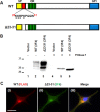
Figure 1.
Figure 1.
Schematic of Δ23–31 PrP, and expression in transfected cells. A, PrP residues 23–31, shown in WT PrP, were deleted to make Δ23–31 PrP. SP, N-terminal signal peptide; OR, octapeptide repeats; GPI, C-terminal GPI addition signal. B, Vector (lanes 1, 4), WT PrP (lanes 2, 5), or Δ23–31 PrP (lanes 3, 6) were expressed by transient transfection of N2a cells. Both PrP constructs carried an epitope tag for monoclonal antibody 3F4. Equal amounts of total protein were either left untreated (lanes 1–3) or were treated with PNGase F (lanes 4–6) before blotting with 3F4 antibody. Molecular size markers are shown in kilodaltons. C, WT PrP (FLAG-tagged) and Δ23–31 PrP (3F4-tagged) were coexpressed in BHK cells. Staining for FLAG (red) (i) and 3F4 (green) (ii) completely overlap in the merged image (yellow) (iii). DAPI staining of the nucleus is shown in blue. Scale bar, 25 μm.
Figure 2.
Figure 2.
Δ23–31 PrP is localized to detergent-resistant microdomains, is defective in endocytosis, and is converted into PrPSc in cultured cells. A, Cell lysates from HEK293 cells stably expressing either WT or Δ23–31 PrP were subjected to density gradient ultracentrifugation in an OptiPrep gradient. Fractions were analyzed by Western blotting using antibodies for PrP (6D11) and flotillin-1, a raft-resident protein. The majority of both flotillin-1 and PrP were found at the interface of the 0 and 30% OptiPrep fractions (lanes 2–4). A small amount of PrP, presumably derived from unlysed cells, can be found in the bottom fractions of the gradient (lanes 10–12). Molecular weight markers are shown in kilodaltons. B, N2a cells were transiently transfected with vector (i, iv), or with vector encoding WT PrP (ii, v) or Δ23–31 PrP (iii, vi). Cells were surface-stained with PrP antibody 3F4 on ice, and then incubated at 37°C to initiate endocytosis. Subsequently, cells were incubated in the absence (i–iii) or presence (iv–vi) of PIPLC. Cells were then fixed, permeabilized, and incubated with a fluorescently tagged secondary antibody (green) before DAPI staining (blue). Staining is observed for both WT and Δ23–31 PrP without PIPLC treatment (ii, iii), while only WT PrP (v), but not Δ23–31 PrP (vi), shows a punctate pattern of intracellular staining after PIPLC treatment. Scale bar, 25 μm. C, Chronically infected ScN2a cells were transiently transfected with vector, or with vector encoding WT (3F4) PrP or Δ23–31 (3F4) PrP. Cell lysates were incubated without (lanes 1–3) or with (lanes 4–6) PK before Western blotting with 3F4 antibody to detect transfected PrP. Both WT PrP (lane 5) and Δ23–31 PrP (lane 6) are converted into PK-resistant forms.
Figure 3.
Figure 3.
Expression and solubility of Δ23–31 PrP in transgenic mice. A, Equal volumes of protein from the brains of mice of the indicated genotypes were either untreated (lanes 1–5) or were treated with PNGase F (lanes 6–10) before blotting with anti-PrP antibody 6D11. Δ23–31 PrP is expressed at six times, four times, and one times in the three transgenic lines (lanes 3–5), compared with the level of WT PrP in a non-Tg mouse (lane 2). The asterisk denotes the endogenous C1 cleavage product. B, Brain lysates from Prn-p0/0 (lanes 1, 2), non-Tg (lanes 3, 4), Tg(Δ23–31) (lanes 5, 6), and Tg(PG14) (lanes 7, 8) mice were ultracentrifuged to separate soluble (S) and insoluble (I) fractions. Like WT, Δ23–31 PrP is found only in the soluble fraction, while PG14 PrP, an aggregation-prone mutant, is partially insoluble.
Figure 4.
Figure 4.
Tg(Δ23–31) mice survive longer than control mice after scrapie inoculation. Survival times were monitored in RML-inoculated mice of the following genotypes: Tga20+/+ (solid black line); Tga20+/0 (dashed black line); non-Tg (gray line); Tg(Δ23–31) (red line); Tg(Δ23–31) (blue line); Tg(Δ23–31) (green line). Each data point represents one animal. A statistical analysis of these data is shown in Table 1.
Figure 5.
Figure 5.
Tg(Δ23–31) mice accumulate less PrPSc than controls at 70 dpi. Brain homogenates from RML-inoculated mice of the indicated genotypes at 70 dpi were treated without (top panel) or with (bottom panel) PK and were then subjected to Western blotting using anti-PrP antibody 6D11.
Figure 6.
Figure 6.
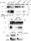
Tg(Δ23–31) mice accumulate greatly reduced amounts of PrPSc. A, Brain homogenates from RML-inoculated mice of the indicated genotypes at the terminal stage were treated without (top panels) or with (bottom panels) PK and were then subjected to Western blotting using anti-PrP antibody 6D11. The average signal intensity for PrP in terminally ill mice of each genotype was quantitated (numbers below the lanes) and is reported as a percentage of the amount of PK-resistant PrP in non-Tg mice. Ages of terminal mice in lanes 1–16 are as follows (in dpi): 69, 71, 76, 86, 159, 159, 112, 140, 142, 146, 147, 150, 157, 168, 301, 304. Actin is shown as a loading control. B, Brain homogenates from terminally ill, RML-infected Tga20+/0 mice and from Tg(Δ23–31) mice (short and long survivors) (+ lanes), and from age-matched, uninoculated control mice (− lanes) were treated with or without PK and Western blotted using anti-PrP antibody D18. Mice were killed at the following times (in dpi): 79, 86, 150, 152, 157, 168, 336, 399, 419, 427 (lanes 1, 2, 4–11, respectively). Uninoculated mice were killed at the following ages (in d): 82, 365 (lanes 3, 12, respectively). C, Brain homogenates from a terminally ill RML-inoculated Tga20+/0 mouse (86 dpi) and a terminally ill Tg(Δ23–31) mouse (140 dpi) were digested with the indicated amounts of PK and subjected to Western blotting with 6D11 antibody to detect PrP. Both short (top) and long (bottom) exposures of the blot are shown.
Figure 7.
Figure 7.
Tg(Δ23–31) and control mice display PrPSc accumulation and spongiform change in similar areas of the brain. A1–C8, Paraffin sections from mice of the indicated genotypes were stained for PrPSc. Representative staining of the thalamus (A1–A8), brainstem (B1–B8), and cortex (C1–C8) is shown. D1–D8, Paraffin sections from mice of the indicated genotypes were stained with hematoxylin and eosin. Representative sections of the brainstem are shown. Panels 1–6 (A–D) are from RML-inoculated mice, and panels 7 and 8 (A–D) are from uninoculated mice. Mice were killed at the following times (in dpi): 75 (A1, B1); 71 (C1); 75 (D1); 89 (A2, B2, C2); 82 (D2); 159 (A3, B3, C3); 150 (D3); 140 (A4, B4, C4); 112 (D4); 136 (A5, B5, C5); 152 (D5); 304 (A6, B6, C6, D6). Uninoculated mice were killed at the following ages (in d): 90 (A7, B7, C7, D7); 159 (A8, B8, C8, D8). Scale bars: A1–C8, 400 μm; D1–D8, 150 μm.
Figure 8.
Figure 8.
Secondary passage of RMLΔ23–31 scrapie into Tg(Δ23–31) mice does not shorten survival times. A, Survival times were monitored in mice of the following genotypes inoculated with RML or RMLΔ23–31: Tg(Δ23–31) (red lines); Tga20+/+ (black lines); non-Tg (gray lines). The circles represent individual mice inoculated with RML, and the squares represent mice inoculated with RMLΔ23–31. A statistical analysis of these data is shown in Table 3. B, Brain homogenates from terminally ill mice of the indicated genotypes inoculated with RML (lanes 1–3) or RMLΔ23–31 (lanes 4–15) were treated without (top panel) or with (bottom panel) PK, and were then subjected to Western blotting using anti-PrP antibody 6D11. Actin is shown as a loading control.
Figure 9.
Figure 9.
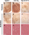
RML strain characteristics are preserved during secondary passage in Tg(Δ23–31) mice. Paraffin sections from terminally ill Tga20+/+, non-Tg, and Tg(Δ23–31) mice inoculated with RMLΔ23–31 display staining for PrPSc in the thalamus (A–C) and the brainstem (D–F). Hematoxylin and eosin staining shows spongiosis in the brainstem of these animals (G–I). Mice were killed at the following times (in dpi): 72 (A, D), 171 (B, E), 132 (C, F), 68 (G), 171 (H), 144 (I). Scale bars: A–F, 400 μm; G–I, 150 μm.
Figure 10.
Figure 10.
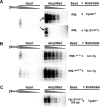
Deletion of residues 23–31 makes PrP an inefficient substrate, but does not compromise its seeding ability. PMCA reactions were run using the indicated seeds and substrates. Experiments were performed at least three times, and a representative example is shown. For each experiment, a nonamplified input sample is shown on the left, and an amplified sample on the right. The lanes represent reactions run with serial sixfold dilutions of the seed. All samples were digested with PK before Western blotting. A, RML from a terminally ill, non-Tg mouse was used to seed conversion of WT PrP (from a Tga20+/0 mouse) and Δ23–31 PrP [from a Tg(Δ23–31) mouse]. B, RMLΔ23–31 [from a terminally ill, RML-inoculated Tg(Δ23–31) mouse] or RML (from a terminally ill Tga20+/0 mouse) was used to seed conversion of WT PrP from a non-Tg mouse. The amounts of the two seeds were equalized, based on Western blotting for PK-resistant PrP. C, Brain homogenate from a healthy Tg(Δ23–31) mouse at 300 d after inoculation with RML was used to seed conversion of WT PrP from a Tga20+/+ mouse.
Figure 11.
Figure 11.
The N-terminal, polybasic domain mediates binding between PrPC and PrPSc. A, Myc-tagged WT or Δ23–28 recombinant PrPs were incubated in the presence or absence of RML-infected brain homogenate before immunoprecipitation with anti-myc antibody 9E10 (+ lanes) or naked beads (− lane). A portion of each sample was then incubated in the absence of PK, and Western blots probed with anti-myc antibody 4A6 (top panel). The remainder of each sample was digested with PK, and Western blots probed with anti-PrP antibody D18 (bottom panel). A sample of the RML used as input was is shown in lane 1 after PK digestion. Δ23–28 PrP pulled down far less PK-resistant PrP than WT PrP (compare lanes 2, 4). Blots shown are representative of at least three independent experiments. B, Model for the role of residues 23–31 in generation of PrPSc. Top panel, When residues 23–31 (represented by yellow plus signs) are present, PrPC (blue) binds strongly to PrPSc (red), leading to efficient conversion. Bottom panel, When these amino acids are deleted, as in Δ23–31 PrP, the PrPC–PrPSc interaction is less favorable, leading to reduced conversion. In this case, PrPSc is still generated but relies upon binding of PrPSc to PrPC at secondary sites. The red explosion shape represents the PK-resistant core of PrPSc. The 23–31 region of PrPSc is not involved in binding to PrPC.

Similar articles

Cited by

References

    1. Abalos GC, Cruite JT, Bellon A, Hemmers S, Akagi J, Mastrianni JA, Williamson RA, Solforosi L. Identifying key components of the PrPC-PrPSc replicative interface. J Biol Chem. 2008;283:34021–34028. - PMC - PubMed
    1. Barry AE, Klyubin I, McDonald JM, Mably AJ, Farrell MA, Scott M, Walsh DM, Rowan MJ. Alzheimer's disease brain-derived amyloid-β-mediated inhibition of LTP in vivo is prevented by immunotargeting cellular prion protein. J Neurosci. 2011;31:7259–7263. - PMC - PubMed
    1. Bell JE, Gentleman SM, Ironside JW, McCardle L, Lantos PL, Doey L, Lowe J, Fergusson J, Luthert P, McQuaid S, Allen IV. Prion protein immunocytochemistry—UK five centre consensus report. Neuropathol Appl Neurobiol. 1997;23:26–35. - PubMed
    1. Bolton DC, Meyer RK, Prusiner SB. Scrapie PrP 27–30 is a sialoglycoprotein. J Virol. 1985;53:596–606. - PMC - PubMed
    1. Borchelt DR, Davis J, Fischer M, Lee MK, Slunt HH, Ratovitsky T, Regard J, Copeland NG, Jenkins NA, Sisodia SS, Price DL. A vector for expressing foreign genes in the brains and hearts of transgenic mice. Genet Anal. 1996;13:159–163. - PubMed

Publication types

MeSH terms

LinkOut - more resources