. 2012 Jun 27;32(26):8879-89.
doi: 10.1523/JNEUROSCI.0558-12.2012.

MicroRNA-124 is a subventricular zone neuronal fate determinant

Affiliations

MicroRNA-124 is a subventricular zone neuronal fate determinant

Malin Åkerblom et al. J Neurosci. .

Abstract

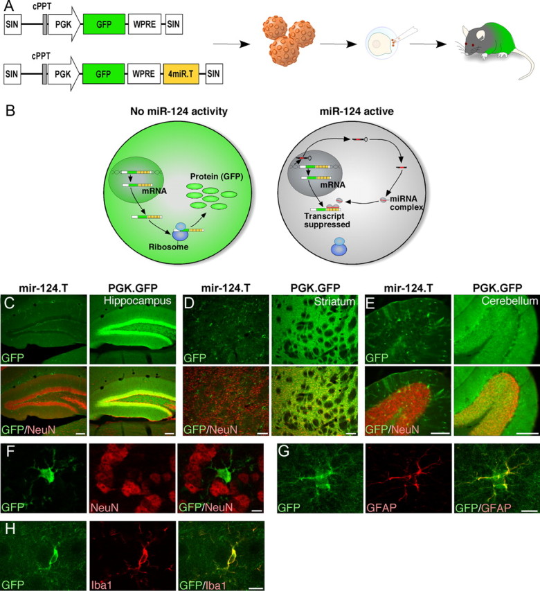
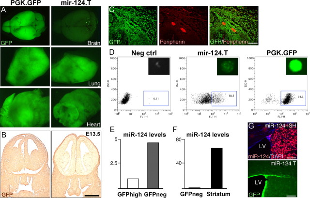
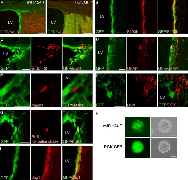
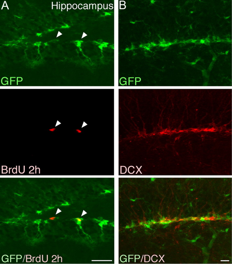
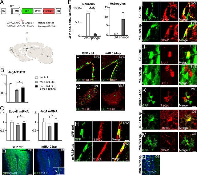
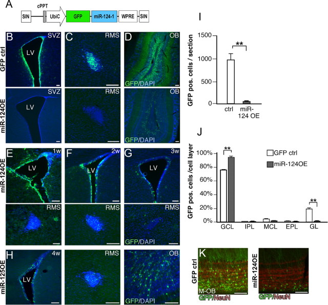
New neurons are continuously generated from neural stem cells with astrocyte properties, which reside in close proximity to the ventricle in the postnatal and adult brain. In this study we found that microRNA-124 (miR-124) dictates postnatal neurogenesis in the mouse subventricular zone. Using a transgenic reporter mouse we show that miR-124 expression is initiated in the rapid amplifying progenitors and remains expressed in the resulting neurons. When we stably inhibited miR-124 in vivo, neurogenesis was blocked, leading to the appearance of ectopic cells with astrocyte characteristics in the olfactory bulb. Conversely, when we overexpressed miR-124, neural stem cells were not maintained in the subventricular zone and neurogenesis was lost. In summary, our results demonstrate that miR-124 is a neuronal fate determinant in the subventricular zone.


Figures

Figure 1.
Figure 1.
miR-124 is a neuron-specific miRNA. A, Illustration of the vectors and experimental approach to generate miR-124.T reporter mice using lentiviral transgenesis. Lentiviral vectors were injected into the perivitelline space of fertilized embryos, which were transplanted into a pseudopregnant mouse. B, The vector-encoded transcript is expressed in both miR-124 expressing cells and nonexpressing cells. In the miR-124-expressing cells, the target site containing transcripts is suppressed, resulting in no GFP expression. CE, GFP expression in miR-124.T reporter mice was found exclusively in nonneuronal cells throughout the brain. Control mice (PGK.GFP) displayed GFP activity in all cells of the brain. Scale bars, 100 μm. FH, Confocal analysis of GFP cells in miR-124.T mice showing that neurons (NeuN) do not express GFP while astrocytes (GFAP) and microglia (IBA1) do. Scale bars, 10 μm. cPPT, Central polypurine tract; SIN, self-inactivating.
Figure 2.
Figure 2.
Characterization of miR.124.T mice. A, Adult miR.124.T mice display low GFP expression in the brain, while other tissues, here exemplified by lung and heart, display similar GFP levels as control animals. B, The developing brain is almost entirely depleted of miR-124 activity at E13.5, as shown by diaminobenzidine-staining for GFP. Scale bar, 1 mm. C, Confocal analysis of E13.5 peripheral ganglion reveals GFP expression in peripheral nervous system neurons labeled with PERIPHERIN. Scale bars, 100 μm. D, Generation of neurospheres from forebrain of E.13.5 miR.124.T embryos. FACS plots showing that in miR.124.T cultures, ∼20% of the cells express GFP at early passages. E, F, FACS sorting of the GFP-expressing cells followed by LNA-qRT-PCR for mature miR-124 reveals that mature miR-124 levels are higher in the GFP negative fraction. G, LNA-ISH for miR-124 reveals high-level expression in neurons (top). There is also an absence of miR-124 expression in the ependymal layer that mirrors the expression pattern in miR-124.T mice (bottom). LV, lateral ventricle. Scale bars, 100 μm. Neg, Negative.
Figure 3.
Figure 3.
miR-124 activity is absent from stem cells but initiated in rapid-amplifying progenitors in the adult SVZ. A, GFP/NeuN expression in the adult SVZ in miR-124 reporter mice demonstrates that miR-124 is absent from parts of the niche. Scale bars, 100 μm. BD, Cells that express GFP, thus lacking miR-124, colabel with S100β and GFAP but only rarely incorporate BrdU in the SVZ. Scale bars, 20 μm. E, F, GFP-expressing cells do not colabel with markers for rapid-amplifying progenitors (MASH1) and migrating neuroblasts (DCX) in miR-124 reporter mice. Scale bars, 20 μm. G, BrdU pulse chase experiment in which the thymidine analog was injected 4 weeks before killing of the animals demonstrates that slowly dividing cells in the SVZ express GFP in miR-124.T mice. Scale bars, 20 μm. H, GFP expression in neurosphere cultures derived from the adult SVZ revealing a small population of green cells in spheres. Scale bar, 100 μm. I, GFP-expressing cells in the miR-124.T mice express the miR-124 target JAG1. Scale bars, 20 μm. LV, Lateral ventricle; w, weeks.
Figure 4.
Figure 4.
GFP expression in hippocampus in adult brain. A, In the DG we found GFP-expressing cells with morphologies resembling what have previously been attributed to progenitor cells. Confocal analysis revealed that a proportion of the GFP-expressing cells incorporated BrdU. It is worth noting that the proportion of BrdU-incorporating cells that expressed GFP was higher in the DG when compared with the SVZ, which reflects differences in the proliferation of progenitors in the two niches. B, GFP-expressing cells did not colabel with DCX. Scale bars, 20 μm.
Figure 5.
Figure 5.
Injection of lentiviral vectors into the ventricles of p3 mice allows efficient targeting of SVZ stem cells. A, B, We found abundant GFP expression in the ventricular zone at both 1 week and 8 weeks after vector injection. Scale bars, 100 μm. C, D, Eight weeks after injection we found GFP-expressing cells colabeling with either GFAP or S100β in the SVZ. Scale bars, 50 μm. E, In the RMS we found GFP-expressing migrating neuroblasts (DCX) 8 weeks after vector injection demonstrating an ongoing migration of GFP-expressing progenitors. Scale bars, 100 μm. F, In the OB we found numerous GFP-expressing neurons that colabeled with the neuronal marker NeuN 8 weeks after vector injection. Scale bars, 100 μm. G, We also performed a pulse chase experiment in which animals were injected with BrdU 4 weeks after vector injection and then killed 4 weeks later, at 8 weeks of age. We found GFP-expressing neurons in the OB that colabeled with BrdU. This demonstrates that cells dividing 4 weeks after vector injection still retain the GFP transgene, showing that slowly dividing stem cells are targeted. Scale bar, 10 μm. w, Weeks.
Figure 6.
Figure 6.
Stable inhibition of miR-124 in the SVZ blocks neurogenesis, leading to the formation of ectopic glial cells in the OB. A, Illustration of the vectors and experimental approach to inhibit miR-124 in the postnatal SVZ. Lentiviral sponge vectors were injected at P3. Illustration shows the migratory route of neuroblasts from the SVZ via the RMS to the OB. B, Luciferase assay in HeLa cells for the miR-124 target gene Jag1 demonstrating the efficiency of the lentiviral constructs. Graph display ratio of Renilla to firefly luciferase activity normalized to cells transfected with the Jag1–3′UTR construct only. Data represent a mean of four independent experiments. Data are presented as mean ± SEM. *p < 0.05, Student's t test. C, Examples of two miR-124 target genes (Evovl1 and Jag1) that display reduced expression in HeLa cells following miR-124 overexpression (black bars). Coexpression of miR-124 sponge rescues the downregulation. Data are presented as mean ± SEM; n = 6. *p < 0.05, Student's t test. Evovl1 is taken from Conaco et al. (2006) and Jag1 from Cheng et al. (2009). D, GFP expression in the OB 4 weeks after control and sponge vector injection. In animals injected with miR-124 sponge vectors, only very few GFP-expressing cells can be detected. OB is counterstained with DAPI. Scale bars, 250 μm. E, Quantification of GFP-expressing neurons and astrocytes in the OB after amplification of the GFP signal using immunohistochemistry. Data are presented as mean ± SEM; n = 3. ***p < 0.001. n.d., not detected. F, G, GFP expression in the SVZ and RMS 4 weeks after vector injection. SVZ is counterstained with s100β and the RMS with DCX. Scale bars: 50 μm (F); 75 μm (G). HJ, GFP-expressing cells in the SVZ 4 weeks after injection of ctrl or miR-124 sponge vector. In animals injected with sponge vector, it was possible to detect GFP cells expressing s100β and GFAP, but these GFP cells did not incorporate BrdU. BrdU was injected 2 h before kill. Scale bars, 10 μm. K, GFP-expressing cells in the RMS 4 weeks after injection of sponge vector colabeled with s100β. Scale bars, 10 μm. L, M, GFP-expressing cells in the OB 4 weeks after sponge injection colabeled with S100β and GFAP. Scale bars, 10 μm. N, GFP-expressing neurons in the OB 4 weeks after injection of a control lentiviral vector expressing a miR-125b sponge sequence. Scale bars, 100 μm. cPPT, Central polypurine tract; SIN, self-inactivating; w, weeks.
Figure 7.
Figure 7.
Overexpression of miR-124 in the postnatal SVZ leads to loss of neural stem cells. A, Illustration of the vector to overexpress miR-124 in the postnatal SVZ. Lentiviral vectors were injected at P3. B, C, Four weeks after the injection of miR-124OE, no GFP-expressing cells can be detected in either SVZ or RMS, suggesting that all stem and progenitor cells overexpressing miR-124 have migrated away and differentiated to OB neurons. D, In animals injected with miR-124OE vectors, OB neurons are generated to a lesser extent when compared with control injections. EG, GFP-expressing cells are gradually lost from the SVZ and RMS after injection of miR-124OE vector. H, Injection of a control lentiviral vector overexpressing the unrelated miR-125b does not cause loss of GFP expression in the SVZ and RMS. I, Quantification of GFP-expressing neurons in the OB. Data are presented as mean ± SEM; n = 3. **p < 0.01. J, Altered integration into the OB after miR-124 overexpression. Data are presented as mean ± SEM; n = 3. *p < 0.05. K, Newborn neurons overexpressing miR-124 integrate primarily in the inner parts of the GCL. LV, Lateral ventricle; M-OB, most medial part of the OB. All scale bars, 100 μm. cPPT, Central polypurine tract; EPL, external plexiform layer; GL, glomerular layer; IPL internal plexiform layer; MCL, mitral cell layer; SIN, self-inactivating; UbiC, ubiquitin C; w, weeks.

References

    1. Ahlenius H, Kokaia Z. Isolation and generation of neurosphere cultures from embryonic and adult mouse brain. Methods Mol Biol. 2010;633:241–252. - PubMed
    1. Ambasudhan R, Talantova M, Coleman R, Yuan X, Zhu S, Lipton SA, Ding S. Direct reprogramming of adult human fibroblasts to functional neurons under defined conditions. Cell Stem Cell. 2011;9:113–118. - PMC - PubMed
    1. Bartel DP. MicroRNAs: target recognition and regulatory functions. Cell. 2009;136:215–233. - PMC - PubMed
    1. Brown BD, Gentner B, Cantore A, Colleoni S, Amendola M, Zingale A, Baccarini A, Lazzari G, Galli C, Naldini L. Endogenous microRNA can be broadly exploited to regulate transgene expression according to tissue, lineage and differentiation state. Nat Biotechnol. 2007;25:1457–1467. - PubMed
    1. Cao X, Pfaff SL, Gage FH. A functional study of miR-124 in the developing neural tube. Genes Dev. 2007;21:531–536. - PMC - PubMed

Publication types

MeSH terms

LinkOut - more resources