. 2012 Jun 27;32(26):8977-87.
doi: 10.1523/JNEUROSCI.6494-11.2012.

Thrombospondin-4 contributes to spinal sensitization and neuropathic pain states

Affiliations

Thrombospondin-4 contributes to spinal sensitization and neuropathic pain states

Doo-Sik Kim et al. J Neurosci. .

Abstract

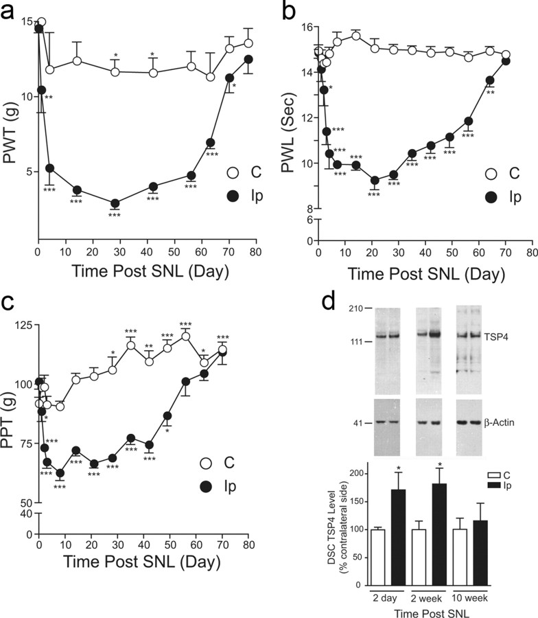
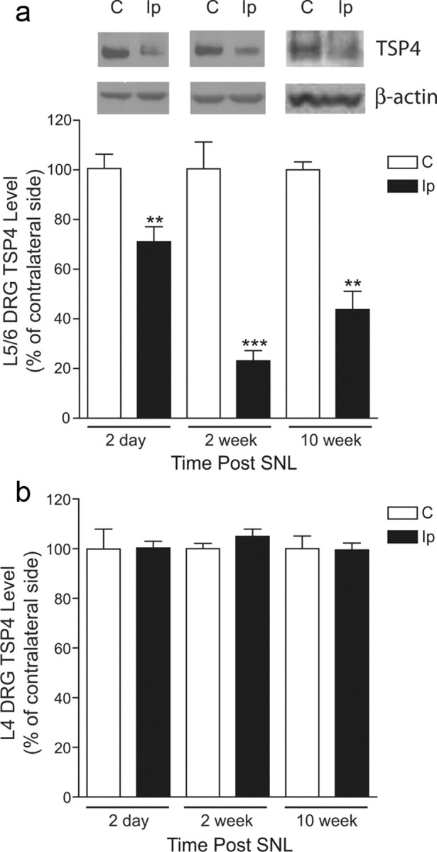
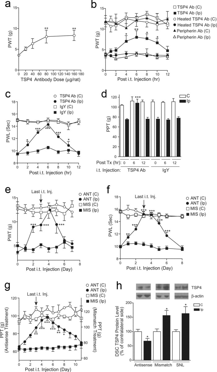
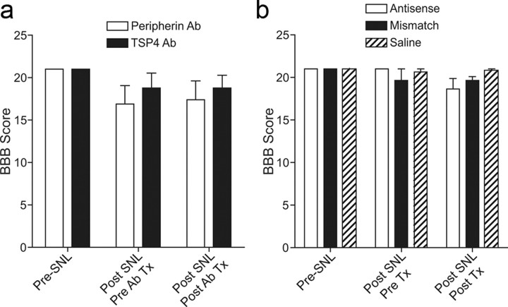
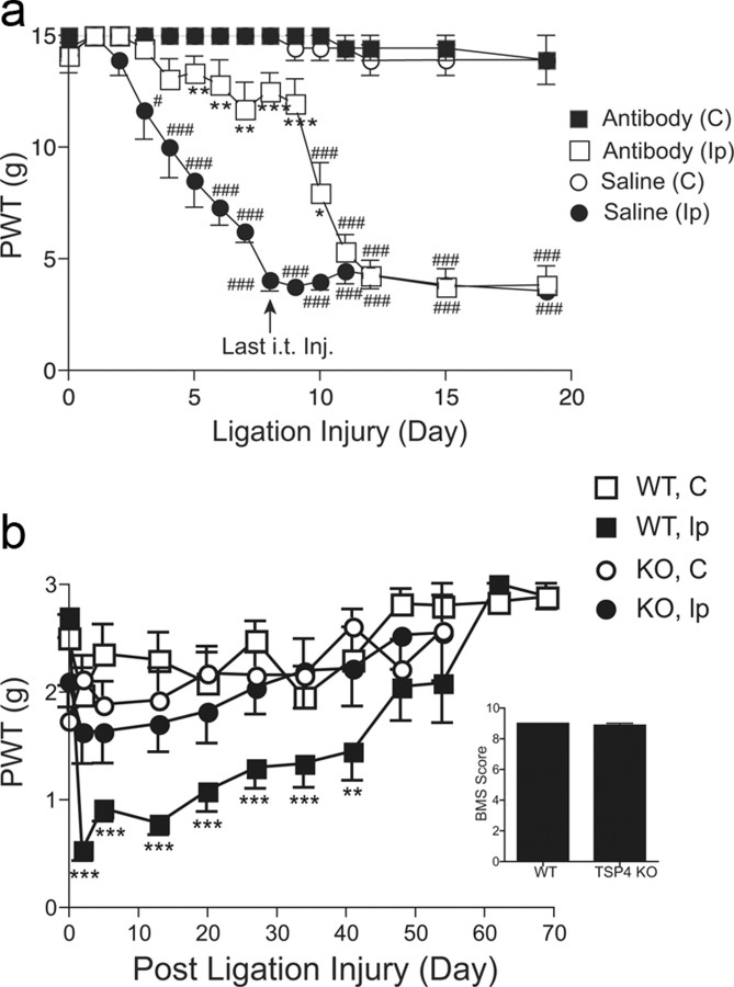
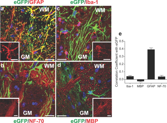
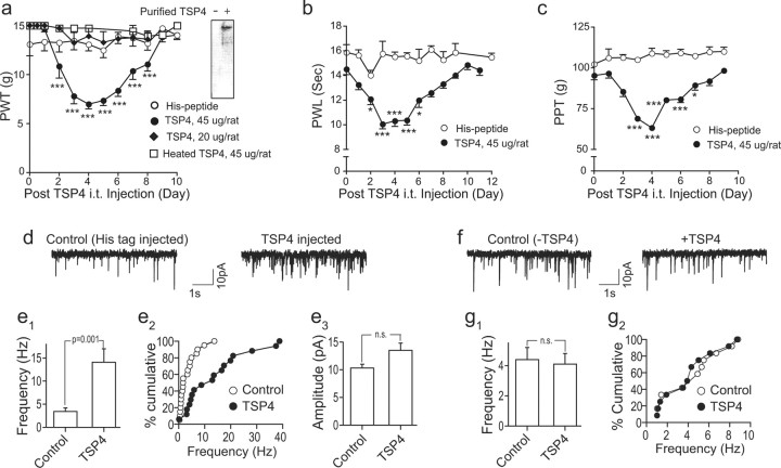
Neuropathic pain is a common cause of pain after nerve injury, but its molecular basis is poorly understood. In a post-gene chip microarray effort to identify new target genes contributing to neuropathic pain development, we report here the characterization of a novel neuropathic pain contributor, thrombospondin-4 (TSP4), using a neuropathic pain model of spinal nerve ligation injury. TSP4 is mainly expressed in astrocytes and significantly upregulated in the injury side of dorsal spinal cord that correlates with the development of neuropathic pain states. TSP4 blockade by intrathecal antibodies, antisense oligodeoxynucleotides, or inactivation of the TSP4 gene reverses or prevents behavioral hypersensitivities. Intrathecal injection of TSP4 protein into naive rats is sufficient to enhance the frequency of EPSCs in spinal dorsal horn neurons, suggesting an increased excitatory presynaptic input, and to cause similar behavioral hypersensitivities. Together, these findings support that injury-induced spinal TSP4 may contribute to spinal presynaptic hypersensitivity and neuropathic pain states. Development of TSP4 antagonists has the therapeutic potential for target-specific neuropathic pain management.


Figures

Figure 1.
Figure 1.
Nerve injury-induced TSP4 upregulation correlated temporally with the development of behavioral hypersensitivities. ac, Unilateral L5/6 SNL injury caused a reduction in PWT to light touch (tactile allodynia) (a), PWL to thermal stimulation (thermal hyperalgesia) (b), and hindpaw pressure withdrawal thresholds (PPT) (mechanical hyperalgesia) (c) in the injury side compared to those in the noninjury side. In the paw pressure testing, there was a time-dependent conditioning adaptive increase in PPT in both groups. All behavioral hypersensitive states recovered gradually after ∼7–10 weeks post-ligation injury. Data presented are the mean ± SEM of at least five rats in each group. *p < 0.05, **p < 0.01, and ***p < 0.001 compared with the pretreatment levels as determined by two-way ANOVA with Bonferroni post-tests. d, Western blot data indicated that L5/6 SNL injury caused TSP4 protein upregulation in dorsal spinal cord (DSC) of the injury side at time points that correlated with the initiation and maintenance of behavioral hypersensitivities. The injury-induced TSP4 upregulation returned to a level similar to that in the noninjury side when injured animals recovered from the behavioral hypersensitive states. Representative Western blot bands are shown on top of each bar group, and estimated molecular weights (kDa) are shown on the left. Data shown are the mean ± SEM of 5–6 rats in each group. *p < 0.05 compared with the contralateral side by Student's t test. C, Contralateral; Ip, ipsilateral.
Figure 2.
Figure 2.
Spinal nerve injury decreased TSP4 protein expression in injured, but not in adjacent uninjured, DRGs. a, Western blot data indicated that L5/6 spinal nerve ligation injury caused the downregulation of TSP4 proteins in L5/6 DRGs of the injury side at all the time points examined. Representative Western blot bands are shown on top of each bar group in the same order as the summarized bar graph data, which are the mean ± SEM of ≥9 rats in each group. **p < 0.01 and ***p < 0.001 compared with the contralateral side by Student's t test. b, In contrast, L5/6 spinal nerve ligation injury did not change TSP4 protein levels in uninjured, adjacent L4 DRG from the injury side. Data presented are the mean ± SEM of nine rats in each group. C, Contralateral; Ip, ipsilateral.
Figure 3.
Figure 3.
Blocking injury-induced spinal TSP4 resulted in reversal of behavioral hypersensitivities. a, Bolus intrathecal injection of TSP4 antibodies dose dependently reversed injury-induced tactile allodynia in SNL rats. Behavioral test on the injury side started 6 h after the bolus injection of TSP4 antibodies. Data shown are the mean ± SEM of 3–6 rats in each group. **p < 0.01 compared with the pretreatment level, as determined by one-way ANOVA with Dunnett's multiple comparison tests. b, Bolus intrathecal injection of TSP4 antibodies (80 μg/rat) reversed injury-induced tactile allodynia in SNL rats without affecting the behavioral thresholds on the noninjury side. The anti-allodynic effect of TSP4 antibodies had an onset time of 4 h, peaked at 6 h, and had a duration of >6 h. Neither heated TSP4 antibodies nor antibodies against another protein, peripherin, showed anti-allodynia effects. Data shown are the mean ± SEM of 6 rats in each group. *p < 0.05 and **p < 0.01 compared with the pretreatment level as determined by two-way ANOVA with Bonferroni post-tests. c, d, Bolus intrathecal injection of TSP4 antibodies (80 μg/rat) reversed injury-induced thermal hyperalgesia (c) and mechanical hyperalgesia (d) in SNL rats without affecting the behavioral thresholds on the noninjury side. The same dose of chicken IgY was used as control. To avoid sensitization of the hindpaw in paw pressure testing, mechanical hyperalgesia was tested in a 6 h interval as shown (d). Data shown are the mean ± SEM of 5–6 rats in each group. *p < 0.05 and ***p < 0.001 compared with the pretreatment level, as determined by two-way ANOVA with Bonferroni post-tests. eg, Daily intrathecal injection of TSP4 antisense (ANT), but not mismatch (MIS), oligodeoxynucleotides (50 μg/rat/d) for 4 d reversed injury-induced tactile allodynia (e), thermal hyperalgesia (f), and mechanical hyperalgesia (g) in SNL rats. Behavioral tests were performed before each daily injection and daily after the last injection. Depending on the modality tested, the anti-nociceptive effect of TSP4 antisense oligodeoxynucleotides had an onset time of 1–3 d, peaked at 3–5 d, and lasted for >2–5 d after the last injection. The baseline PPT value for the mismatch treatment (g, right y-axis) was higher than that in the antisense treatment due to the conditioning adaptive increase in PPT (as shown in Fig. 1c) in the same group of rats that was used for the antisense treatment first, followed by the mismatch treatment at least 48 h apart. Data shown are the mean ± SEM of ≥5 rats in each group. *p < 0.05, **p < 0.01, and ***p < 0.001 compared with the pretreatment level as determined by two-way ANOVA with Bonferroni post-tests. h, Intrathecal TSP4 antisense treatment (50 μg/rat/d for 4 d) blocked injury-induced TSP4 expression in dorsal spinal cord. L5/6 dorsal spinal cord (DSC) samples were collected 1 d after the last oligodeoxynucleotide treatment from SNL rats and were subjected to Western blot analyses. Representative Western blot bands are shown on top of each bar group. Data shown are the mean ± SEM of ≥7 rats in each group. SNL, 6-week SNL rats without treatment; C, Contralateral; Ip, ipsilateral.
Figure 4.
Figure 4.
Locomotor functions were not impaired by intrathecal treatments with TSP4 antibody or antisense oligodeoxynucleotides. a, b, Locomotor functions were analyzed with the Basso, Beattie, Bresnahan (BBB) locomotor rating scale in SNL rats before and after SNL surgery and treatments with antibodies (a) or oligodeoxynucleotides (b) as described. The post-treatment time points were 6 h after bolus antibody injections (a) or overnight after the last intrathecal injection of the 4 d antisense treatments (b); both were correlated with reversals of behavioral hypersensitivities shown in Figure 3b–d and 3e–g, respectively. Data presented are the mean ± SEM of ≥5 rats in each group. Tx, Treatment.
Figure 5.
Figure 5.
Blocking injury induction of spinal TSP4 prevented neuropathic allodynia initiation. a, Intrathecal preemptive treatment with TSP4 antibodies prevented allodynia onset in spinal nerve-ligated rats in a reversible manner. TSP4 antibodies (80 μg/rat/d) were injected directly into L5/6 spinal region immediately before and daily after SNL for 8 d. Behavioral tests were performed before and daily after SNL and before each daily injection. Data presented are the mean ± SEM of 6 rats in each group. *p < 0.05, **p < 0.01, and ***p < 0.001 compared with the saline-injected group. #p < 0.05 and ###p < 0.001 compared with the pretreatment level using two-way ANOVA with Bonferroni post-tests. b, TSP4 null mice had diminished tactile allodynia post SNL. Unilateral L5 SNL was performed in adult male TSP4 null mice (KO) and their age- and sex-matched WT littermates. Behavioral tests were performed blindly before and after SNL. Data presented are the mean ± SEM of ≥7–12 mice in each group. TSP4 null expression did not impair the locomotor functions of the TSP4 KO mice compared with their WT littermates, as analyzed with the Basso Mouse Scale (BMS) for locomotion from ≥8 mice in each group (inset, mean ± SEM). **p < 0.01 and ***p < 0.001 compared with pre-SNL level using two-way ANOVA with Bonferroni post-tests. C, Contralateral; Ip, ipsilateral.
Figure 6.
Figure 6.
SNL did not alter TSP1 and TSP2 protein levels in dorsal spinal cords of WT and TSP4 KO mice. Western blot data indicated that L5 SNL injury (4 weeks) did not alter dorsal spinal cord (DSC) TSP1/2 protein levels significantly in WT and TSP4 KO mice when allodynia was observed in SNL WT but not SNL TSP4 KO mice, as shown in Figure 5b. a, Representative Western blot data. b, Summarized Western blot data presented as the mean ± SEM of 3–4 independent experiments in each group.
Figure 7.
Figure 7.
eGFP immunoreactivity in dorsal spinal cords of SNL TSP4-eGFP mice. a, Overview of an L5 dorsal spinal cord section from a 2-week SNL TSP4-eGFP mouse (representative image from 4 mice). The dotted line outlines the dorsal horn (DH) and dorsal column (DC) areas in both sides. Scale bar, 200 μm. b, Enlarged view of dorsal spinal cord presented in a showing the structure of eGFP-positive cells. Scale bar, 200 μm. ce, SNL injury induced significant increases in the surface area (c) and intensity (d) of eGFP signals in TSP4-eGFP expressing cells in dorsal spinal cord without altering the total number of eGFP-expressing cells (e). Data presented are the mean ± SEM of a total of 24 sections, 6 from each of the 4 SNL mice. ***p < 0.001 compared with contralateral side by Student's t test. C, Contralateral; Ip, ipsilateral.
Figure 8.
Figure 8.
TSP4-eGFP signals in dorsal spinal cord were mainly in astrocytes. ad, Fluorescent immunoreactivity to antibodies of commonly used cell type-specific markers was superimposed with TSP4-eGFP signals in ipsilateral dorsal spinal cord sections from 2-week SNL TSP4-eGFP mice. Representative images from 4 mice are shown. TSP4 promoter-driven eGFP expression signals (green) mainly colocalized with immunoreactivity to antibodies against GFAP, a marker for activated astrocytes (red, a), but not with immunoreactivities to antibodies against NF-70, a marker for neurofilaments (red, b), Iba-1, a marker for microglial cells (red, c), or MBP (red, d). WM, white matter; GM, gray matter. Scale bars, 20 μm. Blue, nuclei. (inset) Enlarged images in each panel showing morphology of each type of immunoreactive cells, respectively. Scale bars: 10 μm. e, Possibility of colocalization of fluorescent signals from eGFP and immunoreactivity to each cell marker antibody was analyzed as described previously (Barlow et al., 2010). Data are presented as Pearson's correlation coefficients from the mean ± SEM derived from pooled data of 12 sections of 4 mice.
Figure 9.
Figure 9.
Increased spinal TSP4 is sufficient to induce dorsal horn neuron sensitization and behavioral hypersensitivities through a chronic mechanism. ac, Bolus intrathecal injection of 45 μg/rat TSP4 into L5/6 spinal regions of naive rats at time 0 led to long-lasting and reversible hindpaw hypersensitivities (averaged from both sides) assessed by blind daily behavioral test, including tactile allodynia (a), thermal hyperalgesia (b), and mechanical hyperalgesia (c). Intrathecal injection of 20 μg/rat TSP4 proteins, heated (45 μg/rat) TSP4 proteins (a), equal molar dose of His-tag peptides (ac) did not cause behavioral hypersensitivities or alter baseline behavioral thresholds. Western blot showing purified TSP4-His fusion proteins detected by TSP4 antibodies (a, inset). Data presented are mean ± SEM of 5–6 rats in each group. *p < 0.05, **p < 0.01, and ***p < 0.001 compared with pretreatment level by two-way ANOVA with Bonferroni post-tests. d, e, Intrathecal injection of TSP4 proteins into L5/6 spinal regions induced spinal neuron sensitization in naive rats that correlated with behavioral hypersensitivities. (d) Representative currents of AMPA receptor-mediated mEPSCs from superficial dorsal horn neuron whole-cell patch recording in L5 spinal cord slices of rats injected intrathecally with equal molar doses of His-tag peptides (Control) or TSP4-His fusion proteins (TSP4; 45 μg/rat) 4 d earlier. Intrathecal TSP4 proteins induced an increase in the average frequency (e1) as well as cumulative frequency distribution (e2), but not amplitude (e3), of mEPSCs. Data presented are summarized mean ± SEM of a total of 17–20 dorsal horn neurons recorded from spinal cord slices of 5 rats in each group. f, g, Acute application of TSP4 proteins to spinal cord slices did not alter mEPSCs in dorsal horn neurons. mEPSCs from naive rat L5 superficial dorsal horn neurons were recorded under identical conditions for spinal cord slice recording (d, e1–e3) except that TSP4 proteins were added to the recording bath for 15 min. Representative traces of mEPSC before and after bath application of 15 μg/mL TSP4 (f), a concentration that is similar to the spinal TSP4 concentration in vivo at the first injection day that caused behavioral and dorsal horn neuron sensitization later as shown in ae, assuming the rate of CSF formation is 2.2 μl/min, approximately 3 mL/d/rat (Cserr, 1965) without considering the turnover of CSF and TSP4 proteins. Summarized graphs showing the lack of acute TSP4 effects on mEPSC average frequency (g1) and cumulative frequency distribution (g2) from rat superficial dorsal horn neurons. Data presented are the mean ± SEM of a total of 12 dorsal horn neurons recorded from spinal cord slices of 3 rats in each group. n.s., Not significant.

Similar articles

Cited by

References

    1. Adams JC. Thrombospondins: multifunctional regulators of cell interactions. Annu Rev Cell Dev Biol. 2001;17:25–51. - PubMed
    1. Adams JC, Lawler J. The thrombospondins. Int J Biochem Cell Biol. 2004;36:961–968. - PMC - PubMed
    1. Arber S, Caroni P. Thrombospondin-4, an extracellular matrix protein expressed in the developing and adult nervous system promotes neurite outgrowth. J Cell Biol. 1995;131:1083–1094. - PMC - PubMed
    1. Barlow AL, Macleod A, Noppen S, Sanderson J, Guérin CJ. Colocalization analysis in fluorescence micrographs: verification of a more accurate calculation of Pearson's correlation coefficient. Microsc Microanal. 2010;16:710–724. - PubMed
    1. Basso DM, Beattie MS, Bresnahan JC. A sensitive and reliable locomotor rating scale for open field testing in rats. J Neurotrauma. 1995;12:1–21. - PubMed

Publication types

MeSH terms