. 2012:7:2699-712.
doi: 10.2147/IJN.S30074. Epub 2012 Jun 1.

Particokinetics: computational analysis of the superparamagnetic iron oxide nanoparticles deposition process

Affiliations

Particokinetics: computational analysis of the superparamagnetic iron oxide nanoparticles deposition process

Walter H Z Cárdenas et al. Int J Nanomedicine. 2012.

Retraction in

Abstract

Background: Nanoparticles in suspension are often utilized for intracellular labeling and evaluation of toxicity in experiments conducted in vitro. The purpose of this study was to undertake a computational modeling analysis of the deposition kinetics of a magnetite nanoparticle agglomerate in cell culture medium.

Methods: Finite difference methods and the Crank-Nicolson algorithm were used to solve the equation of mass transport in order to analyze concentration profiles and dose deposition. Theoretical data were confirmed by experimental magnetic resonance imaging.

Results: Different behavior in the dose fraction deposited was found for magnetic nanoparticles up to 50 nm in diameter when compared with magnetic nanoparticles of a larger diameter. Small changes in the dispersion factor cause variations of up to 22% in the dose deposited. The experimental data confirmed the theoretical results.

Conclusion: These findings are important in planning for nanomaterial absorption, because they provide valuable information for efficient intracellular labeling and control toxicity. This model enables determination of the in vitro transport behavior of specific magnetic nanoparticles, which is also relevant to other models that use cellular components and particle absorption processes.

Keywords: agglomerates; cellular labeling; computational modeling; diffusion; magnetic resonance imaging; magnetite; nanoparticles; sedimentation.


Figures

Figure 1
Figure 1
Representation of process of SPION deposition and cellular internalization. Notes: The result of diffusion and gravitational sedimentation effects guide particle deposition at the bottom of the recipient. Positioning at the boundary of the cellular surfaces is essential for internalization and labeling of the absorbed dose. Abbreviation: SPION, superparamagnetic iron oxide nanoparticle.
Figure 2
Figure 2
Discrete deposition of SPIONs in suspension that reach their terminal settling velocity, vterm. Notes: In the figure, z represents the generic coordinate for depth, where z = 0, and z = L characterizes the top and the bottom of the recipient, respectively. h(t) is the distance particles have fallen down by in a period of time t, computed as vtermt. Abbreviation: SPIONs, superparamagnetic iron oxide nanoparticles.
Figure 3
Figure 3
Transport time of magnetite particle to propagate 1 mm in terms of particle diameter. Notes: Transport is represented for diffusion (red line) and sedimentation (blue line) in an aqueous solution. The 1 mm distance is the mean depth for a culture medium used frequently in in vitro evaluation of intracellular labeling. The intersection point of both types of transport is about 75 nm in diameter for a Fe3O4 particle.
Figure 4
Figure 4
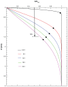
Normalized concentration profiles obtained by the Crank–Nicolson algorithm for resolution of the convection-diffusion equation at different time exposure intervals.
Figure 5
Figure 5
Temporal normalized concentration variability of superparamagnetic iron oxide nanoparticles at z = 1 mm during 24 hours of exposure and considering a mean height of 4 mm.
Figure 6
Figure 6
Dose curves of deposited fraction of magnetite nanoparticles (different sizes) in terms of exposure time. Notes: All curves were obtained considering a mean height of 4 mm. Dashed lines correspond to equation (6) calculation considering the diffusion factor only.
Figure 7
Figure 7
The plotted curves represent the temporal variation in the fraction of dose deposited, with simulated curves for experimental data obtained using magnetic resonance imaging. Notes: The inset shows images acquired by T2-weighted magnetic resonance imaging of suspensions of superparamagnetic iron oxide nanoparticles 50 nm and 200 nm in diameter in Dulbecco’s Modified Eagle’s Medium supplemented with 10% fetal bovine serum. The theoretical curves are the same as those shown in Figure 6.
Figure 8
Figure 8
Variability in fraction of the dose deposited on cellular surfaces with a determined: mean of height, suspension volume, and culture medium for superparamagnetic iron oxide nanoparticles of different diameters.
Figure 9
Figure 9
Fraction of dose deposited per agglomerate of primary superparamagnetic iron oxide nanoparticles (Fe3O4) during 24 hours of exposure with a mean height of 4 mm. Simulated curves for primary nanoparticles 25 nm in diameter (A and B) and 50 nm (C and D). Note: Each line represent a different number of nanoparticles per agglomerate (1, 10, 50, 100, 250, 500, 1000, and 10,000), with varying DF values.
Figure 10
Figure 10
Dose fraction curves for superparamagnetic iron oxide nanoparticles 50 nm in diameter deposited over 6, 12, 18, and 24 hours of exposure. Notes: The concentration profiles increase within duration of exposure and a null m value (blue, orange, cyan and red lines) was applied. Curves with a dispersion factor in terms of time (full black lines) are shown for crescent (mt) and decrescent (−mt) dependencies over time t(h). The value adopted for m was 0.05 h−1.

References

    1. Sousa MH, Rubim JC, Sobrinho PG, Tourinho FA. Biocompatible magnetic fluid precursors based on aspartic and glutamic acid modified maghemite nanostructures. J Magn Magn Mater. 2001;225:67–72.
    1. Tartaj P, Morales MP, Veintemillas-Verdaguer S, Gonzáles-Carreno T, Serna CJ. The preparation of magnetic nanoparticles for applications in biomedicine. J Phys D Appl Phys. 2003;36:R182–R197.
    1. Gupta AK, Wells S. Surface-modified superparamagnetic nanoparticles for drug delivery: preparation, characterization, and cytotoxicity studies. IEEE Trans Nanobioscience. 2004;3:66–73. - PubMed
    1. Kopelman R, Koo Y-EL, Philbert M, et al. Multifunctional nanoparticle platforms for in vivo MRI enhancement and photodynamic therapy of a rat brain cancer. J Magn Magn Mater. 2005;293:404–410.
    1. Bonnemain B. Superparamagnetic agents in magnetic resonance imaging: physicochemical characteristics and clinical applications. J Drug Target. 1998;6:167–174. - PubMed

Publication types