

Antimicrobial applications of nanotechnology: methods and literature
- PMID: 22745541
- PMCID: PMC3383293
- DOI: 10.2147/IJN.S24805
Antimicrobial applications of nanotechnology: methods and literature
Abstract
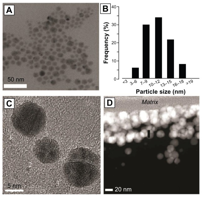
The need for novel antibiotics comes from the relatively high incidence of bacterial infection and the growing resistance of bacteria to conventional antibiotics. Consequently, new methods for reducing bacteria activity (and associated infections) are badly needed. Nanotechnology, the use of materials with dimensions on the atomic or molecular scale, has become increasingly utilized for medical applications and is of great interest as an approach to killing or reducing the activity of numerous microorganisms. While some natural antibacterial materials, such as zinc and silver, possess greater antibacterial properties as particle size is reduced into the nanometer regime (due to the increased surface to volume ratio of a given mass of particles), the physical structure of a nanoparticle itself and the way in which it interacts with and penetrates into bacteria appears to also provide unique bactericidal mechanisms. A variety of techniques to evaluate bacteria viability, each with unique advantages and disadvantages, has been established and must be understood in order to determine the effectiveness of nanoparticles (diameter ≤ 100 nm) as antimicrobial agents. In addition to addressing those techniques, a review of select literature and a summary of bacteriostatic and bactericidal mechanisms are covered in this manuscript.
Keywords: antibacterial; bacteria; biofilm; nanomaterial; nanoparticle; nanotechnology.
Figures
References
-
- Kallen AJ, Mu Y, Bulens S, et al. Health care – associated invasive MRSA infections, 2005–20. JAMA. 2010;304(6):641–648. - PubMed
-
- Jones N, Ray B, Ranjit KT, Manna AC. Antibacterial activity of ZnO nanoparticle suspensions on a broad spectrum of microorganisms. FEMS Microbiol Lett. 2008;279(1):71–76. - PubMed
-
- Sondi I, Salopek-Sondi B. Silver nanoparticles as antimicrobial agent: a case study on E. coli as a model for Gram-negative bacteria. J Colloid Interf Sci. 2004;275(1):177–182. - PubMed
-
- Sonak S, Bhosle NB. A simple method to assess bacterial attachment to surfaces. Biofouling. 1995;9:31–38.
-
- Söderberg TA, Sunzel B, Holm S, Elmros T, Hallmans G, Sjöberg S. Antibacterial effect of zinc oxide in vitro. Scand J Plast Reconstr Surg Hand Surg. 1990;24(3):193–197. - PubMed
Publication types
MeSH terms
Substances
LinkOut - more resources
Full Text Sources
Other Literature Sources
Medical
