. 2012;8(6):882-90.
doi: 10.7150/ijbs.4421. Epub 2012 Jun 16.

Transient and stable GFP expression in germ cells by the vasa regulatory sequences from the red seabream (Pagrus major)

Affiliations

Transient and stable GFP expression in germ cells by the vasa regulatory sequences from the red seabream (Pagrus major)

Fan Lin et al. Int J Biol Sci. 2012.

Abstract

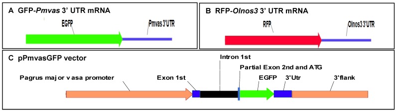
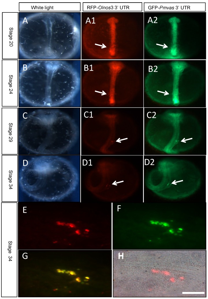
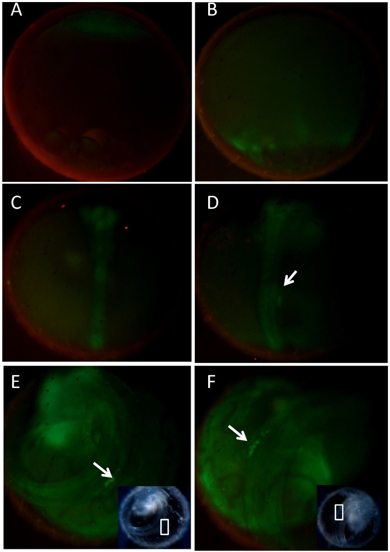
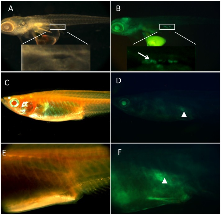
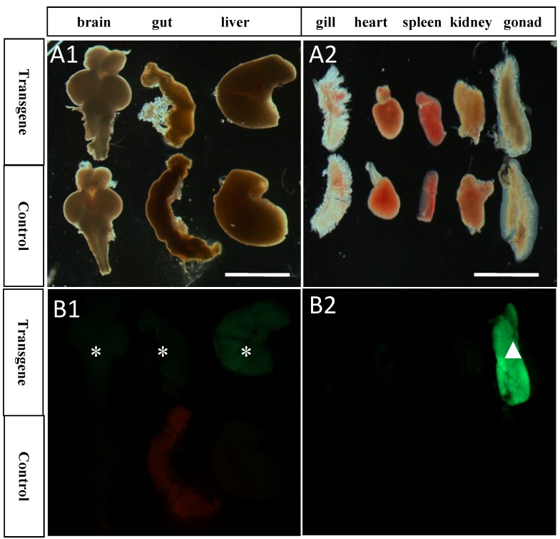
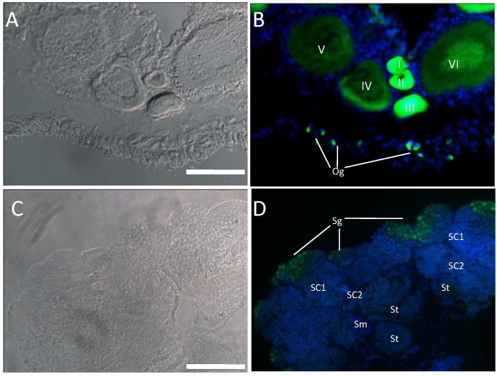
Primordial germ cells (PGCs) are the precursors of gametes responsible for genetic transmission to the next generation. They provide an ideal system for cryopreservation and restoration of biodiversity. Recently, considerable attention has been raised to visualize, isolate and transplant PGCs within and between species. In fish, stable PGC visualization in live embryo and individual has been limited to laboratory fish models such as medaka and zebrafish. One exception is the rainbow trout, which represents the only species with aquaculture importance and has GFP-labeled germ cells throughout development. PGCs can be transiently labeled by embryonic injection of mRNA containing green fluorescence protein gene (GFP) and 3'-untranslated region (3'-UTR) of a maternal germ gene such as vasa, nos1, etc. Stable PGC labeling can be achieved through production of transgenic animals by some transcriptional regulatory sequences from germ genes, such as the vasa promoter and 3'-UTR. In this study, we reported the functional analyses of the red seabream vasa (Pmvas) regulatory sequences, using medaka as a model system. It was showed that injection of GFP-Pmvas3'UTR mRNA was able to label medaka PGCs during embryogenesis. Besides, we have constructed pPmvasGFP transgenic vector, and established a stable transgenic medaka line exhibiting GFP expression in germ cells including PGCs, mitotic and meiotic germ cells of both sexes, under control of the Pmvas transcriptional regulatory sequences. It is concluded that the Pmvas regulatory sequences examined in this study are sufficient for germ cell expression and labeling.

Keywords: GFP; PGCs; Pagrus major; transgene; vasa.


Conflict of interest statement

Competing Interests: The authors have declared that no competing interest exists.

Figures

Fig 1
Fig 1
Structure of chimeric mRNA and transgenic vector for PGCs labeling
Fig 2
Fig 2
Transient PGCs labeling by co-injection of RFP-Olnos3 3'UTR (Red) and GFP-Pmvas 3'UTR (Green) mRNA. PGCs were indicated by white arrows. A-D) embryos of stage 20, stage 24, stage 29 and stage34, respectively, E-H) the gonad region of stage 34 was squashed into cell lever and observed under fluorescence microscopy, Scale bar, 50 µm.
Fig 3
Fig 3
Identification of germline transmitting progeny by genomic PCR. M) DNA marker; P) positive control from pPmvasGFP vector; 1-3) F1 embryos without GFP expression; 4-6) F1 embryos with GFP expression.
Fig 4
Fig 4
Germline Expression of GFP in transgenic medaka before hatching. GFP was monitored by fluorescence microscopy. A-F showed the expression pattern of the transgenic offspring following female transmission. The PGCs were indicated by white arrows. White square box represented the presumptive gonad region under white light. A) stage 10; B) stage 16; C) Stage 24; D) stage 29; E ) stage 34; F) stage 38.
Fig 5
Fig 5
Germline Expression of GFP in transgenic medaka after hatching. A-B) medaka of two days after hatching, with PGCs expression GFP, located at dorsal side of intestine; C-D) medaka of two months after hatching, with gonad expressing GFP; E-F) magnification of corresponding area of C and D, respectively. Tissue with germ cells was encompassed by white square box. PGCs were indicated by white arrow. Gonad with GFP was indicated by white arrowhead.
Fig 6
Fig 6
GFP expression pattern in different tissues of transgenic and control medaka. A1-B1) GFP showed some weak expression in brain, gut and liver of transgenic tissues, while no expression in the control; A2-B2) GFP showed strong expression in gonad, little or no expression in gill, heart, spleen and kidney, while no expression in the control. Asterisks represent tissues with weak GFP expression; White arrowhead represents tissue with strong GFP expression; Scale bar, 2 mm.
Fig 7
Fig 7
GFP expression pattern in germ cells of gonads. Cryosections of ovary (A-B) and testis (C-D) were used to detect endogenous GFP. Nuclei was stained by DAPI (Blue). In ovary, GFP expression was abundant in early stages of oogenesis (oogonia, oocytes of I-III stage) and reduced in late stages oocytes (IV-VI); In testis, Abundant GFP expression was detected in spermatogonia, while little expression was detected in other stages of spermatogenesis. Og, oogonia; I-VI, different stages of oocytes; Sg, spermatogonia; Sc1 and Sc2, primary and secondary spermatocytes; St, spermatids; Sm, sperm; Scale bar, 100 µm.

References

    1. Yoshizaki G, Kobayashi T, Takeuchi T. Primordial germ cell: a novel tool for fish bioengineering. Fish physiol Biochem. 2003;28:453–457.
    1. Xu HY, Li MY, Gui JF, Hong YH. Fish germ cells. Sci China Life Sci. 2010;53:435–446. - PubMed
    1. Okutsu T, Yano A, Nagasawa K. et al. Manipulation of Fish Germ Cell: Visualization, Cryopreservation and Transplantation. J Reprod Dev. 2006;52:685–693. - PubMed
    1. Takeuchi Y. Generation of Live Fry from Intraperitoneally Transplanted Primordial Germ Cells in Rainbow Trout. Biol Reprod. 2003;69:1142–1149. - PubMed
    1. Okutsu T. Testicular germ cells can colonize sexually undifferentiated embryonic gonad and produce functional eggs in fish. Proc Natl Acad Sci. 2006;103:2725–2729. - PMC - PubMed

Publication types

MeSH terms

Substances