. 2012;8(6):901-12.
doi: 10.7150/ijbs.4554. Epub 2012 Jun 22.

Oridonin up-regulates expression of P21 and induces autophagy and apoptosis in human prostate cancer cells

Affiliations

Oridonin up-regulates expression of P21 and induces autophagy and apoptosis in human prostate cancer cells

Xiang Li et al. Int J Biol Sci. 2012.

Abstract

Background: Oridonin (ORI) could inhibit the proliferation and induce apoptosis in various cancer cell lines. However, the mechanism is not fully understood.

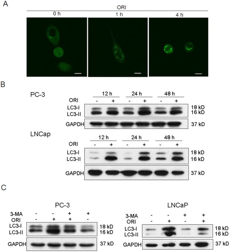
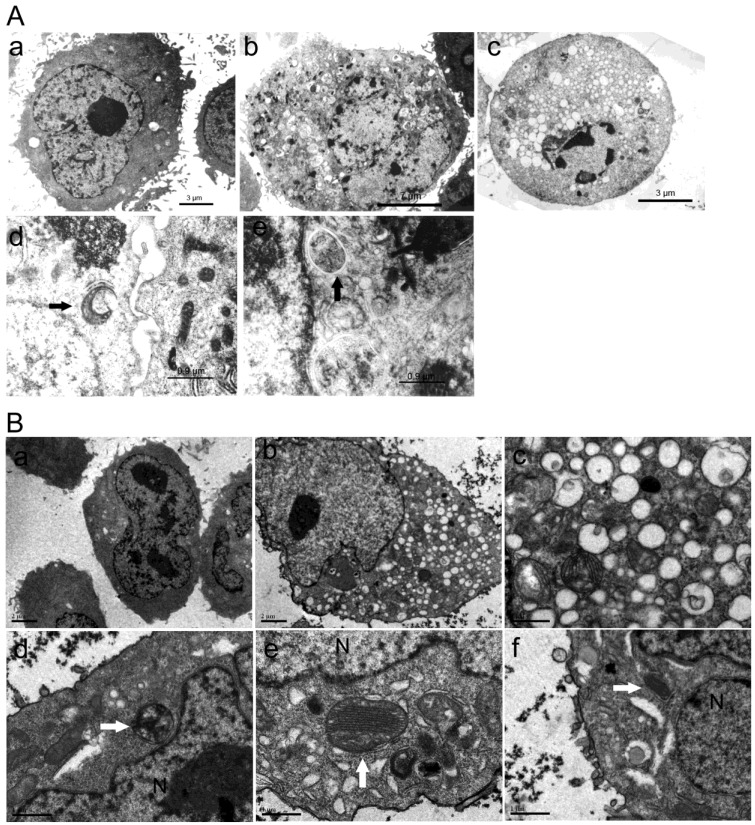
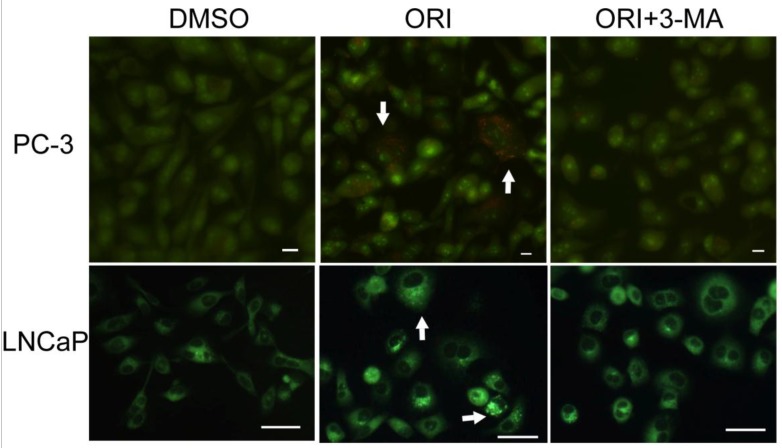
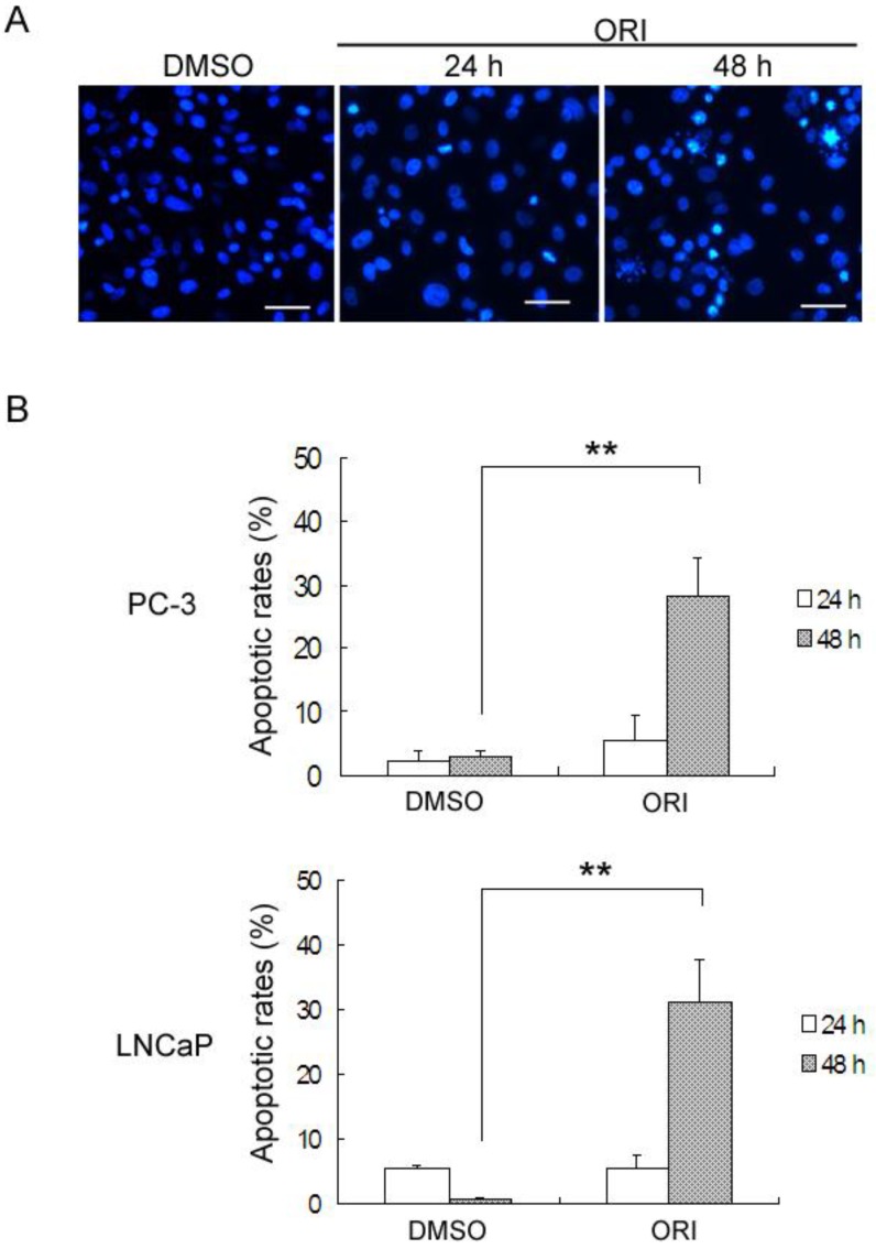
Methods: Human prostate cancer (HPC) cells were cultured in vitro and cell viability was detected by the CCK-8 assay. The ultrastructure changes were observed under transmission electron microscope (TEM). Chemical staining with acridine orange (AO), MDC or DAPI was used to detect acidic vesicular organelles (AVOs) and alternation of DNA. Expression of LC3 and P21 was detected by Western Blot. Apoptotic rates and cell cycle arrest were detected by FACS.

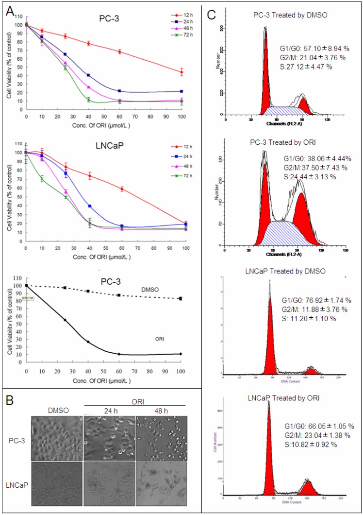
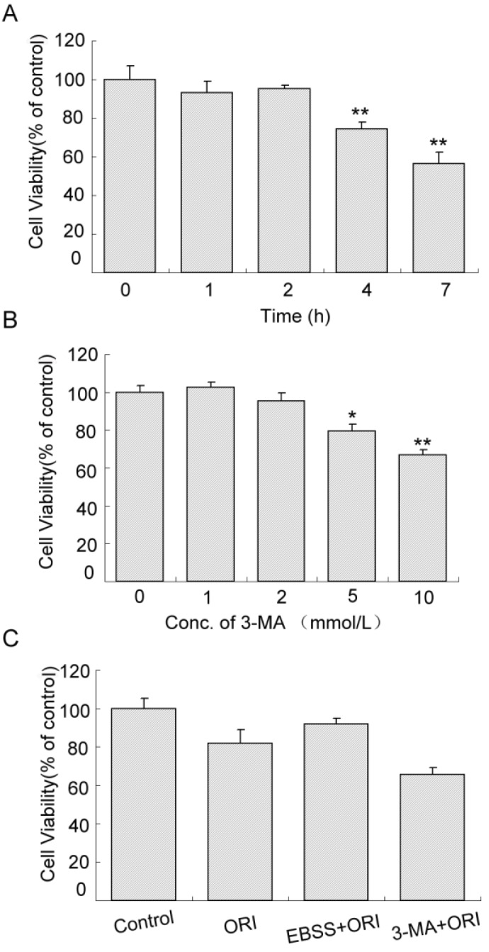
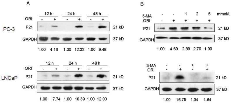
Results: Our study demonstrated that after ORI treatment, the proliferations of human prostate cancer (HPC) cell lines PC-3 and LNCaP were inhibited in a concentration and time-dependent manner. ORI induced cell cycle arrest at the G2/M phase. A large number of autophagosomes with double-membrane structure and acidic vesicular organelles (AVOs) were detected in the cytoplasm of HPC cells treated with ORI for 24 hours. ORI resulted in the conversion of LC3-I to LC3-II and recruitment of LC3-II to the autophagosomal membranes. Autophagy inhibitor 3-methyladenine (3-MA) reduced AVOs formation and inhibited LC3-I to LC3-II conversion. At 48 h, DNA fragmentation, chromatin condensation and disappearance of surface microvilli were detected in ORI-treated cells. ORI induced a significant increase in the number of apoptotic cells (PC-3: 5.4% to 27.0%, LNCaP: 5.3% to 31.0%). Promoting autophagy by nutrient starvation increased cell viability, while inhibition of autophagy by 3-MA promoted cell death. The expression of P21 was increased by ORI, which could be completely reversed by the inhibition of autophagy.

Conclusions: Our findings indicated that autophagy occurred before the onset of apoptosis and protected cancer cells in ORI-treated HPC cells. P21 was involved in ORI-induced autophagy and apoptosis. Our results provide an experimental basis for understand the anti-tumor mechanism of ORI as treatment for prostate cancer.

Keywords: P21.; apoptosis; autophagy; oridonin; prostate cancer.


Conflict of interest statement

Competing Interests: The authors have declared that no competing interest exists.

Figures

Fig 1
Fig 1
ORI inhibited the proliferation of HPC cells. A: Cell viability assay. HPC cells in 96-well plates were treated with different concentrations of ORI or DMSO for different time, and cell viability was determined using the CCK-8 assay (10 μl/well CCK-8 solution for 1 h). B: ORI reduced cell density. Cells in 96-well plate were treated by ORI or DMSO for 24 or 48 h, and observed under phase-contrast microscope (×200). C: Cell cycle was arrested at G2/M phase by ORI. Cells treated with 25 μmol/L ORI or DMSO for 24 h were fixed with ice-cold 75% ethanol (-20°C for 2 h), and stained with PI solution of 50 μg/ml. DNA content was analyzed by FACS. The data in A and C are presented as means ± S.D. from three independent experiments.
Fig 2
Fig 2
ORI treatment induced autophagy in HPC cells. A: LC3II protein was recruited to autophagosomsal membranes. After transfected with tfLC3 plasmid, PC-3 cells in 24-well plates were treated with ORI (25 μmol/L) and examined by confocal microscopy (×400). A punctuate pattern for GFP-LC3 fluorescence was observed. B: ORI increased LC3 expression. Cells were treated with ORI (25 μmol/L) or DMSO for 12, 24 or 48 h, then collected and subject to Western Blot. C: Increase of LC3 level was inhibited by 3-MA. Cells were treated by ORI (25 μmol/L) with or without 3-MA (5 mmol/L) for 24 h, then the expression of LC3 protein were detected by Western Blot. The data in B and C were from three independent experiments.
Fig 3
Fig 3
Formation of autophagosomes was induced by ORI in HPC cells. Cells were treated with ORI (25 μmol/L) or DMSO, and examined under TEM A: Changes of ultra-structure in PC-3 cells induced by ORI. (a) Controls, (b) Cells treated with ORI for 24 h, (c) Cells treated with ORI for 48 h, (d) ORI-induced PAS, (e) ORI-induced autophagosome (arrows). B: Changes of ultrastructure in LNCaP cells. Cells were treated with DMSO or ORI (25 μmol/L) for 24 h. (a) Controls, (b) ORI-treated cells, (c) Cytoplasmic vacuolization, (d) Autophagosome with cytoplasmic material, (e) Autophagosome with ER, (f) PAS with mitochondria (arrows).
Fig 4
Fig 4
Accumulation of AVOs induced by ORI treatment (×200). Cells in 96-well plate were treated by ORI (25 μmol/L) with or without 3-MA (5 mmol/L) for 24 h, stained with AO (5 μg/mL) or MDC (0.05 mmol/L) for 10 min, then observed. Red fluorescent spots of AO and prominent accumulations of MDC were observed in both ORI-treated cells (arrows).
Fig 5
Fig 5
Protective role of autophagy in ORI-treated PC-3 cells. A: Inhibition of proliferation induced by starvation. Cells were cultured in EBSS for different times (1, 2, 4, and 7 h). B: Cytotoxicity of 3-MA. Cells in 96-well plates were treated by 3-MA in different concentrations (1, 2, 5 and 10 mmol/L) for 24 h. C: Autophagy protected cells survival. Cells in 96-well plate were treated with media containing 25 μmol/L ORI, or ORI plus 2 mmol/L 3-MA, or pre-cultured in EBSS for 2 h then treated by ORI in full media for 24 h. Cell viabilities were measured by the CCK-8 assay. The data were presented as means ± S.D. from 3 independent experiments (*P<0.05, **P<0.01).
Fig 6
Fig 6
ORI induced apoptosis in HPC cells after longer treatment. A: Longer treatment with ORI resulted in chromatin condensation and nuclear fragmentation (arrows, ×200). PC-3 cells in 96-well plates were treated with 25 μmol/L ORI for 24 or 48 h, fixed and stained with DAPI (2 μg/mL), and observed for nuclear alterations under the fluorescence microscope. B: The apoptotic rates were evidently increased after longer treatment with ORI. Cells in 6-well plates were treated with ORI (25 μmol/L) for 24 or 48 h, stained with Annexin V-FITC for 10 min and PI for 10 min, then analyzed by FACS. (*P<0.05,**P<0.01).
Fig 7
Fig 7
Inhibition of autophagy blocked the augment of P21 induced by ORI. A: The expression of P21 was up-regulated by ORI. Cells were treated with ORI (25 μmol/L) for 12, 24 or 48 h, and detected by Western Blot. B: ORI-induced augment of P21 protein was blocked by 3-MA. Cells were treated by ORI with or without 3-MA (5 mmol/L), and subjected to Western Blot. The data are from three independent experiments.

References

    1. Crawford ED. Epidemiology of prostate cancer. Urology. 2003;62:3–12. - PubMed
    1. Marks LS, DiPaola RS, Nelson P. et al. PC-SPES: herbal formulation for prostate cancer. Urology. 2002;60:369–75. - PubMed
    1. Cui Q, Tashiro S, Onodera S. et al. Augmentation of oridonin-induced apoptosis observed with reduced autophagy. J Pharmacol Sci. 2006;101:230–9. - PubMed
    1. Cui Q, Tashiro S, Onodera S. et al. Autophagy preceded apoptosis in oridonin-treated human breast cancer MCF-7 cells. Biol Pharm Bull. 2007;30:859–64. - PubMed
    1. Li D, Cui Q, Chen SG. et al. Inactivation of ras and changes of mitochondrial membrane potential contribute to oridonin-induced autophagy in a431 cells. J Pharmacol Sci. 2007;105:22–33. - PubMed

MeSH terms