. 2012;7(6):e39336.
doi: 10.1371/journal.pone.0039336. Epub 2012 Jun 26.

Endothelial expression of TGFβ type II receptor is required to maintain vascular integrity during postnatal development of the central nervous system

Affiliations

Endothelial expression of TGFβ type II receptor is required to maintain vascular integrity during postnatal development of the central nervous system

Kathleen R Allinson et al. PLoS One. 2012.

Erratum in

Abstract

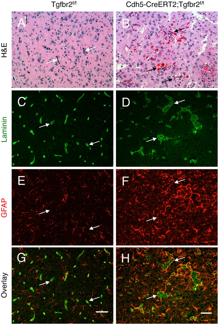
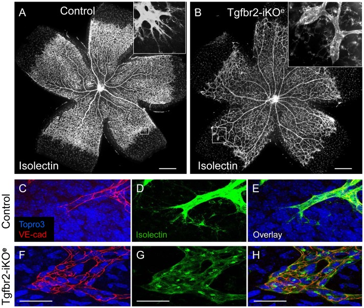
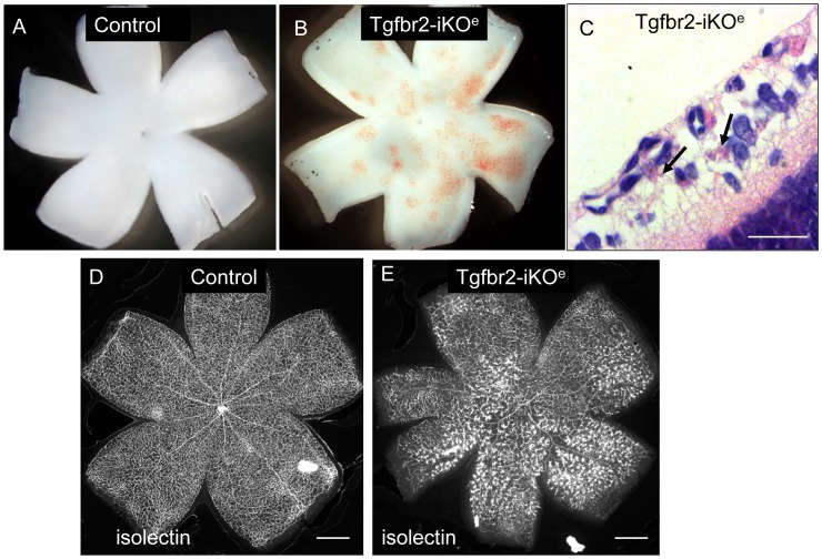
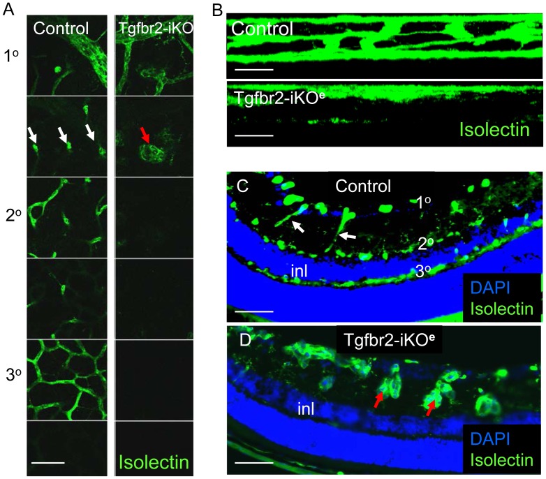
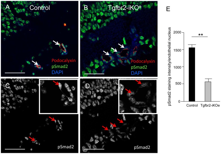
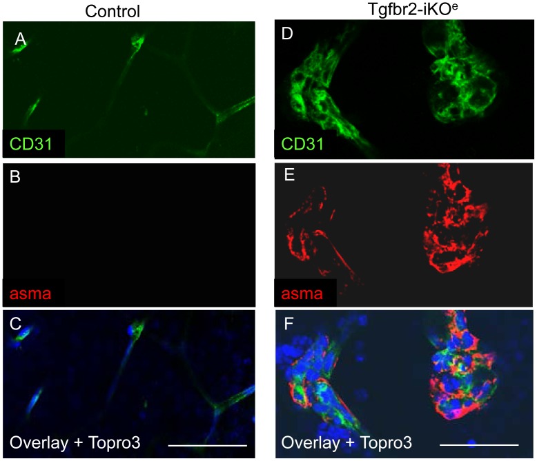
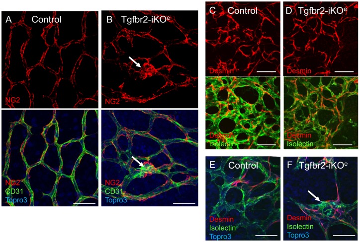
TGFβ signalling in endothelial cells is important for angiogenesis in early embryonic development, but little is known about its role in early postnatal life. To address this we used a tamoxifen inducible Cre-LoxP strategy in neonatal mice to deplete the TypeII TGFβ receptor (Tgfbr2) specifically in endothelial cells. This resulted in multiple micro-haemorrhages, and glomeruloid-like vascular tufts throughout the cerebral cortices and hypothalamus of the brain as well as in retinal tissues. A detailed examination of the retinal defects in these mutants revealed that endothelial adherens and tight junctions were in place, pericytes were recruited and there was no failure of vascular smooth muscle differentiation. However, the deeper retinal plexus failed to form in these mutants and the angiogenic sprouts stalled in their progress towards the inner nuclear layer. Instead the leading endothelial cells formed glomerular tufts with associated smooth muscle cells. This evidence suggests that TGFβ signalling is not required for vessel maturation, but is essential for the organised migration of endothelial cells as they begin to enter the deeper layers of the retina. Thus, TGFβ signalling is essential in vascular endothelial cells for maintaining vascular integrity at the angiogenic front as it migrates into developing neural tissues in early postnatal life.


Conflict of interest statement

Competing Interests: The authors have declared that no competing interests exist.

Figures

Figure 1
Figure 1. Loss of Tgfbr2 in neonatal endothelial cells leads to cerebral vascular pathologies and intracerebral haemorrhage.
Coronal sections through cerebral cortices of P14 control (A) and Tgfbr2–iKOe mice (B) were stained with H&E. Note the abnormal blood vessel morphologies and microhaemorrhage in Tgfbr2 mutant brains. (C-H); Coronal sections through cerebral cortices of P14 control (C, E, G) and Tgfbr2 conditional mutant mice (D, F, H) were double immunofluorescently labelled with anti-laminin (C, D) and anti-GFAP (E, F) to visualize vascular basement membranes and astrocytes, respectively. Note the cerebral blood vessels with glomeruloid-like tufts (D) as well as robust perivascular astrogliosis (F) in Tgfbr2-iKOe mutant sections. A total of 5 mutant and 4 control mice were examined for brain pathologies. Scale bars: 50 µm.
Figure 2
Figure 2. Tgfbr2-iKO mutants show vascular abnormalities in the postnatal retina.
Isolectin stained retinal preparations at postnatal day (P)7 show normal vascular architecture in controls (A), but reduced branching in Tgfbr2-iKOe mutants (B). Frequently, round clusters of endothelial cells (boxed area and inset, B) are seen at the leading edge of the vascular plexus in mutants, at the positions where tip cells normally occur in controls (boxed area and inset, A). High power views of whole mount P9 retinal preparations stained with VE-cadherin and isolectin show the endothelial footprint of a normal tip cell (C,D,E) compared with that of the endothelial cell clusters in the Tgfbr2-iKOe mutant (F,G,H). Scale bars: 500 µm A,B; 50 µm, C–H.
Figure 3
Figure 3. Retinal haemorrhage in Tgfbr2-iKOe neonates upon development of the secondary vascular plexus.
Stereo-images of freshly dissected whole mount P9 retinas show multiple microhaemorrhages in the Tgfbr2-iKOe that are not seen in controls (A,B). H&E stained sections of Tgfbr2-iKOe retina show regions of microhaemorrhage within the retinal tissue (arrows,C). Wholemount view of isolectin stained P14 retinas show multiple glomerular tufts (seen as intensely stained clumps of ECs) in the Tgfbr2-iKOe mutants that are not seen in controls (D,E). This phenotype was seen in over 100 mutant retinas from P8 to P28. Scale bars: 500 µm, C,D; 20 µm C.
Figure 4
Figure 4. Formation of the deeper vascular network is severely impaired in Tgfbr2-iKOe mutant retinas.
Confocal slices of isolectin stained retinas from P14 neonates shows presence of primary, secondary and tertiary networks in controls, but only the primary plexus in Tgfbr2-iKOe mutants. Z-slices showing surface view is shown in panel A whereas side view of vascular plexus is shown in B. Section through a control retina at P21shows the normal organisation of the primary, secondary and tertiary vascular plexus with respect to the surface of the retina and inner nuclear layer (inl). The interconnecting vessels between the primary and secondary plexus are regular small capillaries in controls, indicated by white arrows in Figures A and C. In contrast, this part of the deeper plexus in the Tgfbr2-iKOe retinas contains glomerular tufts (red arrows, A and D). Scale bars: 50 µm A,C,D; 20 µm B.
Figure 5
Figure 5. Reduced Smad2 phosphorylation in ECs in Tgfbr2-iKOe retinas.
Retinal sections (age P14) stained for pSmad2 (green) reveal Smad2 activation in both vascular cells and neural cells. Confocal analysis of podocalyxin staining (red) was used to identify the apical surface of endothelial cells in retinal blood vessels, and DAPI to identify the nuclei. Endothelial cells show reduced levels of pSmad2 activation in the mutant retinas (white arrows, B) compared with controls (white arrows, A). This difference can also be seen in the equivalent monochrome confocal images of pSmad2 staining in the same sections and in the digital zoom image inserts (C,D). Abbreviations: inl, inner nuclear layer. Scale bar: 50 µm. E: Quantitation of pSmad2 staining intensity using NIS-elements software was performed on 58 endothelial nuclei from 3 mutant retinas and 42 nuclei from 3 littermate controls. Endothelial cell nuclei in random fields of view were identified by podocalyxin apical staining. Statistical analysis using a student’s t-test shows a significant reduction of pSmad2 in endothelial cells of Tgfbr2-iKOe mutants compared with controls. ** p<0.001.
Figure 6
Figure 6. Endothelial glomerular tufts in the Tgfbr2-iKOe mutants contain multiple smooth muscle cells.
Immunofluorescent staining of P14 retinal paraffin sections with isolectin-alexa488 and anti-alpha smooth muscle actin (aSMA) conjugated to Cy3 show the typical non-muscularised microvessels of the control retinal plexus (arrows in A and D), whereas high numbers of vascular smooth muscle cells are associated with endothelial glomerular tufts (E,F and H). Scale bar: 50 µm.
Figure 7
Figure 7. Pericytes are associated with endothelial cells in Tgfbr2-iKO mutant retinas.
There were no apparent differences between mutants and controls in the organisation of pericytes on capillary retinal endothelial cells. Pericytes are identified using either NG2 (A,B) or desmin staining (C–F). Pericytes are present in the glomerular tufts in Tgfbr2-iKOe mutant retinas (B, F, arrows). Similar results were seen in a total of 10 mutants and 11 controls aged between P7 and P14. Scale bar: 50 µm.

References

    1. Daneman R, Zhou L, Kebede AA, Barres BA. Pericytes are required for blood-brain barrier integrity during embryogenesis. Nature. 2010;468:562–566. - PMC - PubMed
    1. Armulik A, Genove G, Mae M, Nisancioglu MH, Wallgard E, et al. Pericytes regulate the blood-brain barrier. Nature. 2010;468:557–561. - PubMed
    1. Abbott NJ, Ronnback L, Hansson E. Astrocyte-endothelial interactions at the blood-brain barrier. Nat Rev Neurosci. 2006;7:41–53. - PubMed
    1. Ballabh P, Braun A, Nedergaard M. The blood-brain barrier: an overview: structure, regulation, and clinical implications. Neurobiol Dis. 2004;16:1–13. - PubMed
    1. Roland EH, Hill A. Germinal matrix-intraventricular hemorrhage in the premature newborn: management and outcome. Neurol Clin 21: 833–851, vi-vii. 2003. - PubMed

Publication types

MeSH terms

Substances

LinkOut - more resources