. 2012;7(6):e39636.
doi: 10.1371/journal.pone.0039636. Epub 2012 Jun 22.

Stimulation of the sphenopalatine ganglion induces reperfusion and blood-brain barrier protection in the photothrombotic stroke model

Affiliations

Stimulation of the sphenopalatine ganglion induces reperfusion and blood-brain barrier protection in the photothrombotic stroke model

Haviv Levi et al. PLoS One. 2012.

Abstract

Purpose: The treatment of stroke remains a challenge. Animal studies showing that electrical stimulation of the sphenopalatine ganglion (SPG) exerts beneficial effects in the treatment of stroke have led to the initiation of clinical studies. However, the detailed effects of SPG stimulation on the injured brain are not known.

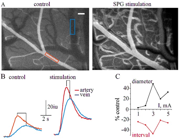
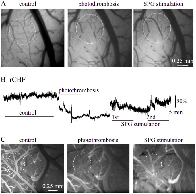
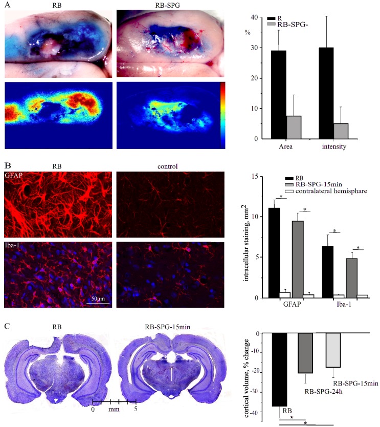
Methods: The effect of acute SPG stimulation was studied by direct vascular imaging, fluorescent angiography and laser Doppler flowmetry in the sensory motor cortex of the anaesthetized rat. Focal cerebral ischemia was induced by the rose bengal (RB) photothrombosis method. In chronic experiments, SPG stimulation, starting 15 min or 24 h after photothrombosis, was given for 3 h per day on four consecutive days. Structural damage was assessed using histological and immunohistochemical methods. Cortical functions were assessed by quantitative analysis of epidural electro-corticographic (ECoG) activity continuously recorded in behaving animals.

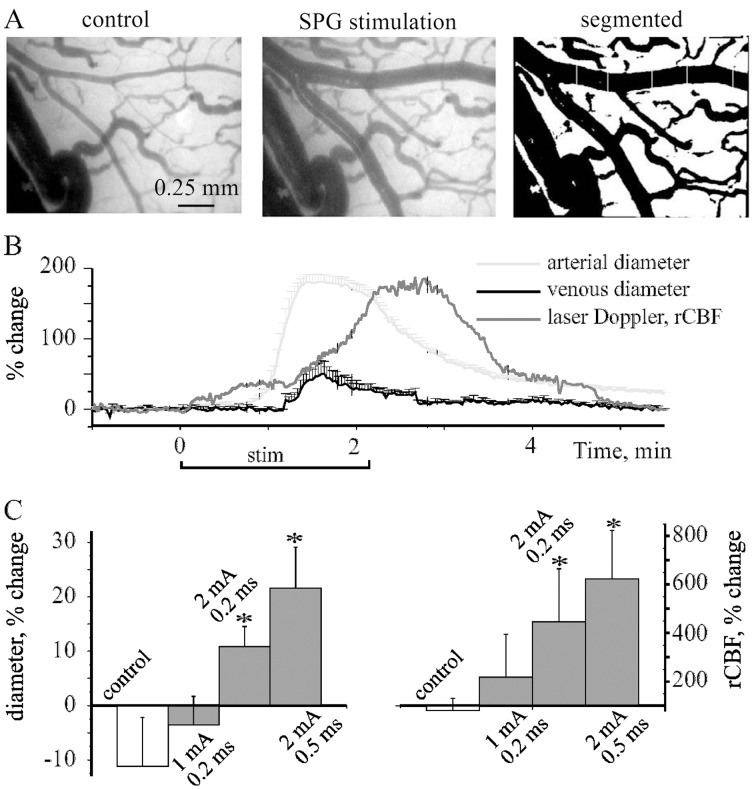
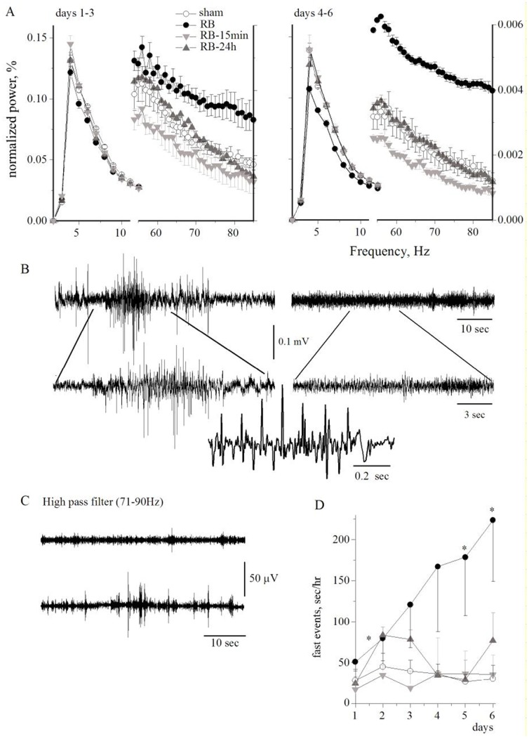
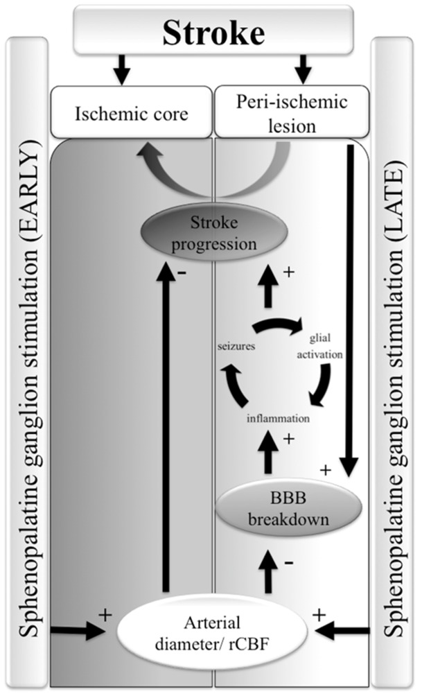
Results: Stimulation induced intensity- and duration-dependent vasodilation and increased cerebral blood flow in both healthy and photothrombotic brains. In SPG-stimulated rats both blood brain-barrier (BBB) opening, pathological brain activity and lesion volume were attenuated compared to untreated stroke animals, with no apparent difference in the glial response surrounding the necrotic lesion.

Conclusion: SPG-stimulation in rats induces vasodilation of cortical arterioles, partial reperfusion of the ischemic lesion, and normalization of brain functions with reduced BBB dysfunction and stroke volume. These findings support the potential therapeutic effect of SPG stimulation in focal cerebral ischemia even when applied 24 h after stroke onset and thus may extend the therapeutic window of currently administered stroke medications.


Conflict of interest statement

Competing Interests: Funding for this research were received from BrainsGate Ltd. This does not alter the authors’ adherence to all the PLoS ONE policies on sharing data and materials.

Figures

Figure 1
Figure 1. SPG stimulation induces vasodilation in the healthy brain A, Left and middle, respectively, representative image of the brain surface under control conditions and during SPG stimulation: right, the same image after processing for diameter measurement.
B, Arterial (red) and venous (blue) changes in diameter (presented as % change from baseline) are shown before, during and after a single stimulation train for the experiment shown in (A). Note that increased rCBF (gray) is following the arterial vasodilation. C, Average % change in arterial diameter (left graph) and rCBF (right graph) during different stimulation protocols (see text). *p<0.05.
Figure 2
Figure 2. Fluorescent angiography during SPG stimulation A, Two representative angiographic images of cortical surface vessels under control conditions (left) and during SPG stimulation (3 mA, 500 µs, right, white bar represents 0.1 mm).
B, Intensity curves (in arbitrary intensity units, iu) for the venous (blue) and arterial (red) compartments for the venous (blue) and arterial (red) compartments (marked in (A)) during control injection (left) and SPG stimulation (right). C, % change in diameter (black) and peak-to-peak (arterial-venous) interval (red) at different stimulation intensities (1–5 mA, 500 µs).
Figure 3
Figure 3. SPG stimulation in the RB-treated cortex.
A, Brain surface images in a typical experiment before (left), and after (middle) photothrombosis and during SPG stimulation (2 mA, 500 µs, right). Note vasodilation and reperfusion of the thrombosed vessels (circled). B, Laser-Doppler recording in the same experiment as in (A), showing reduced rCBF during photothrombosis, which was partially reversible during SPG stimulation. C, Fluorescent angiography from a different rat before (left), after photothrombosis (middle) and during SPG stimulation (2 mA, 500 µs, right).
Figure 4
Figure 4. BBB breakdown, astroglial response and brain damage after SPG stimulation A, Two brain surface images after injection of Evans blue indicates BBB breakdown of RB-treated (left) and RB-SPG-15 min (right) rat brains.
Images below display EB (blue color) intensity, color coded. Both the size of the area with increased EB and the intensity were decreased in SPG-treated rats compared to non-stimulated animals (right graph). B, Immunostaining against the astrocytic marker, GFAP (upper panels) and the microglial marker, Iba-1 (lower panel) in RB-treated (left) and the contra-lateral control hemisphere (right). Bar graphs show area measured with intracellular staining (see Methods). C, Coronal sections of brains from RB (left) and RB-SPG-15 min (right) animals. Bar graphs show change in cortical volume after photothrombosis. *p<0.05.
Figure 5
Figure 5. ECoG Fast Fourier Analysis: A, Averaged power of the normalized ECoG 1-3 (left) and 4–6 (right) days after photothrombosis in low and high frequency ranges (note the different right and left Y axis).
B, Typical ECoG recordings of RB-treated (left) and sham animals (right). C, ECoG recording after high pass filter (71–90 Hz) of RB-treated (lower trace) and sham animals (upper trace). D, Duration of fast activity (sec/h) for the different treated groups. Symbols as in (A). *p<0.05.
Figure 6
Figure 6. A working hypothesis on SPG-induced brain protection after stroke: The ischemic core is surrounded by a peri-ischemic region which is susceptible for an irreversible injury (AKA “stroke progression”).
The peri-ischemic lesion is characterized by dysfunction of the blood-brain barrier (BBB) which induces neuronal hyper-excitability, spreading depolarizations and seizures, inflammation and cellular damage . SPG stimulation at an early, post-insult therapeutic time window induces vasodilation and increased rCBF, sufficient to re-perfuse the ischemic core and to reduce lesion size. When stimulation is initiated at a delayed post-insult therapeutic window (24 h), vasodilation in the peri-ischemic lesion attenuates BBB injury and the associated neuronal hyper-excitability, thus preventing progression of the primary lesion.

References

    1. Ginsberg MD. The new language of cerebral ischemia. Ajnr American Journal Of Neuroradiology. 1997;18:1435–1445. - PMC - PubMed
    1. Dirnagl U, Iadecola C, Moskowitz MA. Pathobiology of ischaemic stroke: an integrated view. Trends in Neurosciences 22: 391–397. Available. 1999. http://www.ncbi.nlm.nih.gov/pubmed/10441299. - PubMed
    1. Kastrup A, Engelhorn T, Beaulieu C, de Crespigny A, Moseley ME. Dynamics of cerebral injury, perfusion, and blood-brain barrier changes after temporary and permanent middle cerebral artery occlusion in the rat. Journal of the neurological sciences 166: 91–99. 1999. Available: http://www.jns-journal.com/article/S0022-510X(99)00121-5/abstract. Accessed 21 February 2012. - PubMed
    1. Shlosberg D, Benifla M, Kaufer D, Friedman A. Blood-brain barrier breakdown as a therapeutic target in traumatic brain injury. Nature reviews Neurology 6: 393–403. Available. 2010. http://www.ncbi.nlm.nih.gov/pubmed/20551947. - PMC - PubMed
    1. Smith WS, Sung G, Starkman S, Saver JL, Kidwell CS, et al. Safety and efficacy of mechanical embolectomy in acute ischemic stroke: results of the MERCI trial. Stroke 36: 1432–1438. Available. 2005. http://www.ncbi.nlm.nih.gov/pubmed/15961709. - PubMed

Publication types