. 2012;7(6):e39659.
doi: 10.1371/journal.pone.0039659. Epub 2012 Jun 26.

Impact of large aggregated uricases and PEG diol on accelerated blood clearance of PEGylated canine uricase

Affiliations

Impact of large aggregated uricases and PEG diol on accelerated blood clearance of PEGylated canine uricase

Chun Zhang et al. PLoS One. 2012.

Abstract

Background: Uricase has proven therapeutic value in treating hyperuricemia but sufficient reduction of its immunogenicity may be the largest obstacle to its chronic use. In this study, canine uricase was modified with 5 kDa mPEG-SPA and the impact of large aggregated uricases and cross-linked conjugates induced by difunctional PEG diol on immunogenicity was investigated.

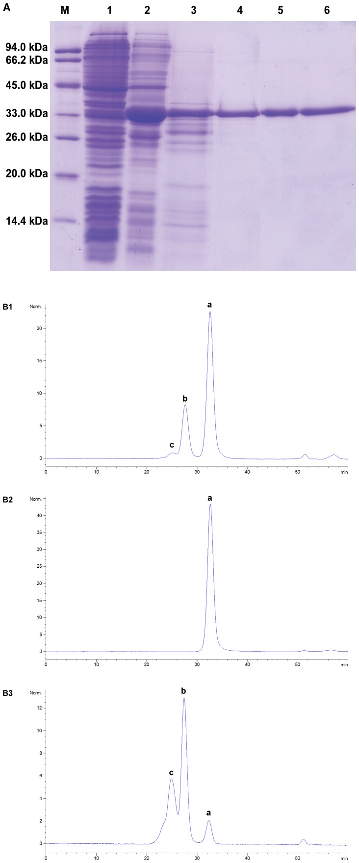
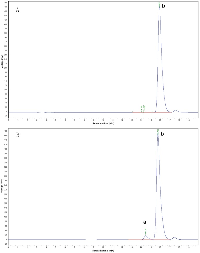
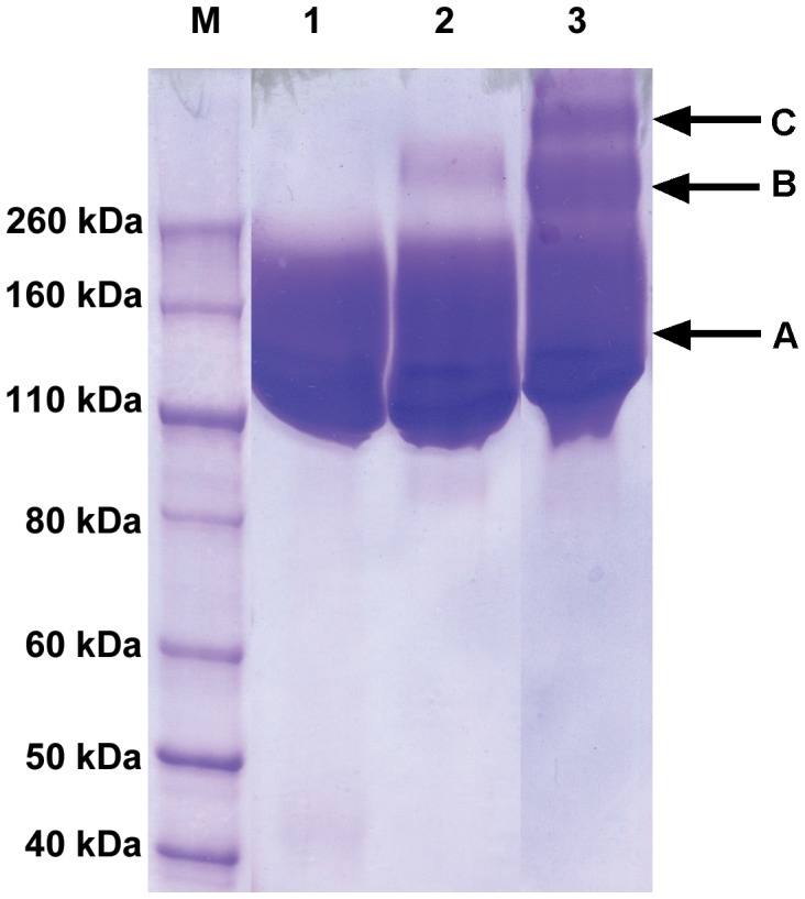
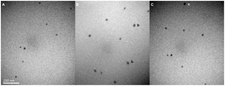
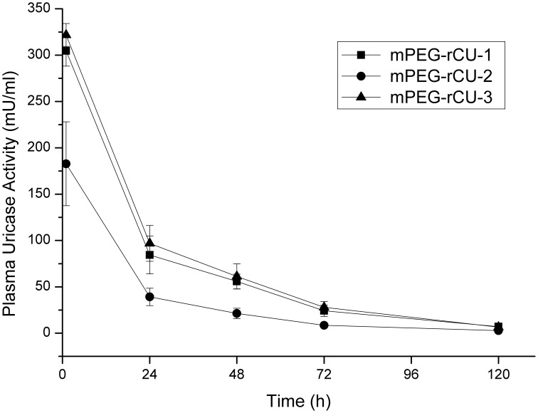
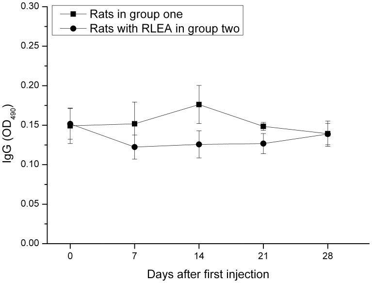
Methods and findings: Recombinant canine uricase was first expressed and purified to homogeneity. Source 15Q anion-exchange chromatography was used to separate tetrameric and aggregated uricase prior to pegylation, while DEAE anion-exchange chromatography was used to remove Di-acid PEG (precursor of PEG diol) from unfractionated 5 kDa mPEG-propionic acid. Tetrameric and aggregated uricases were separately modified with the purified mPEG-SPA. In addition, tetrameric uricases was modified with unfractionated mPEG-SPA, resulting in three types of 5 kDa mPEG-SPA modified uricase. The conjugate size was evaluated by dynamic light scattering and transmission electron microscope. The influence of differently PEGylated uricases on pharmacokinetics and immunogenicity were evaluated in vivo. The accelerated blood clearance (ABC) phenomenon previously identified for PEGylated liposomes occurred in rats injected with PEGylated uricase aggregates. Anti-PEG IgM antibodies, rather than neutralizing antibodies, were found to mediate the ABC.

Conclusions: The size of conjugates is important for triggering such phenomena and we speculate that 40-60 nm is the lower size limit that can trigger ABC. Removal of the uricase aggregates and the PEG diol contaminant and modifying with small PEG reagents enabled ABC to be successfully avoided and sufficient reduction in the immunogenicity of 5 kDa mPEG-modified tetrameric canine uricase.


Conflict of interest statement

Competing Interests: The authors have read the journal’s policy and have the following conflicts: Authors Kai Fan and Xuefeng Ma are employees of Fagen Biomedical Inc. There are no patents, products in development or marketed products to declare. This does not alter the authors' adherence to all the PLoS ONE policies on sharing data and materials, as detailed online in the guide for authors.

Figures

Figure 1
Figure 1. SDS-PAGE and SE-HPLC analyses of rCU during the expression and purification process.
(A) SDS-PAGE analysis lanes: M, standard protein molecular weight markers; 1, crude cell extract before IPTG induction; 2, crude cell extract after IPTG induction for 3 hours; 3, crude extraction in 100 mM sodium carbonate buffer (pH 10.3); 4, purified rCU after ammonium sulfate fractionation; 5, purified rCU after xanthine affinity chromatography; 6, purified rCU after anion exchange chromatography. (B) SE-HPLC analysis: B1 purified rCU after xanthine affinity chromatography; B2 tetrameric wDU eluted with 0.1 M NaCl on a Source 15 Q column; B3 large aggregated wDU eluted with 0.25 M NaCl on a Source 15 Q column; a, b and c correspond to tetramer, octamer and large aggregate, respectively.
Figure 2
Figure 2. SE-HPLC analysis of purified and unfractionated mPEG-PA.
The PEG diol contents were analyzed using a G2000PWXL column with a differential refractive index detector. (A) Purified mPEG-PA; (B) Unfractionated mPEG-PA. a and b correspond to Di-acid PEG and mono-acid mPEG.
Figure 3
Figure 3. Representative chromatogram of mPEG-rCU purification
The three different chromatograms correspond to mPEG-rCU protein (A), unconjugated mPEG (B) and N-hydroxysuccinimide acid (C), respectively.
Figure 4
Figure 4. SDS-PAGE analysis of purified mPEG-rCU.
Lanes 1, 2 and 3 represent mPEG-rCU-1, mPEG-rCU-2 and mPEG-rCU-3. A, B and C correspond to non-crosslinked PEGylated monomeric rCU, crosslinked conjugates between two PEGylated monomeric rCU, and crosslinked conjugates among three and more PEGylated monomeric rCU, respectively.
Figure 5
Figure 5. Representative transmission electron micrograph of mPEG-rCU.
(A), (B) and (C) represent mPEG-rCU-1, mPEG-rCU-2 and mPEG-rCU-3.The magnification is 100000×, scale bar: 200 nm. a, b and c may correspond to PEGylated tetrameric uricase, PEGylated aggregated uricase, and cross-linked conjugates induced by PEG diol, respectively.
Figure 6
Figure 6. Time courses of plasma uricase activity in three groups after intravenous administration of mPEG-rCU (n = 6).
Figure 7
Figure 7. Retention of plasma uricase activity in three groups after different injections of mPEG-rCU.
Plasmas were collected 24 hours after each of four weekly injections of mPEG-rCU. *, ** and *** mean significantly different from the same rats before injection at levels of p<0.05, p<0.01, and p<0.001.
Figure 8
Figure 8. ELISA analysis of IgG antibody against mPEG-rCU.
Serum samples were collected 24 hours before each of four weekly injections of mPEG-rCU. Microtiter plates were coated with 50 µg/ml of mPEG-rCU-1.
Figure 9
Figure 9. ELISA analysis of IgM antibody against mPEG-rCU, rCU, or mPEG-BSA
Serum samples were collected 24 hours before each of four weekly injections of mPEG-rCU. (A) Microtiter plates were coated with 50 µg/ml of mPEG-rCU-1. (B) Microtiter plates were coated with 10 µg/ml of tetrameric rCU. (C) Microtiter plates were coated with 50 µg/ml of mPEG-BSA.

References

    1. Tsukada H, Mochizuki Y, Fujiwara S. The nucleoids of rat liver cell microbodies. Fine structure and enzymes. J Cell Biol. 1966;28:449–460. - PMC - PubMed
    1. Smyth CJ. Disorders associated with hyperuricemia. Arthritis Rheum. 1975;18:713–719. - PubMed
    1. Kissel P, Mauuary G, Royer R, Toussain P. Letter: Treatment of malignant haemopathies and urate oxidase. Lancet. 1975;1:229. - PubMed
    1. Sibony G, North ML, Bergerat JP, Lang JM, Oberling F. [Hyperuricemia resistant to urate oxidase. Role of anti-serum urate oxidase precipitating antibodies]. Presse Med. 1984;13:443. - PubMed
    1. Davis S, Park YK, Abuchowski A, Davis FF. Hypouricaemic effect of polyethyleneglycol modified urate oxidase. Lancet. 1981;2:281–283. - PubMed

Publication types

Substances