

Modulator of Apoptosis 1: A Highly Regulated RASSF1A-Interacting BH3-Like Protein
- PMID: 22745908
- PMCID: PMC3382356
- DOI: 10.1155/2012/536802
Modulator of Apoptosis 1: A Highly Regulated RASSF1A-Interacting BH3-Like Protein
Abstract
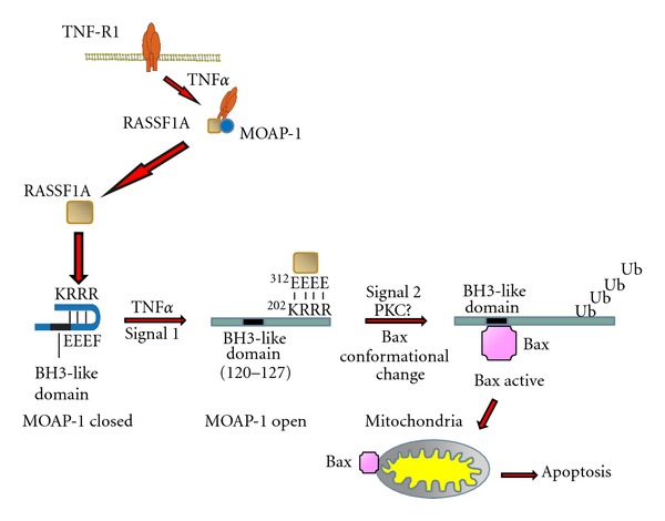
Modulator of apoptosis 1 (MOAP-1) is a BH3-like protein that plays key roles in both the intrinsic and extrinsic modes of cell death or apoptosis. MOAP-1 is part of the Ras association domain family 1A (RASSF1A)/MOAP-1 pro-apoptotic extrinsic signaling pathway that regulates apoptosis by utilizing death receptors such as tumor necrosis factor α (TNFα) or TNF-related apoptosis-inducing ligand (TRAIL) to inhibit abnormal growth. RASSF1A is a bona fide tumor suppressor gene that is epigenetically silenced by promoter-specific methylation in numerous human cancers. MOAP-1 is a downstream effector of RASSF1A that promotes Bax activation and cell death and is highly regulated during apoptosis. We speculate that MOAP-1 and RASSF1A are important elements of an "apoptotic checkpoint" that directly influences the outcome of cell death. The failure to regulate this pro-apoptotic pathway may result in the appearance of cancer and possibly other disorders. Although loss of RASSF1A expression is frequently observed in human cancers, it is currently unknown if MOAP-1 expression may also be affected during carcinogenesis to result in uncontrolled malignant growth. In this article, we will summarize what is known about the biological role(s) of MOAP-1 and how it functions as a downstream effector to RASSF1A.
Figures
Similar articles
-
Modulator of apoptosis 1 (MOAP-1) is a tumor suppressor protein linked to the RASSF1A protein.J Biol Chem. 2015 Oct 2;290(40):24100-18. doi: 10.1074/jbc.M115.648345. Epub 2015 Aug 12. J Biol Chem. 2015. PMID: 26269600 Free PMC article.
-
Dynamics of RASSF1A/MOAP-1 association with death receptors.Mol Cell Biol. 2008 Jul;28(14):4520-35. doi: 10.1128/MCB.02011-07. Epub 2008 May 12. Mol Cell Biol. 2008. PMID: 18474619 Free PMC article.
-
The RASSF1A tumor suppressor activates Bax via MOAP-1.J Biol Chem. 2006 Feb 24;281(8):4557-63. doi: 10.1074/jbc.M512128200. Epub 2005 Dec 12. J Biol Chem. 2006. PMID: 16344548
-
The tumor suppressor RASSF1A in human carcinogenesis: an update.Histol Histopathol. 2005 Apr;20(2):645-63. doi: 10.14670/HH-20.645. Histol Histopathol. 2005. PMID: 15736067 Review.
-
The RASSF1A tumor suppressor.J Cell Sci. 2007 Sep 15;120(Pt 18):3163-72. doi: 10.1242/jcs.010389. J Cell Sci. 2007. PMID: 17878233 Review.
Cited by
-
Tricistronic expression of MOAP-1, Bax and RASSF1A in cancer cells enhances chemo-sensitization that requires BH3L domain of MOAP-1.J Cancer Res Clin Oncol. 2020 Jul;146(7):1751-1764. doi: 10.1007/s00432-020-03231-9. Epub 2020 May 6. J Cancer Res Clin Oncol. 2020. PMID: 32377840 Free PMC article.
-
RASSF1A Site-Specific Methylation Hotspots in Cancer and Correlation with RASSF1C and MOAP-1.Cancers (Basel). 2016 Jun 10;8(6):55. doi: 10.3390/cancers8060055. Cancers (Basel). 2016. PMID: 27294960 Free PMC article.
-
Aberrant Methylation Status of Tumour Suppressor Genes in Ovarian Cancer Tissue and Paired Plasma Samples.Int J Mol Sci. 2019 Aug 23;20(17):4119. doi: 10.3390/ijms20174119. Int J Mol Sci. 2019. PMID: 31450846 Free PMC article.
-
Pnma5 is essential to the progression of meiosis in mouse oocytes through a chain of phosphorylation.Oncotarget. 2017 Jun 9;8(57):96809-96825. doi: 10.18632/oncotarget.18425. eCollection 2017 Nov 14. Oncotarget. 2017. PMID: 29228573 Free PMC article.
-
The tumor suppressor gene, RASSF1A, is essential for protection against inflammation -induced injury.PLoS One. 2013 Oct 16;8(10):e75483. doi: 10.1371/journal.pone.0075483. eCollection 2013. PLoS One. 2013. PMID: 24146755 Free PMC article.
References
-
- El-Kalla M, Onyskiw C, Baksh S. Functional importance of RASSF1A microtubule localization and polymorphisms. Oncogene. 2010;29(42):5729–5740. - PubMed
-
- World Health Organization. The Global Burden of Disease: 2004 Update. 2008.
-
- Vogelstein B. The Cancer Genome. Washington, DC, USA: American Association for Cancer Research; 2010.
-
- Donninger H, Vos MD, Clark GJ. The RASSF1A tumor suppressor. Journal of Cell Science. 2007;120(18):3163–3172. - PubMed
-
- Baksh S, Tommasi S, Fenton S, et al. The tumor suppressor RASSF1A and MAP-1 link death receptor signaling to bax conformational change and cell death. Molecular Cell. 2005;18(6):637–650. - PubMed
LinkOut - more resources
Full Text Sources
Research Materials