

Biomechanics of disc degeneration
- PMID: 22745914
- PMCID: PMC3382964
- DOI: 10.1155/2012/726210
Biomechanics of disc degeneration
Abstract
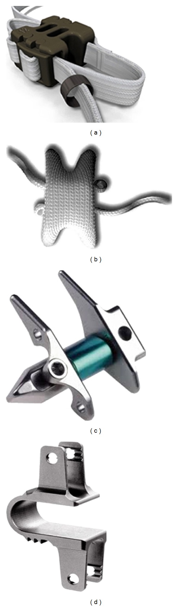
Disc degeneration and associated disorders are among the most debated topics in the orthopedic literature over the past few decades. These may be attributed to interrelated mechanical, biochemical, and environmental factors. The treatment options vary from conservative approaches to surgery, depending on the severity of degeneration and response to conservative therapies. Spinal fusion is considered to be the "gold standard" in surgical methods till date. However, the association of adjacent level degeneration has led to the evolution of motion preservation technologies like spinal arthroplasty and posterior dynamic stabilization systems. These new technologies are aimed to address pain and preserve motion while maintaining a proper load sharing among various spinal elements. This paper provides an elaborative biomechanical review of the technologies aimed to address the disc degeneration and reiterates the point that biomechanical efficacy followed by long-term clinical success will allow these nonfusion technologies as alternatives to fusion, at least in certain patient population.
Figures


Similar articles
-
Motion-preserving technologies for degenerative lumbar spine: The past, present, and future horizons.SAS J. 2011 Sep 1;5(3):75-89. doi: 10.1016/j.esas.2011.05.001. eCollection 2011. SAS J. 2011. PMID: 25802672 Free PMC article.
-
Motion preservation technologies: alternatives to spinal fusion.Am J Orthop (Belle Mead NJ). 2006 Sep;35(9):411-6. Am J Orthop (Belle Mead NJ). 2006. PMID: 17036776 Review.
-
Biomechanics of posterior dynamic stabilization systems.Adv Orthop. 2013;2013:451956. doi: 10.1155/2013/451956. Epub 2013 Mar 31. Adv Orthop. 2013. PMID: 23606975 Free PMC article.
-
External and internal responses of cervical disc arthroplasty and anterior cervical discectomy and fusion: A finite element modeling study.J Mech Behav Biomed Mater. 2020 Jun;106:103735. doi: 10.1016/j.jmbbm.2020.103735. Epub 2020 Mar 22. J Mech Behav Biomed Mater. 2020. PMID: 32321632
-
Biomechanics of the posterior lumbar articulating elements.Neurosurg Focus. 2007 Jan 15;22(1):E1. doi: 10.3171/foc.2007.22.1.1. Neurosurg Focus. 2007. PMID: 17608330 Review.
Cited by
-
Artificial disc replacement in spine surgery.Ann Transl Med. 2019 Sep;7(Suppl 5):S170. doi: 10.21037/atm.2019.08.26. Ann Transl Med. 2019. PMID: 31624736 Free PMC article. Review.
-
Axial T1ρ MRI as a diagnostic imaging modality to quantify proteoglycan concentration in degenerative disc disease.Eur Spine J. 2015 Nov;24(11):2395-401. doi: 10.1007/s00586-014-3582-6. Epub 2014 Sep 19. Eur Spine J. 2015. PMID: 25236594 Free PMC article.
-
Clinical and radiological comparison of posterolateral fusion and posterior interbody fusion techniques for multilevel lumbar spinal stabilization in manual workers.Asian Spine J. 2014 Oct;8(5):571-80. doi: 10.4184/asj.2014.8.5.571. Epub 2014 Oct 18. Asian Spine J. 2014. PMID: 25346809 Free PMC article.
-
Biomechanical Study of Symmetric Bending and Lifting Behavior in Weightlifter with Lumbar L4-L5 Disc Herniation and Physiological Straightening Using Finite Element Simulation.Bioengineering (Basel). 2024 Aug 12;11(8):825. doi: 10.3390/bioengineering11080825. Bioengineering (Basel). 2024. PMID: 39199783 Free PMC article.
-
Degenerative Thoracic Myelopathy: A Scoping Review of Epidemiology, Genetics, and Pathogenesis.Global Spine J. 2024 Jun;14(5):1664-1677. doi: 10.1177/21925682231224768. Epub 2023 Dec 26. Global Spine J. 2024. PMID: 38146739 Free PMC article.
References
-
- Modic MT, Ross JS. Lumbar degenerative disk disease. Radiology. 2007;245(1):43–61. - PubMed
-
- Crow WT, Willis DR. Estimating cost of care for patients with acute low back pain: a retrospective review of patient records. The Journal of the American Osteopathic Association. 2009;109(4):229–233. - PubMed
-
- Schizas C, Kulik G, Kosmopoulos V. Disc degeneration: current surgical options. European Cells & Materials. 2010;20:306–315. - PubMed
-
- Kuslich SD, Ulstrom CL, Michael CJ. The tissue origin of low back pain and sciatica: a report of pain response to tissue stimulation during operations on the lumbar spine using local anesthesia. Orthopedic Clinics of North America. 1991;22(2):181–187. - PubMed
-
- An HS, Anderson PA, Haughton VM, et al. Introduction. Disc degeneration: summary. Spine. 2004;29(23):2677–2678. - PubMed
LinkOut - more resources
Full Text Sources
Miscellaneous
