. 2012 May 31;1(5):557-69.
doi: 10.1016/j.celrep.2012.03.014. Epub 2012 May 3.

Mouse skeletal muscle fiber-type-specific macroautophagy and muscle wasting are regulated by a Fyn/STAT3/Vps34 signaling pathway

Affiliations

Mouse skeletal muscle fiber-type-specific macroautophagy and muscle wasting are regulated by a Fyn/STAT3/Vps34 signaling pathway

Eijiro Yamada et al. Cell Rep. .

Abstract

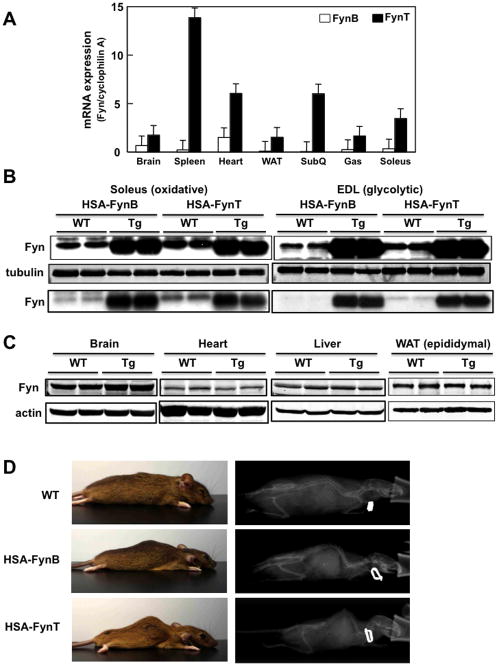
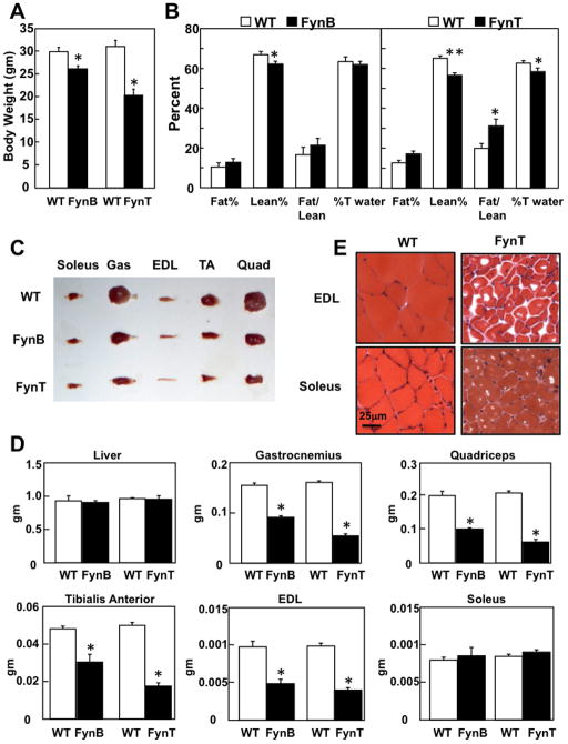
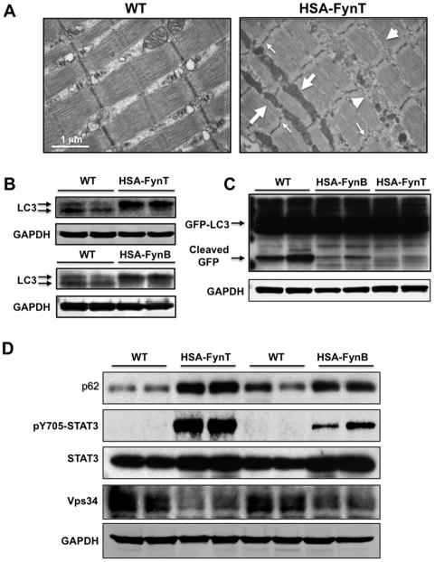
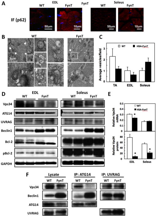
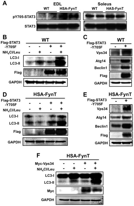
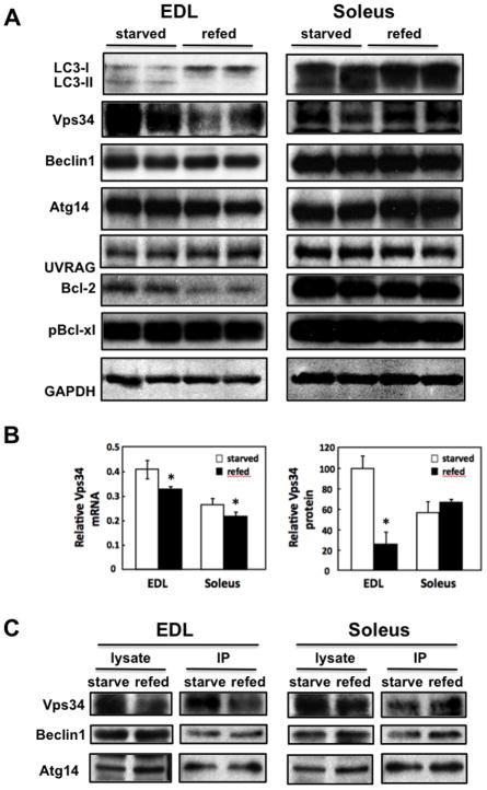
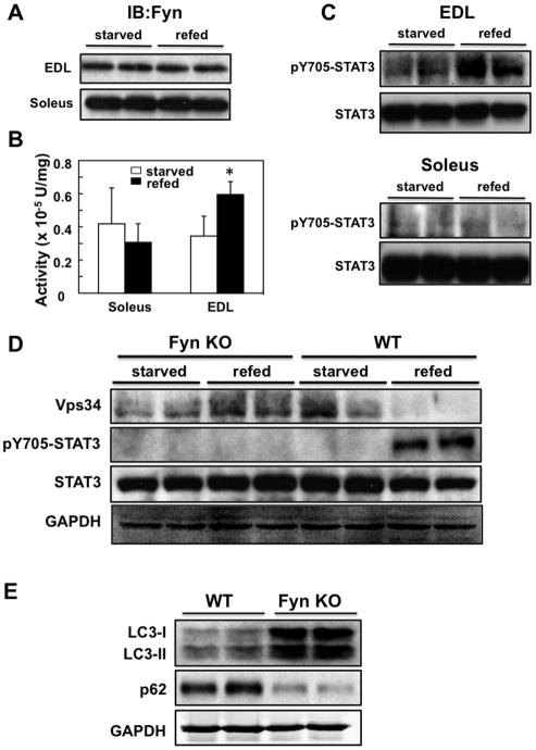
Skeletal muscle atrophy induced by aging (sarcopenia), inactivity, and prolonged fasting states (starvation) is predominantly restricted to glycolytic type II muscle fibers and typical spares oxidative type I fibers. However, the mechanisms accounting for muscle fiber-type specificity of atrophy have remained enigmatic. In the current study, although the Fyn tyrosine kinase activated the mTORC1 signaling complex, it also induced marked atrophy of glycolytic fibers with relatively less effect on oxidative muscle fibers. This was due to inhibition of macroautophagy via an mTORC1-independent but STAT3-dependent reduction in Vps34 protein levels and decreased Vps34/p150/Beclin1/Atg14 complex 1. Physiologically, in the fed state endogenous Fyn kinase activity was increased in glycolytic but not oxidative skeletal muscle. In parallel, Y705-STAT3 phosphorylation increased with decreased Vps34 protein levels. Moreover, fed/starved regulation of Y705-STAT3 phosphorylation and Vps34 protein levels was prevented in skeletal muscle of Fyn null mice. These data demonstrate a Fyn/STAT3/Vps34 pathway that is responsible for fiber-type-specific regulation of macroautophagy and skeletal muscle atrophy.

Keywords: Fyn; LKB1; STAT3; Vps34; autophagy; mTORC1; muscle atrophy.


Figures

Figure 1
Figure 1. Relative expression level of FynT and FynB in tissues, and generation of skeletal muscle specific FynB and FynT transgenic mice
A) mRNA expression levels of FynB (open bars) and FynT (solid bars) relative to cyclophilin A in brain, spleen, heart, epididymal (WAT) and subcutaneous (SubQ) adipose tissues, gastrocnemius (Gas) and soleus muscle. B) Fyn protein expression in soleus and extensor digitorum longus (EDL) skeletal muscles of wild type (WT), HSA-FynT and HSA-FynB transgenic mice (upper panels). Tubulin was used as internal loading control. A lighter exposure of the Fyn immunoblot (top panel) is shown below the tubulin immunoblot. C) Fyn protein levels in the brain, heart, liver and epididymal adipose tissue of WT and HSA-FynT transgenic (Tg) mice. Actin was used as internal loading control. D) Physical characteristics (left panels) and microCT pictures (right panels) for wild type (WT), HSA-FynB and FynT transgenic mice.
Figure 2
Figure 2. Skeletal muscle atrophic characteristics of HSA-FynT and HSA-FynB transgenic mice
A) Total body weight of HSA-FynB (FynB) and HSA-FynT (FynT) mice and their respective control littermates (WT). *p< 0.005 vs WT. B) Quantification of fat and lean mass measured by NMR in HSA-FynB (left panel) and HSA-FynT (right panel) mice versus their respective control littermates. *p< 0.05,, **p< 0.01 vs WT, respectively C) Selective muscle sizes from WT, HSA-FynB and HSA-FynT mice. D) Total tissue weight (liver, gastrocnemius, quadriceps, tibialis anterior, EDL and soleus muscles) of WT littermates, HSA-FynB and HSA-FynT transgenic mice. Data are expressed as mean ± SEM. *p<0.005 vs WT. E) Hematoxylin &Eosin (H&E) staining of EDL and soleus muscle from WT and HSA-FynT mice.
Figure 3
Figure 3. HSA-Fyn mice display defective macroautophagy
A) Transmission electron microscopy images of tibialis anterior muscle from WT (left) and HSA-FynT (right) mice. Large arrows indicate expanded mitochondria, small arrows indicate Z-line disruption and arrowheads indicate expanded sarcoplasmic reticulum. B) LC3-I and LC3-II protein levels in gastrocnemius muscle of WT and HSA-FynT (lower) compared to WT and HSA-FynB mice (upper) from two independent animals each. C) Tibialis anterior muscle of WT, HSA-FynB and HSA-FynT mice were transfected with GFP-LC3 as described in Experimental Procedures. The free cleaved GFP (lower band) is generated by autophagy from the GFP-LC3 fusion protein (upper bands) was detected by immunoblotting with a GFP mouse monoclonal antibody. Immunoblotting for the GAPDH protein (panels C, D, and E) was used as a loading control. D) Gastrocnemius muscles of WT, HSA-FynT and HSA-FynB mice were immunobotted with p62, phosphoY705-STAT3, total STAT3 and vps34 antibodies from two independent animals. GAPDH was used as internal loading control.
Figure 4
Figure 4. Autophagy is differently regulated in fast-twitch glycolytic type II fibers (EDL) and slow-twitch oxidative type I fiber (soleus) of HSA-FynT mice
A) Representative p62 immunofluorescence and DAPI staining (blue) images from EDL and soleus muscle of WT and HSA-FynT mice. B) Representative transmission electron microscopic images of tibialis anterior muscles from wild type and HSA-FynT (FynT) mice. Examples of autophagic vacuoles (denoted by arrows in the low magnification field) are show at higher magnification in the inserts. C) Quantification of autophagic vacuoles (AV) by transmission electron microscopic in tibialis anterior (TA), EDL and soleus muscles from wild type (open bars) and HSA-FynT (filled bars) mice. The average number of autophagic vacuoles per field was quantified by morphological analyses of at least 15 electron microscopy images from 3 independent mice. D) Beclin1 and Beclin1 complex-related proteins (Vps34, Atg14, UVRAG, Bcl2 and phospho-Bcl-2) expression levels in EDL and soleus muscles of WT and HSA-FynT mice. GAPDH was used as internal loading control. These are representative immunoblots independently performed 3 times E) mRNA expression levels (upper) and protein levels (lower) of Vps34 relative to Hrpt1 or GAPDH in EDL and soleus muscles of WT and HSA-FynT mice. F) Vps34 presence in Beclin1 complex-1 (containing Atg14) and Beclin1 complex-2 (containing UVRAG) in quadriceps muscle of WT and HSA-FynT mice was determined by immunoprecipitation (IP) using either an Atg14 or UVRAG antibody followed by immunoblotting with Vps34, Beclin1, Atg14 and UVRAG antibodies. These are representative immunoblots independently performed 3 times.
Figure 5
Figure 5. STAT3 is tyrosine phosphorylated in EDL but not soleus muscle of HSA-FynT mice and the dominant negative form of STAT3 (STAT3-Y705F) rescues autophagy deficiency in HSA-FynT mice
A) EDL and soleus muscle isolated from WT and HSA-FynT mice were immunoblotted with phosphoY705-STAT3 and total STAT3 antibodies from two independent animals. B) Tibialis anterior muscles of WT mice were transfected with either an empty vector (−) or the vector containing the dominant interfering (+) STAT3 mutant (Flag-STAT3-Y705F) and autophagic flow was determined as described in Experimental Methods. C) Beclin1 complex related proteins expression levels in the tibialis anterior muscles of WT mice transfected with empty vector (−) or Flag-STAT3-Y705F (+). D) Tibialis anterior muscles of HSA-FynT mice were transfected with empty vector (−) or the vector containing the dominant interfering (+) STAT3 mutant (Flag-STAT3-Y705F) followed by determination of autophagic flow. E) Beclin1 complex related proteins expression levels in the tibialis anterior muscles of HSA-FynT mice transfected with an empty vector (−) or Flag-STAT3-Y705F (+). These are representative immunoblots independently performed 4 times for panel A and 3 times for panels B–E. F) Tibialis anterior muscles of HSA-FynT mice were transfected with empty vector (−) or the vector containing the Vps34 (Myc-Vps34) followed by determination of autophagic flow.
Figure 6
Figure 6. Beclin1 and Beclin1 complex-related proteins are differentially regulated in the EDL and soleus muscles of WT animals in the refed state
A) WT mice were food restricted for 30 h (starved) and then allowed free access to food for 5 h (refed). The EDL and soleus muscles were isolated and immunoblotted for LC3 in two independent animals. These are representative immunoblots independently performed 3 times. B) mRNA expression levels (left) and protein levels (right) of Vps34 relative to Hrpt1 or GAPDH in EDL and soleus muscles of starved and refed WT mice. C) The EDL and soleus muscle from starved and refed WT mice (lysates) were immunoprecipitated (IP) with an ATG14 antibody and immunoblotted for Vps34, Beclin1 and Atg14. These are representative immunoblots independently performed 3 times.
Figure 7
Figure 7. Autophagy is differentially regulated in the EDL and soleus muscles of WT mice in the starved and refed state
A) EDL and soleus muscle from starved and refed WT mice were immunoblotted for Fyn protein levels in two independent animals. B) Fyn protein kinase activity in the EDL and soleus muscles of starved and refed WT mice was determined as described in Experimental Procedures from 5 independent mice. *p< 0.05 vs. starved. C) EDL (upper) and soleus muscle (lower) from starved and refed WT mice were immunoblotted for pY705-STAT3 and total STAT3 protein levels in two independent animals. D) Quadriceps muscle of FynKO and WT mice were immunoblotted with Vps34, phosphoY705-STAT3 and total STAT3 from two independent animals. GAPDH was used as internal loading control. E) LC3-I, LC3-II and p62 protein levels in quadriceps muscle of FynKO and WT mice from two independent animals each. These are representative immunoblots independently performed 3 times in panels A, C, D and E)

Similar articles

Cited by

References

    1. Alessi DR, Sakamoto K, Bayascas JR. LKB1-dependent signaling pathways. Annu Rev Biochem. 2006;75:137–163. - PubMed
    1. Backer JM. The regulation and function of Class III PI3Ks: novel roles for Vps34. Biochem J. 2008;410:1–17. - PubMed
    1. Bassel-Duby R, Olson EN. Signaling pathways in skeletal muscle remodeling. Annu Rev Biochem. 2006;75:19–37. - PubMed
    1. Bastie C, Zong H, Xu J, Busa B, Judex S, Kurland IJ, Pessin JE. Role of the Src Kinase Family Member Fyn in the Integrative Metabolic Regulation of Peripheral Tissue Fatty Acid Oxidation. Cell Metab. 2007;5 - PubMed
    1. Bechet D, Tassa A, Taillandier D, Combaret L, Attaix D. Lysosomal proteolysis in skeletal muscle. Int J Biochem Cell Biol. 2005;37:2098–2114. - PubMed

Publication types

MeSH terms

Substances

LinkOut - more resources