. 2013 Jan 1;18(1):36-50.
doi: 10.1089/ars.2011.4457. Epub 2012 Aug 16.

Redox-sensitive oxidation and phosphorylation of PTEN contribute to enhanced activation of PI3K/Akt signaling in rostral ventrolateral medulla and neurogenic hypertension in spontaneously hypertensive rats

Affiliations

Redox-sensitive oxidation and phosphorylation of PTEN contribute to enhanced activation of PI3K/Akt signaling in rostral ventrolateral medulla and neurogenic hypertension in spontaneously hypertensive rats

Kay L H Wu et al. Antioxid Redox Signal. .

Abstract

Aims: The activity of phosphoinositide 3-kinase (PI3K)/serine/threonine protein kinase (Akt) is enhanced under hypertension. The phosphatase and tensin homolog deleted on chromosome 10 (PTEN) is a negative regulator of PI3K signaling, and its activity is redox-sensitive. In the rostral ventrolateral medulla (RVLM), which is responsible for the maintenance of blood pressure, oxidative stress plays a pivotal role in neurogenic hypertension. The present study evaluated the hypothesis that redox-sensitive inactivation of PTEN results in enhanced PI3K/Akt signaling in RVLM, leading to neurogenic hypertension.

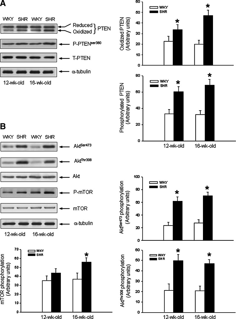
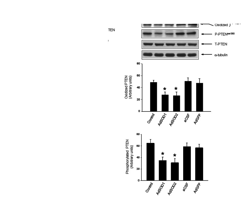
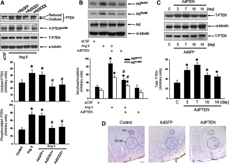
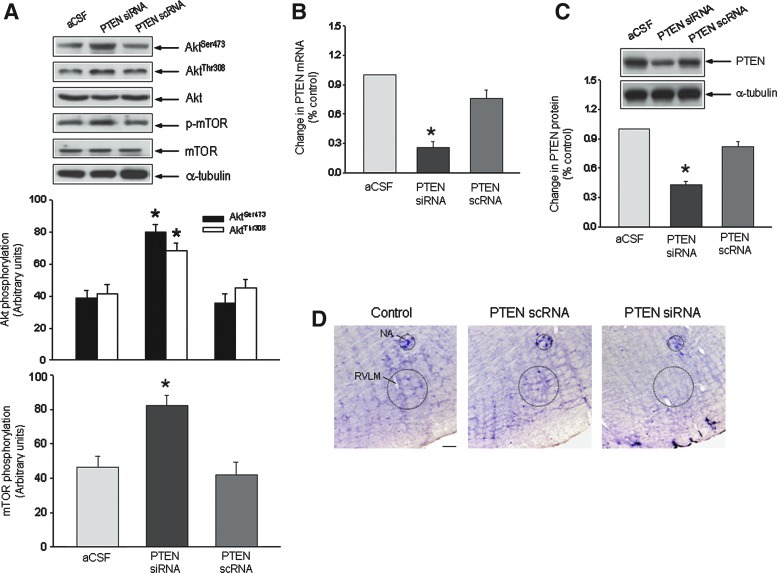
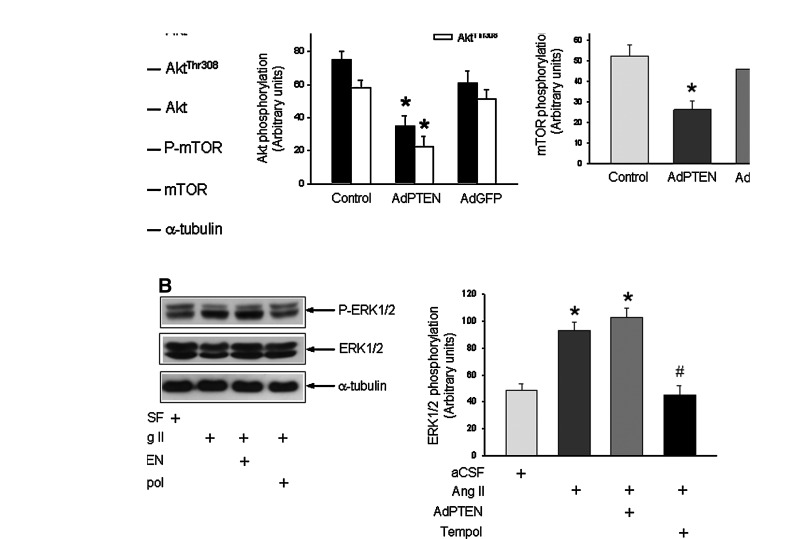
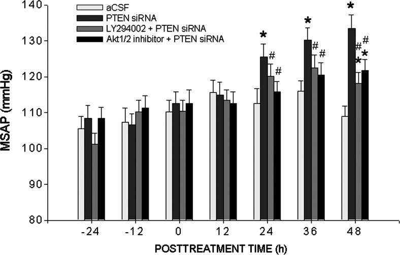
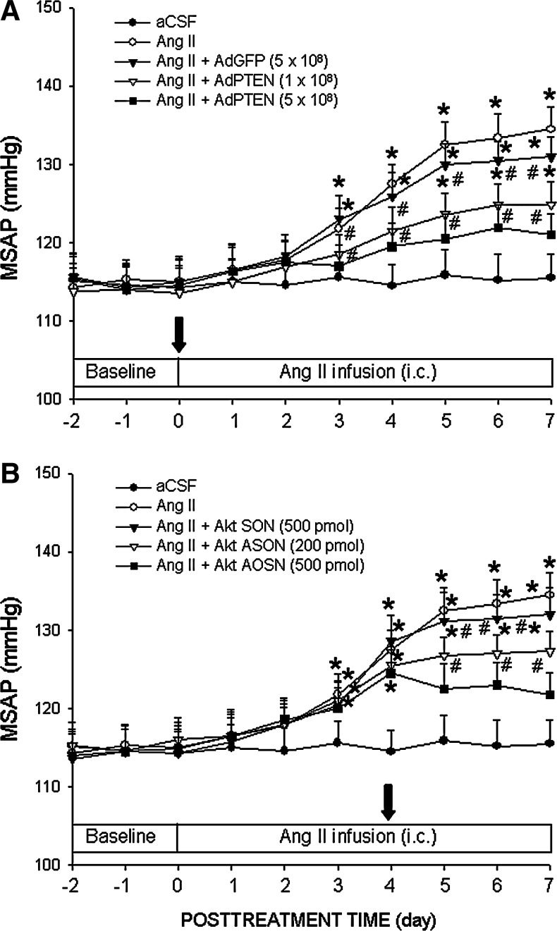
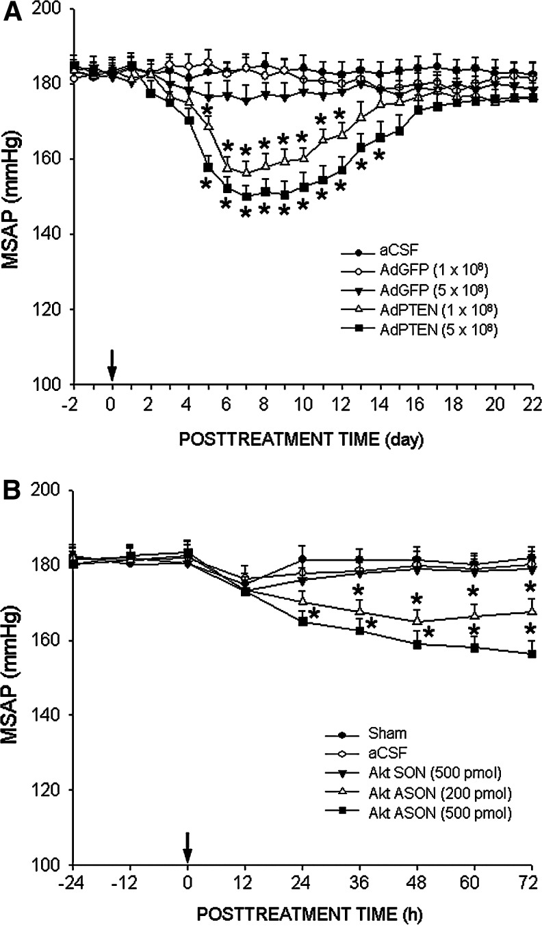
Results: Compared to age-matched normotensive Wistar-Kyoto (WKY) rats, PTEN inactivation in the form of oxidation and phosphorylation were greater in RVLM of spontaneously hypertensive rats (SHR). PTEN inactivation was accompanied by augmented PI3K activity and PI3K/Akt signaling, as reflected by the increase in phosphorylation of Akt and mammalian target of rapamycin. Intracisternal infusion of tempol or microinjection into the bilateral RVLM of adenovirus encoding superoxide dismutase significantly antagonized the PTEN inactivation and blunted the enhanced PI3K/Akt signaling in SHR. Gene transfer of PTEN to RVLM in SHR also abrogated the enhanced Akt activation and promoted antihypertension. Silencing PTEN expression in RVLM with small-interfering RNA, on the other hand, augmented PI3K/Akt signaling and promoted long-term pressor response in normotensive WKY rats.

Innovation: The present study demonstrated for the first time that the redox-sensitive check-and-balance process between PTEN and PI3K/Akt signaling is engaged in the pathogenesis of hypertension.

Conclusion: We conclude that an aberrant interplay between the redox-sensitive PTEN and PI3k/Akt signaling in RVLM underpins neural mechanism of hypertension.


Figures

FIG. 1.
FIG. 1.
Increased oxidation and phosphorylation of phosphatase and tensin homolog deleted on chromosome 10 (PTEN) and enhanced phosphoinositide 3-kinase (PI3K)/serine/threonine protein kinase (Akt) activity in rostral ventrolateral medulla (RVLM) of spontaneously hypertensive rat (SHR). (A) Representative Western blots and densitometric analysis of protein level of reduced or oxidized PTEN, or phosphorylated PTEN (P-PTEN) or total PTEN (T-PTEN), detected from RVLM of normotensive Wistar-Kyoto (WKY) rats or SHR at age of 12 or 16 weeks. Tissue samples from RVLM were collected by micropunches, and the extracted protein was analyzed using Western blotting as described under the Materials and Methods section. (B) Representative Western blots and densitometric analysis of protein level of phosphorylated Akt at Ser473 and Thr308 or mammalian target of rapamycin (mTOR) (P-mTOR), total Akt or mTOR, or α-tubulin, detected from RVLM of normotensive WKY rats or SHR at age of 12 or 16 weeks. Values are mean±standard error of mean of four independent experiments on samples pooled from six to eight animals in each group. *p<0.05 versus age-matched WKY rat group in the Student's t-test.
FIG. 2.
FIG. 2.
Oxidation and phosphorylation of PTEN in RVLM of SHR are mediated by NADPH oxidase- and mitochondria-derived reactive oxygen species (ROS). (A) Representative Western blots and densitometric analysis of protein level of reduced or oxidized PTEN, P-PTEN or T-PTEN, or α-tubulin, detected from RVLM of SHR (at age of 16 weeks) on day 7 after intracisternal (i.c.) infusion of artificial cerebrospinal fluid (aCSF), antisense oligonucleotide (ASON) or sense oligonucleotide (SON) against p47phox subunit of the NADPH oxidase, a mobile electron carrier in the inner membrane of mitochondria, coenzyme Q10 (CoQ10). Infusion of test agents into the cisterna magna was performed as described under the Materials and Methods section. (B) Representative Western blots and densitometric analysis of protein level of reduced or oxidized PTEN, P-PTEN or T-PTEN, or α-tubulin, detected from RVLM of SHR (at age of 16 weeks) on day 7 after microinjection bilaterally into RVLM of adenovirus encoding superoxide dismutase 1 or 2 (AdSOD1 or AdSOD2), or adenoviral vector encoding green fluorescent protein (AdGFP). In vivo gene delivery of AdSOD1 or AdSOD2 to the bilateral RVLM was performed as described under the Materials and Methods section. Values are mean±standard error of mean of four independent experiments on samples pooled from six to eight animals in each group. *p<0.05 versus sham-control group in the post hoc Scheffé multiple range analysis.
FIG. 3.
FIG. 3.
Increased tissue level of ROS in RVLM induces oxidation and phosphorylation of PTEN and activation of PI3K in normotensive WKY rats. (A) Representative Western blots and densitometric analysis of protein level of reduced or oxidized PTEN, P-PTEN or T-PTEN, or α-tubulin, detected from RVLM of WKY rats (at age of 16 weeks) on day 7 after i.c. infusion of aCSF or angiotensin II (Ang II), alone or with additional microinjection bilaterally into RVLM of AdSOD1, AdSOD2. (B) Representative Western blots and densitometric analysis of protein level of phosphorylated Akt or total Akt, or α-tubulin, detected from RVLM of WKY rats (at age of 16 weeks) on day 7 after i.c. infusion of aCSF or Ang II, alone or with additional microinjection bilaterally into RVLM of adenoviral vector encoding PTEN (AdPTEN). In vivo gene delivery of AdSOD1, AdSOD2 or AdPTEN to the RVLM was performed immediately after implantation of osmotic minipumps for i.c. infusion of Ang II as described under the Materials and Methods section. Also shown are representative Western blots and densitometric analysis of temporal changes in protein level of T-PTEN (C) in RVLM of WKY rats after microinjection bilaterally into RVLM of AdPTEN (top panel) or AdGFP (bottom panel). (D) Representative photomicrographs showing PTEN-immunoreactive cells in RVLM of WKY rats subjected to microinjection bilaterally into RVLM of AdGFP or AdPTEN. Values are mean±standard error of mean of four independent experiments on samples pooled from five to eight animals in each group. *p<0.05 versus aCSF or sham-control (C) group, and #p<0.05 versus Ang II group in the post hoc Scheffé multiple range analysis. Dotted circle in (D) depicts anatomical confines of nucleus ambiguous (NA) or RVLM. Scale bar: 100 μm. (To see this illustration in color, the reader is referred to the web version of this article at www.liebertpub.com/ars.)
FIG. 4.
FIG. 4.
Suppression of PTEN increases PI3K/Akt signaling in RVLM of normotensive WKY rats. (A) Representative Western blots and densitometric analysis of protein level of phosphorylated Akt or mTOR, total Akt or mTOR, or α-tubulin, detected from RVLM of WKY rats (at age of 16 weeks) 48 h after microinjection bilaterally into RVLM of PTEN small-interfering RNA (siRNA) or scrambled siRNA (scRNA). Microinjection into the bilateral RVLM of siRNA or scRNA was carried out as described under the Materials and Methods section. Real-time polymerase chain reaction (PCR) analysis of PTEN mRNA (B), or densitometric analysis of protein level of PTEN (C) from RVLM of WKY rats (at age of 16 weeks), detected 48 h after microinjection bilaterally into RVLM of PTEN siRNA or scRNA. Tissue samples were collected from RVLM and mRNA or protein level was determined using real time PCR or Western blotting. (D) Representative photomicrographs showing PTEN-immunoreactive cells in RVLM of SHR subjected to microinjection bilaterally into RVLM of PTEN scRNA or siRNA. Values are mean±standard error of mean of four independent experiments on samples pooled from six to eight animals in each group. *p<0.05 versus aCSF group in the post hoc Scheffé multiple range analysis. Dotted circle in (D) depicts anatomical confine of NA or RVLM. Scale bar: 100 μm. (To see this illustration in color, the reader is referred to the web version of this article at www.liebertpub.com/ars.)
FIG. 5.
FIG. 5.
Overexpression of PTEN blunted the enhanced PI3K/Akt signaling in RVLM of SHR. (A) Representative Western blots and densitometric analysis of protein level of phosphorylated Akt or mTOR, total Akt or mTOR, or α-tubulin, detected from RVLM of SHR (at age of 16 weeks) on day 7 after microinjection bilaterally into RVLM of AdPTEN or AdGFP. Tissue samples were collected from RVLM and protein expression was determined using Western blotting as described under the Materials and Methods section. (B) Representative Western blots and densitometric analysis of protein level of total and phosphorylated ERK1/2, or α-tubulin, detected from RVLM of WKY rats (at age of 16 weeks) on day 7 after i.c. infusion of Ang II, alone or with additional microinjection into bilateral RVLM of AdPTEN or coinfusion of tempol into the cisterna magna. Note that in vivo gene delivery of AdPTEN to the RVLM significantly reversed the Ang II-induced phosphorylation of Akt or mTOR, but not the induced phosphorylation of ERK1/2. Values are mean±standard error of mean of four independent experiments on samples pooled from six to seven animals in each group. *p<0.05 versus aCSF or sham-control group, and #p<0.05 versus Ang II group in the post hoc Scheffé multiple range analysis.
FIG. 6.
FIG. 6.
Silencing PTEN in RVLM promotes pressor responses in normotensive WKY rats. Temporal changes in mean systemic arterial pressure (MSAP) in WKY rats that received microinjection bilaterally into RVLM of PTEN siRNA, alone or with additional i.c. infusion of PI3K inhibitor, LY294002, or Akt kinase inhibitor. SAP was recorded for 48 h under conscious conditions by radiotelemetry. Values are mean±standard error of mean recorded from six to eight animals per group. *p<0.05 versus aCSF group and #p<0.05 versus PTEN siRNA group at corresponding time points in the post hoc Scheffé multiple range analysis.
FIG. 7.
FIG. 7.
Overexpression of PTEN or gene knockdown of Akt in RVLM attenuates Ang II-induced long-term pressor response in normotensive WKY rats. Temporal changes in MSAP of WKY rats that received i.c. infusion of Ang II, alone or with additional microinjection bilaterally into RVLM of AdPTEN or AdGFP (A) or Akt ASON or SON (B). AdPTEN or AdGFP was microinjected into the bilateral RVLM immediately following implantation of osmotic minipumps for i.c. infusion of Ang II as described under the Materials and Methods section. Akt ASON or SON was microinjected into the bilateral RVLM on day 4 after i.c. infusion of Ang II. SAP was recorded in animals under conscious condition by radiotelemetry. Values are mean±standard error of mean recorded from five to nine animals per group. *p<0.05 versus aCSF group and #p<0.05 versus Ang II group at corresponding time points in the post hoc Scheffé multiple range analysis.
FIG. 8.
FIG. 8.
Overexpression of PTEN or gene knockdown of Akt in RVLM promotes antihypertension in SHR. Temporal changes in MSAP of SHR following microinjection into the bilateral RVLM of AdPTEN or AdGFP (A), or Akt ASON or SON (B). AdPTEN, AdGFP, Akt ASON or Akt SON was microinjected into the bilateral RVLM on day 0, and SAP was recorded in animals under conscious condition by radiotelemetry. Changes in MSAP of SHR after overexpression of PTEN in RVLM were recorded for 22 days; and those after Akt ASON treatment was recorded for 72 h. Values are mean±standard error of mean recorded from five to eight animals per group. *p<0.05 versus aCSF group at corresponding time points in the post hoc Scheffé multiple range analysis.

Similar articles

Cited by

References

    1. Blanco-Aparicio C. Renner O. Leal JF. Carnero A. PTEN, more than the AKT pathway. Carcinogenesis. 2007;28:1379–1386. - PubMed
    1. Chan JYH. Wang LL. Chao YM. Chan SHH. Downregulation of basal iNOS at the rostral ventrolateral medulla is innate in SHR. Hypertension. 2003;41:563–570. - PubMed
    1. Chan SHH. Hsu KS. Huang CC. Wang LL. Ou CC. Chan JYH. NADPH oxidase-derived superoxide anion mediates angiotensin II-induced pressor effect via activation of p38 mitogen-activated protein kinase in the rostral ventrolateral medulla. Circ Res. 2005;97:772–780. - PubMed
    1. Chan SHH. Tai MH. Li CY. Chan JYH. Reduction in molecular synthesis or enzyme activity of superoxide dismutases and catalase contributes to oxidative stress and neurogenic hypertension in spontaneously hypertensive rats. Free Radic Biol Med. 2006;40:2028–2039. - PubMed
    1. Chan SHH. Wang LL. Wang SH. Chan JYH. Differential cardiovascular responses to blockade of nNOS or iNOS in rostral ventrolateral medulla of the rat. Br J Pharmacol. 2001;133:606–614. - PMC - PubMed

Publication types

MeSH terms