. 2012 Dec;221(6):577-89.
doi: 10.1111/j.1469-7580.2012.01537.x. Epub 2012 Jul 3.

Characterization of the annulus fibrosus-vertebral body interface: identification of new structural features

Affiliations

Characterization of the annulus fibrosus-vertebral body interface: identification of new structural features

Y S Nosikova et al. J Anat. 2012 Dec.

Abstract

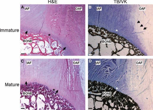
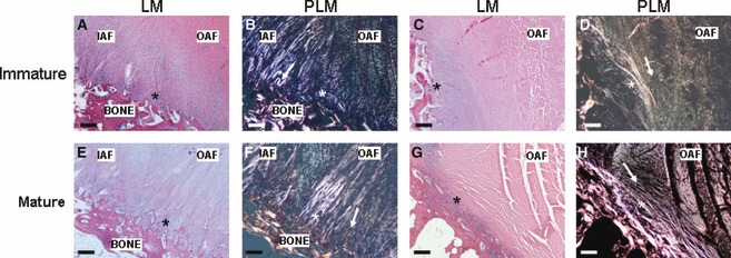
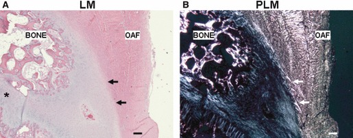
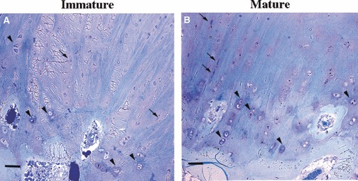
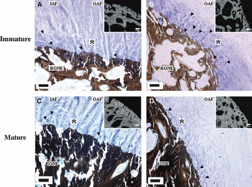
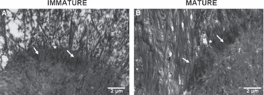
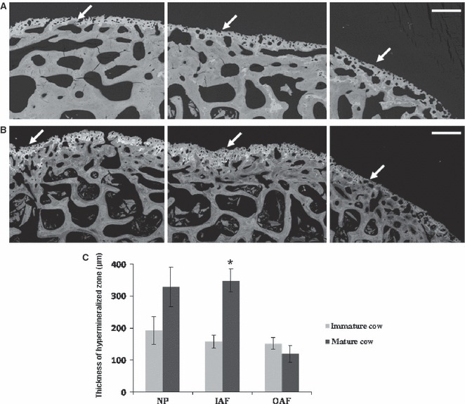
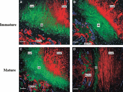
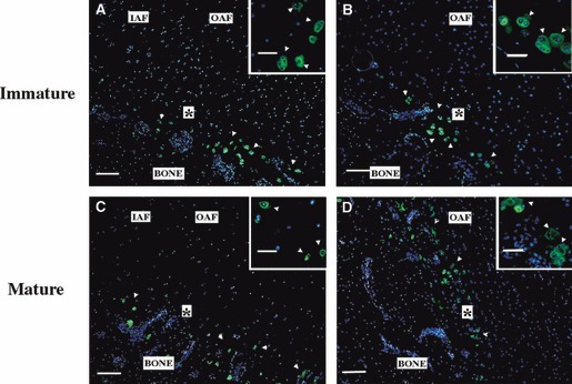
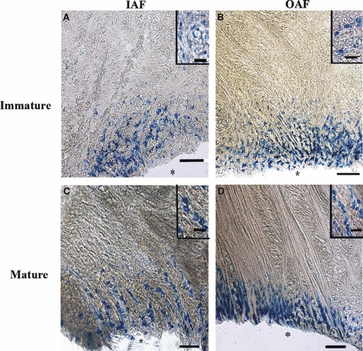
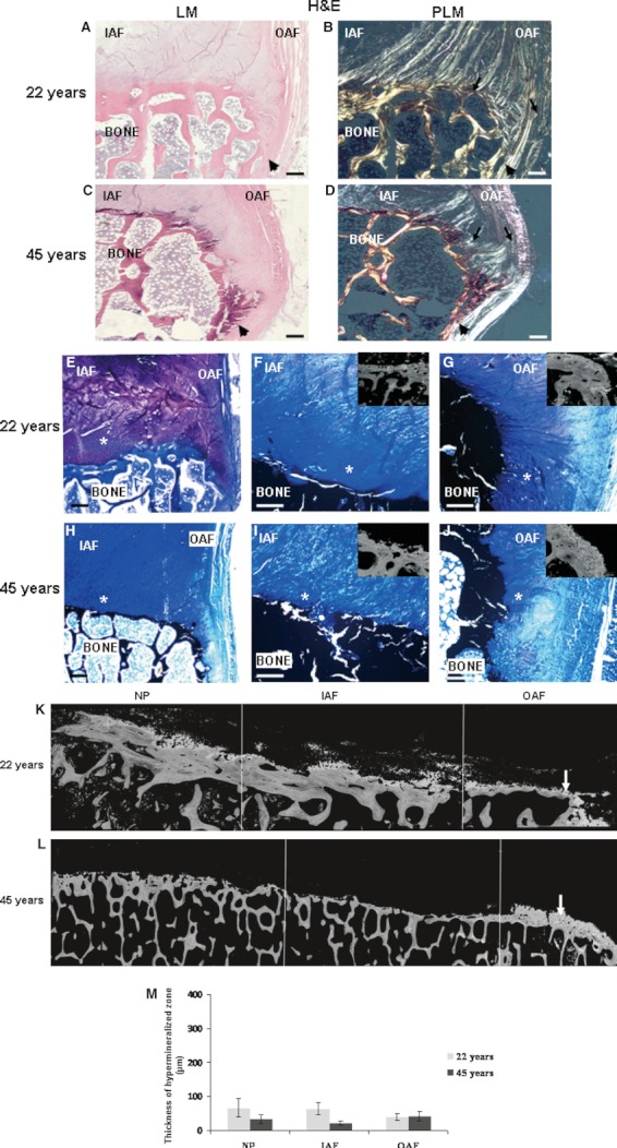
Current surgical treatments for degenerative intervertebral disc disease do not restore full normal spinal movement. Tissue engineering a functional disc replacement may be one way to circumvent this limitation, but will require an integration of the different tissues making up the disc for this approach to be successful. Hence, an in-depth characterization of the native tissue interfaces, including annulus insertion into bone is necessary, as knowledge of this interface is limited. The objective of this study was to characterize the annulus fibrosus-vertebral bone (AF-VB) interface in immature (6-9 months old) and mature (18-24 months old) bovine discs, as well as to define these structures for normal adult human (22 and 45 years old) discs. Histological assessment showed that collagen fibers in the inner annulus, which are predominantly type II collagen, all appear to insert into the mineralized endplate zone. In contrast, some of the collagen fibers of the outer annulus, predominantly type I collagen, insert into this endplate, while other fibers curve laterally, at an ∼ 90° angle, to the outer aspect of the bone, and merge with the periosteum. This is seen in both human and bovine discs. Where the AF inserts into the calcified zone of the AF-VB interface, it passes through a chondroid region, rich in type II collagen and proteoglycans. Annulus cells (elongated cells that are not surrounded by proteoglycans) are present at this interface. This cartilage zone is evident in both human and bovine discs. Type X collagen and alkaline phosphatase are localized to the interface region. Age-associated differences in bovine spines are observed when examining the interface thickness and the matrix composition of the cartilaginous endplate, as well as the thickness of the mineralized endplate. These findings will assist with the design of the AF-VB interface in the tissue engineered disc.


Figures

Fig. 1
Fig. 1
Photomicrographs of coronal sections of a representative bovine immature (A, B) and mature (C, D) IVD [(A and C): hematoxylin and eosin (H&E); (B and D): toluidine blue and von Kossa (TB/VK)]. Arrowheads indicate the proteoglycans in the outer annulus interlamellar space. Arrow indicates the chondroid matrix at the AF–VB interface (scale bar: 250 μm; IAF, inner annulus fibrosus; OAF, outer annulus fibrosus; *AF–VB interface).
Fig. 2
Fig. 2
Photomicrographs of coronal sections of representative bovine immature (A–D) and mature (E–H) discs at the inner and outer (A, B, E, F), and outer aspect of the outer (C, D, G, H) annulus insertion into the calcified region [hematoxylin and eosin (H&E) light (LM) (A, C, E, G) and polarized light (PLM) (B, D, F, H)] (scale bar: 125 μm; IAF, inner annulus fibrosus; OAF, outer annulus fibrosus; *AF–VB interface; ↑indicating direction of collagen fibers).
Fig. 3
Fig. 3
Photomicrographs of coronal sections of a representative bovine immature outer annulus lamellae insertion into the outer aspect of VB distant to the disc [H&E light microscopy (LM) (A) and polarized light microscopy (PLM) (B)]. Arrows indicate the outer annulus collagen merging with the cartilage. *Growth plate within the VB (scale bar: 250 μm; OAF, outer annulus fibrosus).
Fig. 4
Fig. 4
Photomicrographs of undecalcified sections (500 nm thick) of the OAF–VB interface of a representative immature (A) and mature (B) cow (toluidine blue). The arrowheads indicate chondrocytes and arrows indicate annulus cells (scale bar: 35 μm).
Fig. 5
Fig. 5
Photomicrographs of coronal undecalcified sections of representative bovine immature (A, B) and mature (C, D) discs at the inner and outer (A, C), and the most outer aspect of the outer (B, D) annulus insertion into the calcified region (toluidine blue and von Kossa). Arrowheads indicate the location of chondrocytes (scale bar: 125 μm; IAF, inner annulus fibrosus; OAF, outer annulus fibrosus; *AF–VB interface). The inserts (scale bar: 200 μm) represent the corresponding interface region as visualized by BSE. The hypermineralized zone appears more white than the bone.
Fig. 6
Fig. 6
Electron micrographs of sections of the OAF–VB interface following decalcification of a representative immature (A) and mature (B) cow. The arrows indicate the insertion of collagen fibers into electron-dense material (*tidemark).
Fig. 7
Fig. 7
Backscatter images of immature (A) and mature (B) bovine disc interface with the vertebral bone. Arrows indicate hypermineralized zone at the interface. (C) Quantification of the thickness of the hypermineralized zone across each disc region. The data are presented as the mean ± SEM (n = 6 and 15 discs of immature and mature cows, respectively; scale bar: 500 μm; IAF, inner annulus fibrosus; NP, nucleus pulposus; OAF, outer annulus fibrosus).
Fig. 8
Fig. 8
Distribution of types I (red) and II (green) collagen in representative images of immature (A, B) and mature (C, D) bovine samples at the inner and outer (A, C), and the most outer aspect of the outer (B, D) annulus insertion into the calcified region, as visualized by immunostaining. Cell nuclei are blue (DAPI; scale bar: 125 μm; IAF, inner annulus fibrosus; OAF, outer annulus fibrosus; *AF–VB interface).
Fig. 9
Fig. 9
Distribution of type X collagen (green) in a representative bovine immature (A, B) and mature (C, D) inner and outer (A, C), and the most outer aspect of the annulus insertion into the calcified region (B, D), as visualized by immunostaining. Cell nuclei are blue (DAPI). The inserts show cells at the interface at higher magnification. Arrowheads indicate clusters of cells surrounded by type X collagen at the AF–VB interface (scale bar: 125 μm and insert: 35 μm; *AF–VB interface; IAF, inner annulus fibrosus; OAF, outer annulus fibrosus).
Fig. 10
Fig. 10
Alkaline phosphatase activity (blue) is present in immature (A, B) and mature (C, D) discs at the inner (A, C) and outer (B, D) annulus insertion site. Inserts show cells at the interface at higher magnification. *Where tissue was removed from the calcified zone (scale bar: 125 μm; insert: 35 μm; IAF, inner annulus fibrosus; OAF, outer annulus fibrosus).
Fig. 11
Fig. 11
(A–D) Photomicrographs of coronal disc sections of representative 22- (A, B, E–G) and 45- (C, D, H–J) year-old human discs. Arrows indicate the direction of the outer annulus fibers. Arrowheads indicate the outer annulus collagen insertion to the periosteum of the bone. (A–D) Hematoxylin and eosin (H&E); (E–J): toluidine blue/von Kossa; scale bar: 250 μm). (F, G, I, J) Higher magnification images of (E) and (H) of the inner and outer (F, I) and the most outer aspect of the outer (G, J) annulus insertion into calcified zone of a 22- (F, G) and 45- (I, J) years-old human (scale bar: 125 μm). The inserts (scale bar: 200 μm) represent the corresponding interface region visualized by BSE–SEM. PLM, polarized light microscopy. *AF–VB interface. (K–L) Backscatter images of disc interface with the vertebral bone. Arrows indicate the hypermineralized zone (scale bar: 500 μm). (M) Quantification of the thickness of the hypermineralized zone of the human endplate. The data are presented as mean ± SEM (n = 3) (IAF, inner annulus fibrosus; NP, nucleus pulposus; OAF, outer annulus fibrosus).

Similar articles

Cited by

References

    1. Adams MA, Roughley PJ. What is intervertebral disc degeneration, and what causes it? Spine. 2006;31:2151–2161. - PubMed
    1. Aigner T, Gresk-otter KR, Fairbank JC, et al. Variation with age in the pattern of type X collagen expression in normal and scoliotic human intervertebral discs. Calcif Tissue Int. 1998;63:263–268. - PubMed
    1. Alini M, Eisenstein SM, Ito K, et al. Are animal models useful for studying human disc disorders/degeneration? Eur Spine J. 2008;17:2–19. - PMC - PubMed
    1. Attia M, Santerre JP, Kandel RA. The response of annulus fibrosus cell to fibronectin-coated nanofibrous polyurethane-anionic dihydroxyoligomer scaffolds. Biomaterials. 2011;32:450–460. - PubMed
    1. Ayad S, Abedin MZ, Grundy SM, et al. Isolation and characterisation of an unusual collagen from hyaline cartilage and intervertebral disc. FEBS Lett. 1981;123:195–199. - PubMed

Publication types

LinkOut - more resources