. 2012 Jul 2:9:156.
doi: 10.1186/1742-2094-9-156.

Clusters of activated microglia in normal-appearing white matter show signs of innate immune activation

Affiliations

Clusters of activated microglia in normal-appearing white matter show signs of innate immune activation

Jack van Horssen et al. J Neuroinflammation. .

Abstract

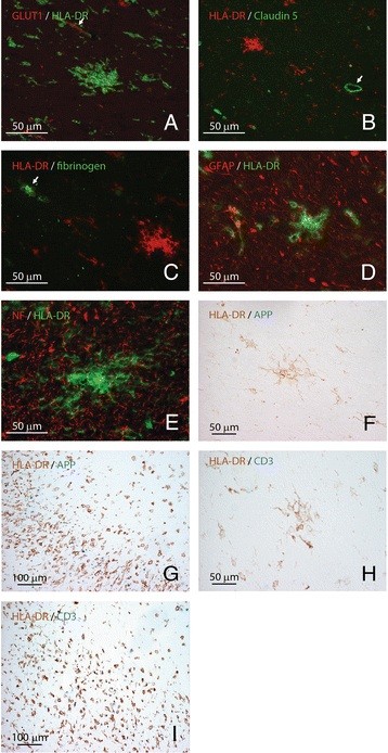
Background: In brain tissues from multiple sclerosis (MS) patients, clusters of activated HLA-DR-expressing microglia, also referred to as preactive lesions, are located throughout the normal-appearing white matter. The aim of this study was to gain more insight into the frequency, distribution and cellular architecture of preactive lesions using a large cohort of well-characterized MS brain samples.

Methods: Here, we document the frequency of preactive lesions and their association with distinct white matter lesions in a cohort of 21 MS patients. Immunohistochemistry was used to gain further insight into the cellular and molecular composition of preactive lesions.

Results: Preactive lesions were observed in a majority of MS patients (67%) irrespective of disease duration, gender or subtype of disease. Microglial clusters were predominantly observed in the vicinity of active demyelinating lesions and are not associated with T cell infiltrates, axonal alterations, activated astrocytes or blood-brain barrier disruption. Microglia in preactive lesions consistently express interleukin-10 and TNF-α, but not interleukin-4, whereas matrix metalloproteases-2 and -9 are virtually absent in microglial nodules. Interestingly, key subunits of the free-radical-generating enzyme NADPH oxidase-2 were abundantly expressed in microglial clusters.

Conclusions: The high frequency of preactive lesions suggests that it is unlikely that most of them will progress into full-blown demyelinating lesions. Preactive lesions are not associated with blood-brain barrier disruption, suggesting that an intrinsic trigger of innate immune activation, rather than extrinsic factors crossing a damaged blood-brain barrier, induces the formation of clusters of activated microglia.


Figures

Figure 1
Figure 1
Preactive lesions are composed of clusters of HLA-DR-positive microglia. In NAWM, i.e., in the absence of apparent myelin loss (A, E: proteolipid protein) preactive lesions are defined as circumscribed nodules of activated microglia expressing HLA-DR (B, C) and CD68 (D). Preactive lesions are predominantly observed in blocks containing active lesions (E: proteolipid protein; F, G: HLA-DR). Figure 1G represents a magnification of the outlined square in (F). In some cases microglial nodules are surrounded by a halo devoid of microglia (H: HLA-DR). Original magnifications: A, B: 20×; C, D: 40×; E, F: 4×; G, H: 40×.
Figure 2
Figure 2
Correlation of preactive lesions with the occurrence of white matter lesions. Presence of preactive lesions (PAL) in tissue blocks in NAWM (A) or blocks containing active and chronic active lesions (B). The numbers of blocks of NAWM (A) or active lesions (B) were expressed as percentage of all blocks. Pearson correlation testing was performed for respective groups. The presence of preactive lesions negatively correlates with the number of blocks containing no lesions (p = 0.0128; R2 0.28), whereas the presence of preactive lesions positively correlates with the number of active lesions (p = 0.049; R2 0.19). Frequency of preactive lesions were determined in respective lesions and results compared using one-way ANOVA followed by Tukey’s multiple comparison test. A tendency exists that preactive lesions are more frequently observed in blocks containing active and chronic active lesions compared to blocks containing no lesion or chronic inactive lesions; however, this failed being statistically significant (C).
Figure 3
Figure 3
Cellular organization of preactive lesions. Preactive lesions were not associated with the cerebrovasculature (A: HLA-DR in green, glucose transporter protein-1 in red, arrows indicate vessels). No alterations were observed in the expression of the tight junction molecule claudin-5 (B: HLA-DR in red, claudin-5 in green, arrow indicates vessel), and fibrinogen immunoreactivity (C: HLA-DR in red, fibrinogen-FITC in green) was restricted to the lumen of the cerebrovasculature in areas with preactive lesions (C, arrow). Analysis on the expression of glial fibrillary acidic protein (D: HLA-DR in green, glial fibrillary protein in red) and neurofilament (E: HLA-DR in green, neurofilament in red), common markers for astrocytes and axons, respectively, demonstrated no signs of astrogliosis or axonal changes in or surrounding preactive lesions. Intra-axonal amyloid precursor protein accumulation, indicative of acute axonal injury, was absent in preactive lesions (F: APP in blue, HLA-DR in brown), but present in the rim of a chronic active MS lesions (G: APP in blue, HLA-DR in brown). T cell infiltrates were not associated with preactive lesions (H: CD3 in blue, HLA-DR in brown), but restricted to highly inflammatory areas (I: CD3 in blue, HLA-DR in brown). Original magnifications: A, C-F and H: 40×; B, G and I: 20×.
Figure 4
Figure 4
Microglia associated with preactive lesions express distinct inflammatory mediators. Interleukin-4 is present in the cerebrovasculature (A) and not in preactive lesions (B, arrow), (A-C; interleukin-4 in green, HLA-DR in red). The cytokine interleukin-10 was expressed by microglia in preactive lesions (d-F; interleukin-10 in green, HLA-DR in red). Both matrix metalloproteinase-2 (G-I; MMP-2 in green, HLA-DR in red) and −9 (J-L; MMP-9 in green, HLA-DR in red) were virtually absent in preactive lesions. TNF-α was strongly expressed by microglia in preactive lesions (M-O; TNF-α in green, HLA-DR in red). Original magnifications A-C: 10×; d-O: 40×.
Figure 5
Figure 5
Microglia associated with preactive lesions express NADPH oxidase-2 subunits. Clusters of HLA-DR immunopositive microglia (B and C, HLA-DR) in NAWM (PLP, A) express various NADPH oxidase-2 subunits, including gp91phox (D), p22phox (E) and p47phox (F) in consecutive sections. Figure 1C-F represents a magnification of the outlined square in Figure 1B. Original magnifications A, B: 4×, C-F: 40×.

References

    1. Bo L, Vedeler CA, Nyland HI, Trapp BD, Mørk SJ. Subpial demyelination in the cerebral cortex of multiple sclerosis patients. J Neuropathol Exp Neurol. 2003;62:723–732. - PubMed
    1. Barnett MH, Prineas JW. Relapsing and remitting multiple sclerosis: pathology of the newly forming lesion. Ann Neurol. 2004;5:458–468. - PubMed
    1. Amor S, Puentes F, Baker D, van der Valk P. Inflammation in neurodegenerative diseases. Immunology. 2010;129:154–169. doi: 10.1111/j.1365-2567.2009.03225.x. - DOI - PMC - PubMed
    1. Lassmann H, Suchanek G, Ozawa K. Histopathology and the blood-cerebrospinal fluid barrier in multiple sclerosis. Ann Neurol. 1994;36:S42–S46. doi: 10.1002/ana.410360713. - DOI - PubMed
    1. Hendriks JJ, Teunissen CE, de Vries HE, Dijkstra CD. Macrophages and neurodegeneration. Brain Res Brain Res Rev. 2005;48:185–195. - PubMed

Publication types