

Gray matter abnormalities in social anxiety disorder: primary, replication, and specificity studies
- PMID: 22748614
- PMCID: PMC3465490
- DOI: 10.1016/j.biopsych.2012.05.022
Gray matter abnormalities in social anxiety disorder: primary, replication, and specificity studies
Abstract
Background: Despite increasing evidence that neuroanatomical abnormalities underlie pathological anxiety, social anxiety disorder (SAD)-although among the most common of anxiety disorders-has received little attention. With magnetic resonance imaging, we: 1) examined gray matter (GM) differences between generalized SAD and healthy control groups; 2) retested the findings in an independent clinical sample; and 3) tested for specificity by contrasting the SAD group to a separate group of panic disorder (PD) subjects.
Methods: The primary SAD group (n = 16) was required to meet DSM-IV criteria for SAD, with onset by age 30 years; control subjects (n = 20) had no lifetime history of anxiety. The replication sample included 17 generalized SAD and 17 control subjects. The PD comparison group (n = 16) was required to have no lifetime SAD. Images were acquired on a 1.5-Tesla GE Signa magnetic resonance imaging scanner with a three-dimensional T1-weighted spoiled gradient recalled pulse sequence. Morphological differences were determined with voxel-based morphometry, in SPM8.
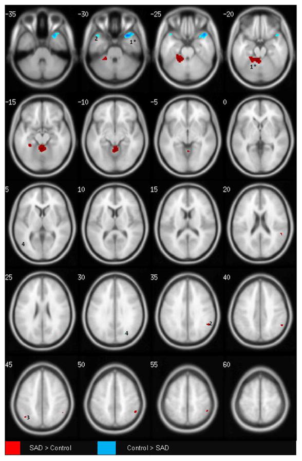
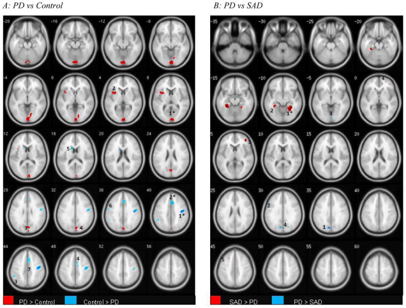
Results: After adjusting for age, gender, and total intracranial volume, SAD (as compared with control) subjects had greater GM in the left parahippocampal and middle occipital, and bilateral supramarginal and angular cortices, and left cerebellum; and lower GM in bilateral temporal poles and left lateral orbitofrontal cortex. Cerebellar, parahippocampal, and temporal pole differences were observed in both samples, survived whole brain corrections, and were not observed in the PD group, pointing to relative specificity to SAD.
Conclusions: These findings parallel the functional literature on SAD and suggest structural abnormalities underlying the functional disturbances.
Copyright © 2013 Society of Biological Psychiatry. Published by Elsevier Inc. All rights reserved.
Figures
Similar articles
-
Gray Matter Abnormalities in Non-comorbid Medication-naive Patients with Major Depressive Disorder or Social Anxiety Disorder.EBioMedicine. 2017 Jul;21:228-235. doi: 10.1016/j.ebiom.2017.06.013. Epub 2017 Jun 15. EBioMedicine. 2017. PMID: 28633986 Free PMC article.
-
Decreased gray matter volume of the medial orbitofrontal cortex in panic disorder with agoraphobia: a preliminary study.Prog Neuropsychopharmacol Biol Psychiatry. 2013 Aug 1;45:195-200. doi: 10.1016/j.pnpbp.2013.04.014. Epub 2013 Apr 28. Prog Neuropsychopharmacol Biol Psychiatry. 2013. PMID: 23628432
-
Neuroanatomical deficits in drug-naïve adult patients with generalized social anxiety disorder: a voxel-based morphometry study.Psychiatry Res. 2013 Oct 30;214(1):9-15. doi: 10.1016/j.pscychresns.2013.06.002. Epub 2013 Aug 13. Psychiatry Res. 2013. PMID: 23953332
-
Distinct grey matter volume alterations in adult patients with panic disorder and social anxiety disorder: A systematic review and voxel-based morphometry meta-analysis.J Affect Disord. 2021 Feb 15;281:805-823. doi: 10.1016/j.jad.2020.11.057. Epub 2020 Nov 13. J Affect Disord. 2021. PMID: 33243552
-
Structural magnetic resonance imaging in anxiety disorders: an update of research findings.Braz J Psychiatry. 2008 Sep;30(3):251-64. doi: 10.1590/s1516-44462008000300013. Braz J Psychiatry. 2008. PMID: 18833427 Review.
Cited by
-
A pilot study of gray matter volume changes associated with paroxetine treatment and response in social anxiety disorder.Psychiatry Res. 2015 Mar 30;231(3):279-85. doi: 10.1016/j.pscychresns.2015.01.008. Epub 2015 Jan 19. Psychiatry Res. 2015. PMID: 25659476 Free PMC article.
-
Functional magnetic resonance imaging during emotion recognition in social anxiety disorder: an activation likelihood meta-analysis.Front Hum Neurosci. 2013 Jan 17;6:347. doi: 10.3389/fnhum.2012.00347. eCollection 2012. Front Hum Neurosci. 2013. PMID: 23335892 Free PMC article.
-
OFC and its connectivity with amygdala as predictors for future social anxiety in adolescents.Dev Cogn Neurosci. 2020 Aug;44:100804. doi: 10.1016/j.dcn.2020.100804. Epub 2020 Jun 8. Dev Cogn Neurosci. 2020. PMID: 32716853 Free PMC article.
-
The common traits of the ACC and PFC in anxiety disorders in the DSM-5: meta-analysis of voxel-based morphometry studies.PLoS One. 2014 Mar 27;9(3):e93432. doi: 10.1371/journal.pone.0093432. eCollection 2014. PLoS One. 2014. PMID: 24676455 Free PMC article.
-
Cerebellar Neural Circuits Involving Executive Control Network Predict Response to Group Cognitive Behavior Therapy in Social Anxiety Disorder.Cerebellum. 2017 Jun;16(3):673-682. doi: 10.1007/s12311-017-0845-x. Cerebellum. 2017. PMID: 28155138
References
-
- Diagnostic and Statistical Manual of Psychiatric Disorders. Washington, DC: American Psychiatric Association; 1994.
-
- Stein DJ, Craske MG, Friedman MJ, Phillips KA. Meta-structure issues for the DSM-5: how do anxiety disorders, obsessive-compulsive and related disorders, post-traumatic disorders, and dissociative disorders fit together? Curr Psychiatry Rep. 2011;13(4):248–50. - PubMed
-
- Jefferys D. Social phobia. The most common anxiety disorder. Aust Fam Physician. 1997;26(9):1061, 4–7. - PubMed
Publication types
MeSH terms
Grants and funding
LinkOut - more resources
Full Text Sources
Other Literature Sources
Medical
