Ligand-specific interactions modulate kinetic, energetic, and mechanical properties of the human β2 adrenergic receptor
- PMID: 22748765
- PMCID: PMC4506644
- DOI: 10.1016/j.str.2012.05.010
Ligand-specific interactions modulate kinetic, energetic, and mechanical properties of the human β2 adrenergic receptor
Abstract
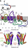
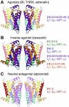
G protein-coupled receptors (GPCRs) are a class of versatile proteins that transduce signals across membranes. Extracellular stimuli induce inter- and intramolecular interactions that change the functional state of GPCRs and activate intracellular messenger molecules. How these interactions are established and how they modulate the functional state of GPCRs remain to be understood. We used dynamic single-molecule force spectroscopy to investigate how ligand binding modulates the energy landscape of the human β2 adrenergic receptor (β2 AR). Five different ligands representing either agonists, inverse agonists or neutral antagonists established a complex network of interactions that tuned the kinetic, energetic, and mechanical properties of functionally important structural regions of β2 AR. These interactions were specific to the efficacy profile of the ligands investigated and suggest that the functional modulation of GPCRs follows structurally well-defined interaction patterns.
Copyright © 2012 Elsevier Ltd. All rights reserved.
Figures


Comment in
-
Ligands stabilize specific GPCR conformations: but how?Structure. 2012 Aug 8;20(8):1289-90. doi: 10.1016/j.str.2012.07.009. Structure. 2012. PMID: 22884104 No abstract available.
Similar articles
-
Influence of lipid composition on the structural stability of g-protein coupled receptor.Chem Pharm Bull (Tokyo). 2013;61(4):426-37. doi: 10.1248/cpb.c12-01059. Chem Pharm Bull (Tokyo). 2013. PMID: 23546002
-
Conserved binding mode of human beta2 adrenergic receptor inverse agonists and antagonist revealed by X-ray crystallography.J Am Chem Soc. 2010 Aug 25;132(33):11443-5. doi: 10.1021/ja105108q. J Am Chem Soc. 2010. PMID: 20669948 Free PMC article.
-
Analysis of full and partial agonists binding to beta2-adrenergic receptor suggests a role of transmembrane helix V in agonist-specific conformational changes.J Mol Recognit. 2009 Jul-Aug;22(4):307-18. doi: 10.1002/jmr.949. J Mol Recognit. 2009. PMID: 19353579 Free PMC article.
-
Effect of fenoterol stereochemistry on the β2 adrenergic receptor system: ligand-directed chiral recognition.Chirality. 2011;23 Suppl 1(Suppl 1):E1-6. doi: 10.1002/chir.20963. Epub 2011 May 26. Chirality. 2011. PMID: 21618615 Free PMC article. Review.
-
β2-Adrenoceptor signalling bias in asthma and COPD and the potential impact on the comorbidities associated with these diseases.Curr Opin Pharmacol. 2018 Jun;40:142-146. doi: 10.1016/j.coph.2018.04.012. Epub 2018 May 12. Curr Opin Pharmacol. 2018. PMID: 29763833 Review.
Cited by
-
Olfactory Receptors Modulate Physiological Processes in Human Airway Smooth Muscle Cells.Front Physiol. 2016 Aug 4;7:339. doi: 10.3389/fphys.2016.00339. eCollection 2016. Front Physiol. 2016. PMID: 27540365 Free PMC article.
-
Minireview: More than just a hammer: ligand "bias" and pharmaceutical discovery.Mol Endocrinol. 2014 Mar;28(3):281-94. doi: 10.1210/me.2013-1314. Epub 2014 Jan 16. Mol Endocrinol. 2014. PMID: 24433041 Free PMC article. Review.
-
Structural Insight into G Protein-Coupled Receptor Signaling Efficacy and Bias between Gs and β-Arrestin.ACS Pharmacol Transl Sci. 2019 Apr 3;2(3):148-154. doi: 10.1021/acsptsci.9b00012. eCollection 2019 Jun 14. ACS Pharmacol Transl Sci. 2019. PMID: 32259053 Free PMC article.
-
Molecular modeling studies give hint for the existence of a symmetric hβ₂R-Gαβγ-homodimer.J Mol Model. 2013 Oct;19(10):4443-57. doi: 10.1007/s00894-013-1923-8. Epub 2013 Aug 8. J Mol Model. 2013. PMID: 23925512
-
Analysis of L-DOPA and droxidopa binding to human β2-adrenergic receptor.Biophys J. 2021 Dec 21;120(24):5631-5643. doi: 10.1016/j.bpj.2021.11.007. Epub 2021 Nov 10. Biophys J. 2021. PMID: 34767786 Free PMC article.
References
-
- Bai TR. Beta 2 adrenergic receptors in asthma: a current perspective. Lung. 1992;170:125–141. - PubMed
-
- Ballesteros JA, Jensen AD, Liapakis G, Rasmussen SG, Shi L, Gether U, Javitch JA. Activation of the beta 2-adrenergic receptor involves disruption of an ionic lock between the cytoplasmic ends of transmembrane segments 3 and 6. J. Biol. Chem. 2001;276:29171–29177. - PubMed
-
- Barnes PJ. Beta-adrenoceptors on smooth muscle, nerves and inflammatory cells. Life Sci. 1993;52:2101–2109. - PubMed
-
- Bippes CA, Muller DJ. High-resolution atomic force microscopy and spectroscopy of native membrane proteins. Rep. Prog. Phys. 2011;74 - PubMed
Publication types
MeSH terms
Substances
Grants and funding
LinkOut - more resources
Full Text Sources
Research Materials

