Environmental conditions modulate neurotoxic effects of psychomotor stimulant drugs of abuse
- PMID: 22748829
- PMCID: PMC3687356
- DOI: 10.1016/B978-0-12-386986-9.00006-5
Environmental conditions modulate neurotoxic effects of psychomotor stimulant drugs of abuse
Abstract
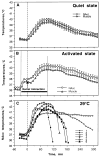
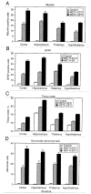
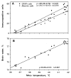
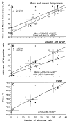
Psychomotor stimulants such as methamphetamine (METH), amphetamine, and 3,4-metylenedioxymethamphetamine (MDMA or ecstasy) are potent addictive drugs. While it is known that their abuse could result in adverse health complications, including neurotoxicity, both the environmental conditions and activity states associated with their intake could strongly enhance drug toxicity, often resulting in life-threatening health complications. In this review, we analyze results of animal experiments that suggest that even moderate increases in environmental temperatures and physiological activation, the conditions typical of human raves parties, dramatically potentiate brain hyperthermic effects of METH and MDMA. We demonstrate that METH also induces breakdown of the blood-brain barrier, acute glial activation, brain edema, and structural abnormalities of various subtypes of brain cells; these effects are also strongly enhanced when the drug is used at moderately warm environmental conditions. We consider the mechanisms underlying environmental modulation of acute drug neurotoxicity and focus on the role of brain temperature, a critical homeostatic parameter that could be affected by metabolism-enhancing drugs and environmental conditions and affect neural activity and functions.
Copyright © 2012 Elsevier Inc. All rights reserved.
Figures


Similar articles
-
Not just the brain: methamphetamine disrupts blood-spinal cord barrier and induces acute glial activation and structural damage of spinal cord cells.CNS Neurol Disord Drug Targets. 2015;14(2):282-94. doi: 10.2174/1871527314666150217121354. CNS Neurol Disord Drug Targets. 2015. PMID: 25687701 Free PMC article.
-
MDMA, Methylone, and MDPV: Drug-Induced Brain Hyperthermia and Its Modulation by Activity State and Environment.Curr Top Behav Neurosci. 2017;32:183-207. doi: 10.1007/7854_2016_35. Curr Top Behav Neurosci. 2017. PMID: 27677782 Free PMC article.
-
Leakage of the blood-brain barrier followed by vasogenic edema as the ultimate cause of death induced by acute methamphetamine overdose.Int Rev Neurobiol. 2019;146:189-207. doi: 10.1016/bs.irn.2019.06.010. Epub 2019 Jul 9. Int Rev Neurobiol. 2019. PMID: 31349927 Free PMC article. Review.
-
Acute methamphetamine intoxication: brain hyperthermia, blood-brain barrier, brain edema, and morphological cell abnormalities.Int Rev Neurobiol. 2009;88:65-100. doi: 10.1016/S0074-7742(09)88004-5. Int Rev Neurobiol. 2009. PMID: 19897075 Free PMC article. Review.
-
MDMA ("ecstasy"), methamphetamine and their combination: long-term changes in social interaction and neurochemistry in the rat.Psychopharmacology (Berl). 2004 May;173(3-4):318-25. doi: 10.1007/s00213-004-1786-x. Epub 2004 Mar 17. Psychopharmacology (Berl). 2004. PMID: 15029472
Cited by
-
Methamphetamine and HIV-1-induced neurotoxicity: role of trace amine associated receptor 1 cAMP signaling in astrocytes.Neuropharmacology. 2014 Oct;85:499-507. doi: 10.1016/j.neuropharm.2014.06.011. Epub 2014 Jun 17. Neuropharmacology. 2014. PMID: 24950453 Free PMC article.
-
Recent advances in methamphetamine neurotoxicity mechanisms and its molecular pathophysiology.Behav Neurol. 2015;2015:103969. doi: 10.1155/2015/103969. Epub 2015 Mar 12. Behav Neurol. 2015. PMID: 25861156 Free PMC article. Review.
-
Not just the brain: methamphetamine disrupts blood-spinal cord barrier and induces acute glial activation and structural damage of spinal cord cells.CNS Neurol Disord Drug Targets. 2015;14(2):282-94. doi: 10.2174/1871527314666150217121354. CNS Neurol Disord Drug Targets. 2015. PMID: 25687701 Free PMC article.
-
Amphetamine withdrawal differentially affects hippocampal and peripheral corticosterone levels in response to stress.Brain Res. 2016 Aug 1;1644:278-87. doi: 10.1016/j.brainres.2016.05.030. Epub 2016 May 18. Brain Res. 2016. PMID: 27208490 Free PMC article.
-
Methamphetamine-induced toxicity: an updated review on issues related to hyperthermia.Pharmacol Ther. 2014 Oct;144(1):28-40. doi: 10.1016/j.pharmthera.2014.05.001. Epub 2014 May 14. Pharmacol Ther. 2014. PMID: 24836729 Free PMC article. Review.
References
-
- Alberts DS, Sonsalla PK. Methamphetamine-induced hyperthermia and dopaminergic neurotoxicity in mice: pharmacological profile of protective and nonprotective agents. J Pharmacol Exp Ther. 1995;275:1104–1114. - PubMed
-
- Ali SF, Newport GD, Holson RR, Slikker W, Bowyer JF. Low environmental temperatures or pharmacological agents that produce hypothermia decrease methamphetamine neurotoxicity in mice. Brain Res. 1994;658:33–38. - PubMed
-
- Arai H, Uto Y, Ogawa X, Sato K. Effect of low temperature on glutamate-induced intracellular calcium accumulation and cell death in cultured hippocampal neurons. Neurosci Lett. 1993;163:132–134. - PubMed
-
- Bekay L, Lee JC, Lee GC, Peng GR. Experimental cerebral concussion: An electron microscopic study. J Neurosur. 1977;47:525–531. - PubMed
-
- Bondarenko A, Chesler M. Rapid astrocyte death induced by transient hypoxia, acidosis, and extracellular ion shifts. Glia. 2001;34:134–142. - PubMed
Publication types
MeSH terms
Substances
Grants and funding
LinkOut - more resources
Full Text Sources
Other Literature Sources
Medical

