Review
. 2012:110:1-24.
doi: 10.1016/B978-0-12-387665-2.00001-8.

Dynamics of lesion processing by bacterial nucleotide excision repair proteins

Affiliations
Review

Dynamics of lesion processing by bacterial nucleotide excision repair proteins

Neil M Kad et al. Prog Mol Biol Transl Sci. 2012.

Abstract

Single-molecule approaches permit an unrivalled view of how complex systems operate and have recently been used to understand DNA-protein interactions. These tools have enabled advances in a particularly challenging problem, the search for damaged sites on DNA. DNA repair proteins are present at the level of just a few hundred copies in bacterial cells to just a few thousand in human cells, and they scan the entire genome in search of their specific substrates. How do these proteins achieve this herculean task when their targets may differ from undamaged DNA by only a single hydrogen bond? Here we examine, using single-molecule approaches, how the prokaryotic nucleotide excision repair system balances the necessity for speed against specificity. We discuss issues at a theoretical, biological, and technical level and finally pose questions for future research.


Figures

F<sc>ig</sc>. 1
Fig. 1
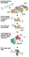
Structural model of bacterial nucleotide excision repair mediated by six proteins. The process of NER is a complex multiprotein cascade of events. Each step requires the recruitment of another protein to the lesion, with UvrB remaining at the lesion site as it interacts with each component of the reaction. Remarkably, despite this central role, UvrB is incapable of binding to the lesion site directly, requiring loading by UvrA2.
F<sc>ig</sc>. 2
Fig. 2
Potential modes of DNA damage searching by UvrA and UvrAB repair proteins. A number of mechanisms by which proteins can search for lesions are depicted. Hopping is distinguished from jumping by the distance over which the translocation occurs; however, in both cases, the protein remains within close proximity of the DNA. Sliding suggests that the protein remains in constant contact with the DNA, making it difficult to separate from hopping. If the protein dissociates from DNA into bulk solution, then a 3D search is employed to find the target site. Directed motion requires the input of energy in the form of nucleotide.
F<sc>ig</sc>. 3
Fig. 3
Oblique angle fluorescence permits a high signal to noise view of DNA tightropes. To generate OAF (top right), a standard TIRF (bottom right) optical path is steered to a subcritical angle, resulting in a far-field illumination beam emerging at a steep angle. Although this was achieved using a custom-built system (left), this is possible using off-the-shelf systems with little difficulty.
F<sc>ig</sc>. 4
Fig. 4
Quantum dot conjugation strategies. To ensure that there is no cross talk between different quantum dots, a differential conjugation strategy is important. Two approaches that were used for UvrA and UvrB conjugations are highlighted. UvrA (left) was bound to quantum dots using a short peptide sequence extension, which is biotinylated either endogenously or more efficiently using biotin ligase. UvrB could be differentially labeled by using an HA tag, which can be labeled using an antibody sandwich strategy.
F<sc>ig</sc>. 5
Fig. 5
Summary of motion of UvrA2 and UvrA2B2 on DNA. (A) UvrA2 has been shown to perform a 3D search with an average bound life time of 7 s on DNA. UvrA2 was also observed jumping from DNA helix to another helix over distances of 1–2 μm (not shown). (B) UvrB collapses UvrA’s search mode from 3D to 1D sliding and increases its average lifetime on DNA to 40 s. Three different sliding modes were observed for UvrA2B2. (C) The nature of how UvrC finds its way to the UvrB–DNA preincision complex is not known. The low concentrations of UvrC in the cell would make a 3D search very inefficient if UvrC remained statically bound for longer than 1 s.

References

    1. Peng Y, Wang H, Santana dos Santos L, Kisker C, Van Houten B. Chapter 13. Nucleotide excision repair from bacteria to humans: Structure-function studies. In: Penning TM, editor. Chemical carcinogenesis, current cancer research. Springer Science + Business Media, LLC; New York, NY: 2011.
    1. Jaciuk M, Nowak E, Skowronek K, Tanska A, Nowotny M. Structure of UvrA nucleotide excision repair protein in complex with modified DNA. Nat Struct Mol Biol. 2011;18:191–7. - PMC - PubMed
    1. Croteau DL, DellaVecchia MJ, Perera L, Van Houten B. Cooperative damage recognition by UvrA and UvrB: identification of UvrA residues that mediate DNA binding. DNA Repair (Amst) 2008;7:392–404. - PMC - PubMed
    1. Croteau DL, DellaVecchia MJ, Wang H, Bienstock RJ, Melton MA, Van Houten B. The C-terminal zinc finger of UvrA does not bind DNA directly but regulates damage-specific DNA binding. J Biol Chem. 2006;281:26370–81. - PMC - PubMed
    1. Wagner K, Moolenaar GF, Goosen N. Role of the insertion domain and the zinc-finger motif of Escherichia coli UvrA in damage recognition and ATP hydrolysis. DNA Repair (Amst) 2011;10:483–96. - PubMed

LinkOut - more resources