Review
. 2012 Sep;98(3):279-301.
doi: 10.1016/j.pneurobio.2012.06.008. Epub 2012 Jun 26.

High-frequency neural activity and human cognition: past, present and possible future of intracranial EEG research

Affiliations
Review

High-frequency neural activity and human cognition: past, present and possible future of intracranial EEG research

Jean-Philippe Lachaux et al. Prog Neurobiol. 2012 Sep.

Abstract

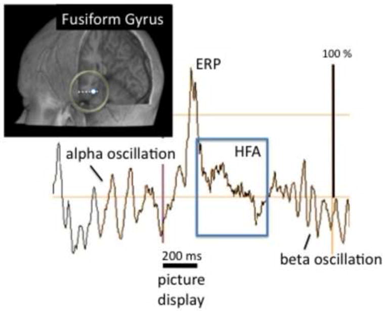
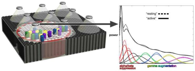
Human intracranial EEG (iEEG) recordings are primarily performed in epileptic patients for presurgical mapping. When patients perform cognitive tasks, iEEG signals reveal high-frequency neural activities (HFAs, between around 40 Hz and 150 Hz) with exquisite anatomical, functional and temporal specificity. Such HFAs were originally interpreted in the context of perceptual or motor binding, in line with animal studies on gamma-band ('40 Hz') neural synchronization. Today, our understanding of HFA has evolved into a more general index of cortical processing: task-induced HFA reveals, with excellent spatial and time resolution, the participation of local neural ensembles in the task-at-hand, and perhaps the neural communication mechanisms allowing them to do so. This review promotes the claim that studying HFA with iEEG provides insights into the neural bases of cognition that cannot be derived as easily from other approaches, such as fMRI. We provide a series of examples supporting that claim, drawn from studies on memory, language and default-mode networks, and successful attempts of real-time functional mapping. These examples are followed by several guidelines for HFA research, intended for new groups interested by this approach. Overall, iEEG research on HFA should play an increasing role in cognitive neuroscience in humans, because it can be explicitly linked to basic research in animals. We conclude by discussing the future evolution of this field, which might expand that role even further, for instance through the use of multi-scale electrodes and the fusion of iEEG with MEG and fMRI.


Figures

Figure 1
Figure 1. Task-induced high-frequency activity can be seen in raw iEEG signals
A picture (face stimulus) was flashed foveally for 200 ms while the patient fixated the center of a computer screen. The signal is a raw bipolar recording from the fusiform gyrus (expressed in % of the maximal amplitude value during the time window of interest). HFA is clearly visible in the raw trace (square box), together with the event-related potential evoked by the stimulus, and alpha and beta oscillations (respectively before and after stimulus presentation).
Figure 2
Figure 2. Conceptual schematic of how iEEG broadband gamma activity increases could result from modulation of band-limited gamma oscillations
Area of cerebral cortex is recorded by array of surface electrodes on the left. The power spectrum for one iEEG site recorded under resting vs. active conditions is schematized on the right (arrow). The field of view of each recording site (inverted cones) includes many neuronal assemblies (cylinders) with different functional response sensitivities (larger size and smaller number for illustration). Neuronal assemblies activated by a task (colored cylinders) are synchronized and generate membrane potential oscillations at different band-limited gamma frequencies that depend on the resonant properties of each assembly. Membrane potential oscillations with different center frequencies (colored bands on right corresponding to cylinders to left) collectively contribute to signals recorded by iEEG electrodes and their summation gives a broadband shape to gamma responses in the power spectrum. Note that many more assemblies and bands than can be represented here would be needed to produce the shape of commonly observed spectral responses. Also, different sets of neural assemblies and bands may be involved during different trials of the same task, depending on stimulus properties and a variety of initial conditions. Assemblies not immediately engaged in task-related cortical processing are represented by black (and white) cylinders. Task-related power suppression in alpha/beta frequencies (red circular area on left and red bands on right) occurs in a wider area and theoretically reflects thalamocortical gating mechanisms that permit or facilitate cortical processing in assemblies with similar but distinct response sensitivities. Reproduced with permission from Elsevier.
Figure 3
Figure 3. Cross-frequency coupling during multi-item working memory
(A) During maintenance of multiple items (trial-unique novel faces), hippocampal oscillations in the beta/gamma frequency range occur predominantly during a specific phase range of lower-frequency oscillations in the theta range. These data support a computer model of working memory according to which multiple items can be simultaneously maintained by a multiplexing of cycles of high-frequency activity during specific phases of low-frequency oscillations. This effect is more pronounced during the delay period of the working memory task (B) as compared to an inter-trial baseline (C) as well as compared to surrogate data (not shown). Figure modified from Axmacher et al., 2010a.
Figure 4
Figure 4. High-frequency “ripple” oscillations support memory consolidation in the human brain
(A) Individual trial of high frequency activity in unfiltered hippocampal raw data (top, gray trace) and in the same trial band-pass filtered between 80-140 Hz (bottom). (B) Grand-average time-frequency representation of these data across 11 patients. (C) Ripple-triggered grand average of unfiltered data reveal locking of ripples to a low-frequency oscillation reminiscent of (but slower than) a physiological sharp wave in rodents. (D) Inter-individual correlation between the number of ripples in rhinal cortex and memory for items learned prior to a sleep period (p = 0.005). Figure modified from Axmacher et al., 2008.
Figure 5
Figure 5. Timing of HFA dissociates between four distinct functional roles during visual search
The four panels show single-trial HFA during visual search in four anatomical sites (FEF: frontal eye field; DLPFC: dorso-lateral prefrontal cortex). Color codes band-limited power of iEEG signals in the [50 Hz-150 Hz] frequency range, expressed in % increase or decrease relative to the average value across the entire experiment. Stimulus onset occurs at 0 ms, yellow dots indicate reaction time. The task required that patients find a target (tilted letter ‘T’) embedded in an array of distractors (tilted ‘L’s with random orientation). S5, S6, S8 and S10 are four different patients. The four sites are characterized by dissimilar response timing, indicative of clearly distinct functional roles.
Figure 6
Figure 6. Event-related causal (ERC) interactions between iEEG sites with HFA during an auditory word repetition task
Three sequential time intervals of the task are shown: auditory word perception (stimulus onset to median stimulus duration, left panel), word retrieval and response preparation (between median stimulus offset and median response onset, middle panel), and spoken response (including 750 msec following the median response latency, right panel). Arrows indicate the directions and intensities of statistically significant increases in event-related causal (ERC) interactions between recording sites. The width and color of each arrow both represent linearly the magnitude of its integral ERC flow. Color scale (on left) has the same range for all ERCs, scaled from minimum to maximum (10% of the smallest ERCs not shown). The integral (sum) of ERC outflows is illustrated by semi-transparent purple circles. The radius of each circle is proportional to the normalized sum of statistically significant event-related increases in causal interactions directed outwardly from the site (originating at the site). Plots of event-related iEEG HFA are shown for select iEEG sites (insets). Only statistically significant power increases and decreases are plotted. Stimulus onset is at 0 seconds. Vertical markers indicate the times for median stimulus offset (o) and the median response onset (r). Results of electrocortical stimulation mapping (ESM) are represented by colored bars between pairs of electrode sites: Red - involuntary tongue movement, Purple - impaired spoken picture naming and auditory sentence comprehension (modified Token Test). F indicates frontal iEEG sites, and B indicates basal temporal sites. Composite of illustrations reproduced from Korzeniewska et al. (2011) with permission from Elsevier.

Similar articles

Cited by

References

    1. Allison T, McCarthy G, Nobre A, Puce A, Belger A. Human extrastriate visual cortex and the perception of faces, words, numbers, and colors. Cereb Cortex. 1994;4:544–554. - PubMed
    1. Anastassiou CA, Perin R, Markram H, Koch C. Ephaptic coupling of cortical neurons. Nat Neurosci. 2011;14(2):217–23. - PubMed
    1. Aoki F, Fetz EE, Shupe L, Lettich E, Ojemann GA. Increased gamma-range activity in human sensorimotor cortex during performance of visuomotor tasks. Clin Neurophysiol. 1999;110:524–537. - PubMed
    1. Axmacher N, Cohen MX, Fell J, Haupt S, Dümpelmann M, Elger CE, Schlaepfer TE, Lenartz D, Sturm V, Ranganath C. Intracranial EEG correlates of expectancy and memory formation in the human hippocampus and nucleus accumbens. Neuron. 2010b;65(4):541–9. - PubMed
    1. Axmacher N, Elger CE, Fell J. Ripples in the medial temporal lobe are relevant for human memory consolidation. Brain. 2008;131:1806–1817. - PubMed

Publication types