Review
. 2012 Sep 1;369(1):54-64.
doi: 10.1016/j.ydbio.2012.06.016. Epub 2012 Jun 27.

Genetic and cellular mechanisms regulating anterior foregut and esophageal development

Affiliations
Review

Genetic and cellular mechanisms regulating anterior foregut and esophageal development

Ian J Jacobs et al. Dev Biol. .

Abstract

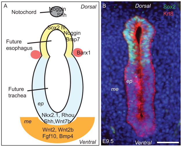
Separation of the single anterior foregut tube into the esophagus and trachea involves cell proliferation and differentiation, as well as dynamic changes in cell-cell adhesion and migration. These biological processes are regulated and coordinated at multiple levels through the interplay of the epithelium and mesenchyme. Genetic studies and in vitro modeling have shed light on relevant regulatory networks that include a number of transcription factors and signaling pathways. These signaling molecules exhibit unique expression patterns and play specific functions in their respective territories before the separation process occurs. Disruption of regulatory networks inevitably leads to defective separation and malformation of the trachea and esophagus and results in the formation of a relatively common birth defect, esophageal atresia with or without tracheoesophageal fistula (EA/TEF). Significantly, some of the signaling pathways and transcription factors involved in anterior foregut separation continue to play important roles in the morphogenesis of the individual organs. In this review, we will focus on new findings related to these different developmental processes and discuss them in the context of developmental disorders or birth defects commonly seen in clinics.


Figures

Figure 1
Figure 1
Dorsal-ventral patterning of the E9.5 anterior foregut. (A) Schematic section through the unseparated anterior foregut tube showing high levels of Sox2, Noggin, Bmp7 in the dorsal epithelium, which will give rise to the esophagus. Conversely, the transcription factor Nkx2.1 and signaling molecules Shh and Wnt7b, along with the Rho GTPase family member Rhou, are highly expressed in the ventral epithelium, which will contribute to the formation of the trachea. The homeobox gene Barx-1 is expressed predominantly in the mesenchyme demarcating the separation site of the dorsal and ventral foregut. Wnt2, Wnt2b, Fgf10 and Bmp4 are enriched in the ventral mesenchyme and are important for gene expression in the underlying epithelium. Mutation of Sox2, Nkx2.1 or Rhou or defects in the Shh, Wnt or Bmp signaling pathways leads to abnormal foregut development, including the formation of esophageal atresia with/without tracheoesophageal fistula (EA/TEF). (B) Immunostained section through the E9.5 foregut tube showing high levels of Sox2 protein in the dorsal epithelium. The cytoskeleton protein Keratin 8 (Krt8) is expressed in both the dorsal and ventral pseudostratified epithelium. Nuclei are counterstained with DAPI. Scale bar: 50μm. ep, epithelium; me, mesenchyme.
Figure 2
Figure 2
Dynamic signaling activities in the early anterior foregut tube before and after separation. (A–D) Wnt signaling indicated by the BAT-Gal reporter line. (A) Wnt activity is high in the ventral foregut epithelium at E9.5 as shown by X-gal staining. (B–D) Wnt signaling activities are observed in the epithelium lining both tubes after the anterior foregut separates into the trachea and esophagus at E11.5. (C,D) Sagittal sections. (E) Bmp signaling activity (as reported by Bmp reporter line BRE-LacZ) is high in the epithelium and mesenchyme of the ventral side of the unseparated foregut, consistent with the presence of high levels of Bmp4 and absence of the antagonist Noggin, as shown in Figure 1A. Scale bar: 50μm. eso, esophagus; tra, trachea; lun, lung; duo, duodenum; sto, stomach; ep, epithelium; me, mesenchyme.
Figure 3
Figure 3
Conversion of simple columnar to stratified squamous epithelium in the esophagus involving dynamic Bmp signaling activities. (A) Immunostained cross sections of E11.0 and E12.5 trachea and esophagus. Krt8 is expressed in the epithelium of both the esophagus and trachea after their formation from the foregut. Sox2 remains highly expressed in the epithelium of the dorsal, foregut-derived esophagus at E11.0 and E12.5. Note the conversion from single to multi-layered epithelium in the esophagus from E11.0 to E12.5, shown by co-immunostaining with p63 and Sox2 antibodies. (B) Schematic of the stratification and differentiation of esophageal epithelium through a two-stage Bmp signaling pattern. Stratification of the epithelium from E11.0 to E14.5 correlates with Noggin-mediated suppression of Bmp signaling. Ectopic Bmp activity in Noggin null and Shh-Cre; Rosa26caBmpr1a mutants inhibits the stratification process. Differentiation of the top layers of epithelium at E14.5-P9.0 requires activation of the Bmp signaling pathway while basal progenitor cells remain negative for Bmp signaling. Deletion of Bmpr1a in Shh-Cre; Bmpr1aloxp/loxp mutants inhibits the differentiation of suprabasal cells. Scale bar: 50μm. eso, esophagus; tra, trachea.
Figure 4
Figure 4
Wnt signaling activity as shown by the BAT-Gal reporter in the E13.5 esophagus and stomach. The stratified epithelium in the esophagus and forestomach is strongly positive for X-gal staining. Sporadic X-gal positive cells are also present in the mesenchyme of both the esophagus and forestomach. Note that part of the mesenchyme in the hindstomach is also positive for X-gal staining. Scale bar: 50μm. ep, epithelium; me, mesenchyme; fst, forestomach; eso, esophagus; hst, hindstomach; duo, duodenum.

References

    1. Abdulnour-Nakhoul S, Nakhoul NL, Wheeler SA, Haque S, Wang P, Brown K, Orlando G, Orlando RC. Characterization of esophageal submucosal glands in pig tissue and cultures. Dig Dis Sci. 2007;52:3054–3065. - PubMed
    1. Ai X, Kitazawa T, Do AT, Kusche-Gullberg M, Labosky PA, Emerson CP., Jr SULF1 and SULF2 regulate heparan sulfate-mediated GDNF signaling for esophageal innervation. Development. 2007;134:3327–3338. - PubMed
    1. Angers S, Moon RT. Proximal events in Wnt signal transduction. Nat Rev Mol Cell Biol. 2009;10:468–477. - PubMed
    1. Arnold K, Sarkar A, Yram MA, Polo JM, Bronson R, Sengupta S, Seandel M, Geijsen N, Hochedlinger K. Sox2(+) adult stem and progenitor cells are important for tissue regeneration and survival of mice. Cell Stem Cell. 2011;9:317–329. - PMC - PubMed
    1. Aziz Q, Thompson DG. Brain-gut axis in health and disease. Gastroenterology. 1998;114:559–578. - PubMed

Publication types

Substances

LinkOut - more resources