. 2012 Oct;143(4):1073-83.e22.
doi: 10.1053/j.gastro.2012.06.036. Epub 2012 Jun 27.

Deactivation of hepatic stellate cells during liver fibrosis resolution in mice

Affiliations

Deactivation of hepatic stellate cells during liver fibrosis resolution in mice

Juliane S Troeger et al. Gastroenterology. 2012 Oct.

Abstract

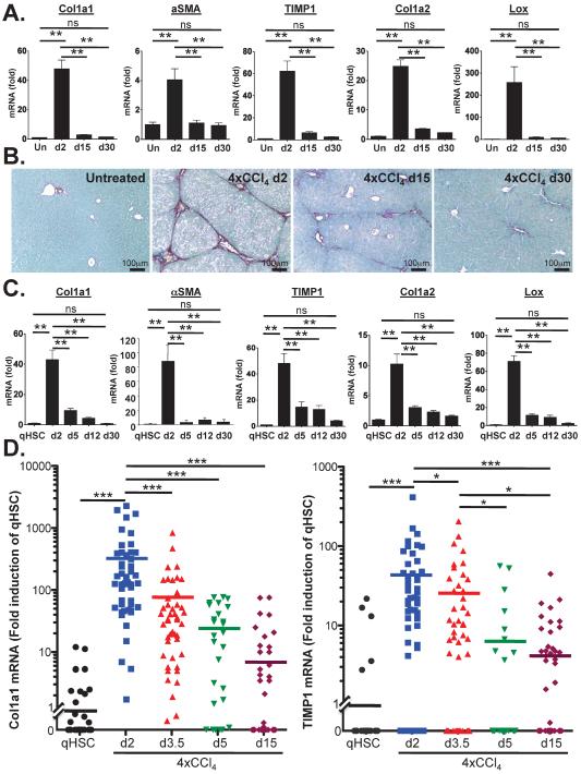
Background & aims: Activated hepatic stellate cells (HSCs), the main fibrogenic cell type in the liver, undergo apoptosis after cessation of liver injury, which contributes to resolution of fibrosis. In this study, we investigated whether HSC deactivation constitutes an additional mechanism of liver fibrosis resolution.

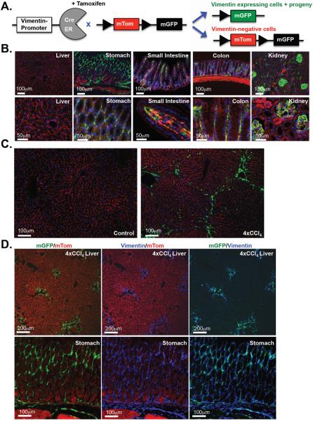
Methods: HSC activation and deactivation were investigated by single-cell PCR and genetic tracking in transgenic mice that expressed a tamoxifen-inducible CreER under control of the endogenous vimentin promoter (Vimentin-CreER).

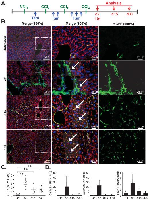
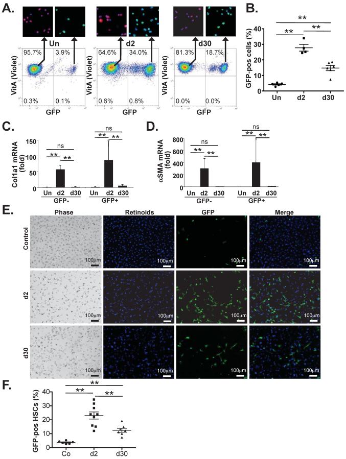
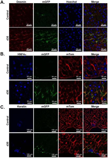
Results: Single-cell quantitative polymerase chain reaction demonstrated activation of almost the entire HSC population in fibrotic livers, and a gradual decrease of HSC activation during fibrosis resolution, indicating deactivation of HSCs. Vimentin-CreER marked activated HSCs, demonstrated by a 6- to 16-fold induction of a membrane-bound green fluorescent protein (mGFP) Cre-reporter after injection of carbon tetrachloride, in liver and isolated HSCs, and a shift in localization of mGFP-marked HSCs from peri-sinusoidal to fibrotic septa. Tracking of mGFP-positive HSCs revealed the persistence of 40%-45% of mGFP expression in livers and isolated HSCs 30-45 days after carbon tetrachloride was no longer administered, despite normalization of fibrogenesis parameters; these findings confirm reversal of HSC activation. After fibrosis resolution, mGFP expression was observed again in desmin-positive peri-sinusoidal HSCs; no mGFP expression was detected in hepatocytes or cholangiocytes, excluding mesenchymal-epithelial transition. Notably, reverted HSCs remained in a primed state, with higher levels of responsiveness to fibrogenic stimuli.

Conclusions: In mice, reversal of HSC activation contributes to termination of fibrogenesis during fibrosis resolution, but results in higher responsiveness of reverted HSCs to recurring fibrogenic stimulation.


Figures

Figure 1
Figure 1. Analysis of CCl4-induced HSC activation and deactivation by single cell qPCR
A–B. Fibrogenic gene expression (A.) and picrosirius red staining (B.) was determined in livers of untreated Balb/c mice (n=4) or mice treated with 4 injections of CCl4 (0.5 μl/g) and sacrificed 2 days (n=6), 15 days (n=5) or 30 days (n=8) after the last CCl4 injection. Shown is a fold induction of mRNA levels compared to untreated controls (A) and representative pictures (B). C. Determination of fibrogenic gene expression in HSCs isolated from untreated Balb/c mice (n=4) or from Balb/c mice treated with 4 injections of CCl4 (0.5 μl/g) and sacrificed either 2 days (n=3), 5 days (n=3), 12 days (n=3) or 30 days (n=5) after the last CCl4 injection. D. Activation and deactivation was determined in single-cell FACS-sorted HSCs isolated from Balb/c untreated mice (n=58) or at various time points after 4 injections of CCl4 (for d2 n=49, for d3.5 n=45, for d5 n=29, for d15 n=53). Shown is one of three representative HSC isolations for each time point. *p <0.05, ** p<0.01, *** p<0.001
Figure 2
Figure 2. Vimentin-CreER marks vimentin-expressing cells in the intestinal tract and in the fibrotic liver
A. Schematic diagram showing mTom/mGFP reporter gene expression in the absence and presence of tamoxifen-inducible Cre-mediated recombination. B. Vim-CreER BAC-transgenic mice were crossed to mTom-mGFP Cre reporter mice and injected 6× with tamoxifen to visualize Cre-mediated recombination. Shown are representative sections of the liver, stomach, small intestine, colon and kidney with Cre-mediated recombination visible as green mGFP positive cells. C. Shown are representative sections of normal liver after 6 injections of tamoxifen and liver treated with 6 injections of tamoxifen and 4 injections of CCl4. D. Livers from mice treated with 4 injections of CCl4 or stomach from untreated mice were stained with anti-vimentin antibody and analyzed by confocal microscopy to demonstrate overlap between Vimentin-CreER induced mGFP expression (green) and endogenous vimentin expression (blue).
Figure 3
Figure 3. Hepatic myofibroblasts de-activate during regression of CCl4-induced liver fibrosis
A. Schematic diagram showing the timing of tamoxifen and CCl4 injections, and sacrifice of VimCreER mice. B. mGFP and mTom expression in livers of untreated or CCl4-treated mice were visualized by confocal microscopy 2, 15 and 30 days after the last CCl4 injection. The middle and right panel show higher magnification representing the area marked by dotted white lines in the left panel. C. mGFP expression was quantified and expressed as percentage of total area for mice receiving no CCl4 (n=10), and CCl4-treated mice at day 2 (n=6), day 15 (n=7) or day 30 (n=7) after the last CCl4 injection. D. Fibrogenic gene expression in whole liver from Vim-CreER mice was performed by qPCR for mice receiving no CCl4 (n=7), and CCl4-treated mice at day 2 (n=4), day 15 (n=3) or day 30 (n=3) after the last CCl4 injection. ** p<0.01
Figure 4
Figure 4. Hepatic stellate cells de-activate after cessation of liver injury
Vimentin-CreER mice were treated with 6 tamoxifen and 4 CCl4 injections and HSCs were isolated 2 and 30 days after the last CCl4 injection. HSCs from control mice receiving corn oil and tamoxifen were isolated at a time corresponding to 2 days after the last CCl4 injection. A–B. mGFP expression was determined by flow-cytometric analysis of Vitamin A-autofluorescent HSCs. Shown are representative FACS images of each time point (A.) and quantification (B.) of HSCs from untreated mice (n=4), HSCs isolated 2 days (n=4) and 30 days (n=6) after the last CCl4 injection. Inserts show HSCs from each sorted cell population confirming cells as vitamin A-positive HSCs expressing mTom but not mGFP, or mGFP but not mTom. C–D. qPCR for Col1a1 (C.) and αSMA (D.) was performed in mGFP-negative HSCs and mGFP-positive HSCs from untreated mice (n=5 each), mGFP-negative HSCs and mGFP-positive HSCs isolated from mice 2 days after 4×CCl4 injection (n=4 each), and mGFP-negative HSCs and mGFP-positive HSCs isolated from mice 30 days after 4×CCl4 injection (n=5 GFP-negative, n=4 GFP-positive). E–F. mGFP expression was determined by fluorescent microscopy of plated HSCs at various time points after CCl4 and in control HSCs. Shown are representative pictures (E.) of vitamin A fluorescence, mGFP expression and an overlay of both, and a quantification (F.) of GFP-positive/Vitamin A-positive HSCs isolated from untreated mice (n=6), HSCs isolated 2 days (n=9) and 30 days (n=7) after the last CCl4 injection. ** p<0.01
Figure 5
Figure 5. Hepatic myofibroblasts do not undergo mesenchymal-epithelial transition
A. To determine whether mGFP-labeled myofibroblast re-differentiate into HSCs, livers were stained for desmin and analyzed by confocal microscopy. Representative images show overlap between desmin (red) and mGFP in untreated mice and CCl4-treated mice 31 days after the last tamoxifen injection. B. To determine whether mGFP-labeled myofibroblasts differentiate into hepatocytes, livers were stained with HNF4α. Confocal microscopy revealed no mGFP expressing cells with HNF4α-positive nuclei (blue). C. To determine whether mGFP-labeled myofibroblasts may differentiate into cholangiocytes, livers were stained with pankeratin antibody. Confocal microscopy revealed no overlap between pankeratin-positive bile ducts (blue) and mGFP (green).
Figure 6
Figure 6. Reverted HSCs are more responsive to fibrogenic stimuli
HSCs were isolated from age-matched mice that were left untreated or mice that received 4 injections of CCl4 (0.5 μl/g i.p.), followed by 45 days without treatment. A–B. Shown is a representative αSMA immunoblot of untreated HSCs (Ctrl) and d45 reverted HSCs (d45) using unplated freshly isolated quiescent HSCs (qHSC), HSCs cultured for 48h in 0.1% FBS-containing media, HSCs cultured for 48h in 10% FBS-containing media, or HSCs cultured treated with PDGF (20 ng/ml) or TGFβ (2 ng/ml) in 0.1% FBS-containing media, and a densitometric analysis (B) of αSMA immunoblots from 5 independent HSC isolations per group, normalized to actin levels. C–D. Shown are qPCR results for fibrogenic markers Col1a1 (C) and Acta2 (D) using the above-described samples of control and d45 reverted HSCs. E. Microarray analysis comparison of qHSCs and reverted HSCs, isolated from mice that received 4 injections of CCl4 (0.5 μl/g i.p.), followed by 45 days without treatment. Shown are all genes with a corrected p-value of <0.05 and a log fold-change of at least 0.67. F. qPCR of genes selected from the above microarray comparing expression levels of HSCs isolated from untreated mice (n=4), d2 after 4×CCl4 treatment (n=3) and d45 after 4×CCl4 treatment (n=4). Shown is a fold in comparison to qHSCs isolated from untreated mice. * p<0.05. ** p<0.01

Comment in

References

    1. Bataller R, Brenner DA. Liver fibrosis. J Clin Invest. 2005;115:209–18. - PMC - PubMed
    1. Friedman SL. Hepatic stellate cells: protean, multifunctional, and enigmatic cells of the liver. Physiol Rev. 2008;88:125–72. - PMC - PubMed
    1. Kluwe J, Wongsiriroj N, Troeger JS, et al. Absence of hepatic stellate cell retinoid lipid droplets does not enhance hepatic fibrosis but decreases hepatic carcinogenesis. Gut. 2011;60:1260–8. - PubMed
    1. Morcos SH, Khayyal MT, Mansour MM, et al. Reversal of hepatic fibrosis after praziquantel therapy of murine schistosomiasis. Am J Trop Med Hyg. 1985;34:314–21. - PubMed
    1. Abdel-Aziz G, Lebeau G, Rescan PY, et al. Reversibility of hepatic fibrosis in experimentally induced cholestasis in rat. Am J Pathol. 1990;137:1333–42. - PMC - PubMed

Publication types

MeSH terms