. 2012 Oct;20(10):1197-207.
doi: 10.1016/j.joca.2012.06.002. Epub 2012 Jun 29.

Intra-articular injection of human mesenchymal stem cells (MSCs) promote rat meniscal regeneration by being activated to express Indian hedgehog that enhances expression of type II collagen

Affiliations

Intra-articular injection of human mesenchymal stem cells (MSCs) promote rat meniscal regeneration by being activated to express Indian hedgehog that enhances expression of type II collagen

M Horie et al. Osteoarthritis Cartilage. 2012 Oct.

Abstract

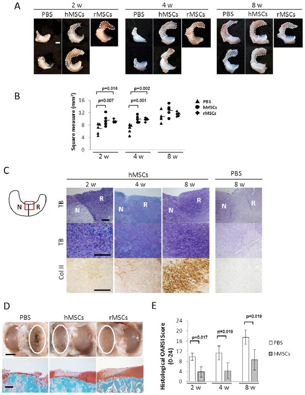
Objective: Meniscal regeneration was previously shown to be enhanced by injection of mesenchymal stem/stromal cells (MSCs) but the mode of action of the MSCs was not established. The aim of this study was to define how injection of MSCs enhances meniscal regeneration.

Design: A hemi-meniscectomy model in rats was used. Rat-MSCs (rMSCs) or human-MSCs (hMSCs) were injected into the right knee joint after the surgery, and PBS was injected into the left. The groups were compared macroscopically and histologically at 2, 4, and 8 weeks. The changes in transcription in both human and rat genes were assayed by species-specific microarrays and real-time RT-PCRs.

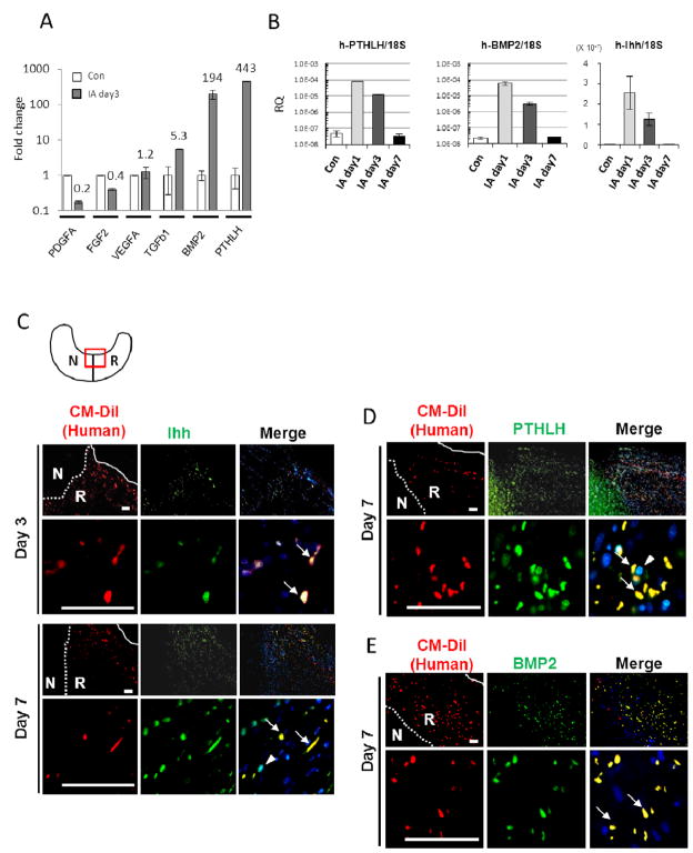
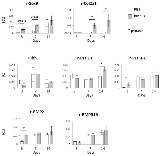
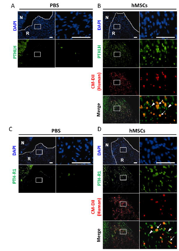
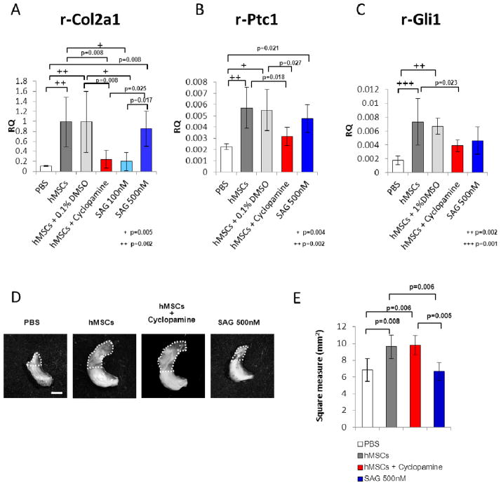
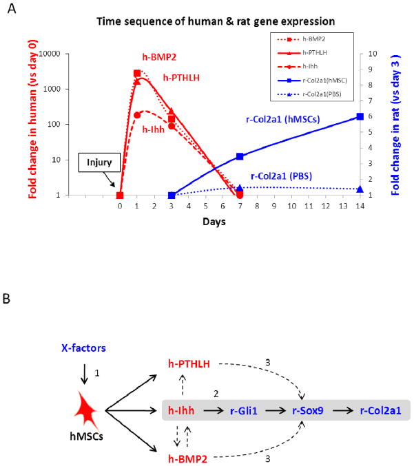
Results: Although the number of hMSCs decreased with time, hMSCs enhanced meniscal regeneration in a manner similar to rMSCs. hMSCs injection increased expression of rat type II collagen (rat-Col II), and inhibited osteoarthritis progression. The small fraction of hMSCs was activated to express high levels of a series of genes including Indian hedgehog (Ihh), parathyroid hormone-like hormone (PTHLH), and bone morphogenetic protein 2 (BMP2). The presence of hMSCs triggered the subsequent expression of rat-Col II. An antagonist of hedgehog signaling inhibited the expression of rat-Col II and an agonist increased expression of rat-Col II in the absence of hMSCs.

Conclusions: Despite rapid reduction in cell numbers, intra-articular injected hMSCs were activated to express Ihh, PTHLH, and BMP2 and contributed to meniscal regeneration. The hedgehog signaling was essential in enhancing the expression of rat-Col II, but several other factors provided by the hMSCs probably contributed to the repair.


Conflict of interest statement

Conflict of interest

DJP is a co-founder of Temple Therapeutics LLC. The other authors state that they have no conflicts of interest.

Figures

Figure 1
Figure 1
Intra-articular injection of hMSC promoted regeneration of rat meniscus and inhibited development of osteoarthritis after the meniscectomy. (A) Representative macroscopic findings of the meniscus 2, 4, and 8 weeks after the injection of PBS (left), hMSCs (middle), or rMSCs (right). The white dotted line indicates the regenerated tissue. Scale bar, 1 mm. (B) Area of the total meniscus injected with PBS, hMSCs, or rMSCs at 2, 4, and 8 weeks. The horizontal lines are mean values; n = 5 for each group (one-way ANOVA followed by Bonferroni post-tests. P values less than 0.017 were considered to be statistically significant.) (C) Representative sections of the meniscus stained with Toluidine Blue (top and middle), and immunostained for type II collagen (bottom) after PBS or hMSCs injection. The staining in the PBS-treated sample was less with toluidine blue and the anti-body for type II collagen. The schema of the meniscus on the left is shown for orientation. Symbols: N, native meniscus; R, regenerated meniscus; TB, Toluidine Blue; Col II, type II collagen. Scale bar, 100 μm. (D) Representative gross photographs (top) and sections (bottom) of the joint surface of the tibia at 8 weeks. The cartilage was stained with India ink to identify fibrillation and erosion. The white circle indicates the medial tibial plateau. The tibia was sectioned coronally and stained with safranin-O and fast green to identify cartilage (red). Scale bars, 2 mm (top) or 200 μm (bottom). (E) Quantification of histological analysis using the OARSI cartilage osteoarthritis histopathology grading system. Values are mean with lower and upper limit of 95 % CI; n = 5 for each group (Mann-Whitney U test). Symbols: OARSI, Osteoarthritis Research Society International.
Figure 2
Figure 2
Intra-articular injected hMSCs were activated to express Ihh, PTHLH, and BMP2. (A) Real-time RT-PCR for human-specific mRNA in regenerated meniscus 3 days after injection. Values are mean with lower and upper limit of 95 % CI of fold increase over Con, normalized by ΔΔCt for human-specific GAPDH; n = 4. Symbols: Con, 20,000 hMSCs added to injured meniscus from PBS injected knee before RNA extraction; IA day3, regenerated meniscus 3 days after intra-articular injection of hMSCs. (B) Time sequence of human gene expression by real-time RT-PCR for human PTHLH, BMP2, and Ihh in regenerated meniscus. Values are mean with lower and upper limit of 95 % CI of the relative quantities (RQ) normalized to 18S rRNA reflecting total levels of both human and rat transcripts. (C-E) Immunohistochemistry of anti-human/rat Ihh (C), PTHLH (D), or BMP2 (E) 3 or 7 days after hMSC injection. Primary antibodies for Ihh and PTHLH reacting with both human and rat were used. Low magnification (top) and higher magnification (bottom) of regenerated meniscus. Right column: merged images (CM-DiI, red; Ihh/PTHLH/BMP2, green; DAPI, blue). The white dotted line indicates border between native and regenerated meniscus, and the white solid line indicates outer edge of regenerated meniscus. CM-DiI labeled hMSCs (red) expressed Ihh, PTHLH, and BMP2 (green) (arrows in merge). Note: CM-DiI negative rat-derived cells surrounding hMSCs also expressed Ihh and PTHLH (arrowhead in merge) at day 7. The schema of the meniscus on the top left (C) is shown for orientation. Symbols: N, native meniscus; R, regenerated meniscus. Scale bars, 50 μm.
Figure 3
Figure 3
hMSCs up-regulated expression of chondrogenic genes in rat cells. Real-time RT-PCR assay for rat-specific mRNA (Sox9, Col2a1, Ihh, PTHLH, PTH-R1, BMP2, and BMPR1A) in the regenerated meniscus 3, 7 and 14 days after PBS or hMSC injection. Values are mean with lower and upper limit of 95 % CI of the relative quantities (RQ) normalized to rat-specific GAPDH; n = 4 for each group. *p< 0.001 (Mann-Whitney U test).
Figure 4
Figure 4
A large number of PTHLH/PTH-R1 positive cells were found in the regenerated tissue after injection of hMSCs. (A–D) Immunohistochemistry of anti-human/rat PTHLH (A, B) or PTH-R1 (C, D) 7 days after the injection of PBS (A, C) or hMSCs (B, D). Low magnification images are shown in the left and higher magnification of the regenerated tissue are shown in the right. Staining of nuclei (DAPI, blue), CM-DiI (red), and PTHLH or PTH-R1 (green) are shown. The white dotted line indicates the border between the native meniscus and regenerated tissue, and the white solid line indicates the outer edge of the regenerated tissue. A large number of PTHLH or PTH-R1 positive cells (green) were observed in the regenerated meniscus after the injection of hMSCs (B, D). In contrast, few PTHLH or PTH-R1 positive cells were present in the PBS injection group (B, D). Note that not only CM-DiI positive human cells (arrows in merge) but also CM-DiI-negative rat cells (arrowheads) expressed PTHLH or PTH-R1.
Figure 5
Figure 5
Effects of an inhibitor and an angonist of hedgehog signaling. (A) Real-time RT-PCR assay for rat-specific Col2a1 mRNA. RNA was recovered from the regenerated meniscus 2 weeks after the various treatments, including injection of PBS, hMSCs, hMSCs plus 0.1% DMSO (as a control of cyclopamine), hMSCs plus cyclopamine (50 μL of a 20 μM in 0.1% DMSO), 100 nM or 500 nM Smoothened agonist (SAG), or fibroblasts. Values are mean with lower and upper limit of 95 % CI of the relative quantities (RQ) normalized to rat-specific GAPDH; n = 4 for each group. + p=0.005, ++ p= 0.002 (one-way ANOVA followed by Bonferroni post-tests. P values less than 0.0033 were considered to be statistically significant.) (B, C) Real-time RT-PCR assay for rat-specific Ptc1 (B) and Gli1 (C), downstream targets of hedgehog signaling. RNA was recovered from the regenerated meniscus 3 days after the various treatments. Values are mean with lower and upper limit of 95 % CI of the relative quantities (RQ) normalized to rat-specific GAPDH; n = 4 for each group. + p=0.004, ++ p= 0.002 in B, ++ p=0.002, +++ p=0.001 in C (one-way ANOVA followed by Bonferroni post-tests. P values less than 0.005 were considered to be statistically significant.) (D) Representative macroscopic photographs of the meniscus 2 weeks after the various treatments. Scale bar: 1 mm. (E) Area of the total meniscus 2 weeks after the various treatments. Values are mean with lower and upper limit of 95 % CI; n = 4 for each group (one-way ANOVA followed by Bonferroni post-tests. P values less than 0.0083 were considered to be statistically significant.)
Figure 6
Figure 6
Possible mechanism of rat meniscal regeneration by intra-articular injection of hMSCs. (A) Summary graph of time sequence of human and rat gene expression changes by species-specific real time RT-PCR. Intra-articular injected hMSCs were activated to express human Ihh, PTHLH, and BMP2 (red) as early as day 1. Values for human genes are expressed as fold increase over values for controls (Con) of cultured hMSCs, normalized by ΔΔCt for 18S to reflect total levels of both human and rat transcripts. Rat Col2a1 gene expression in the regenerated meniscus was increased after the injection of hMSCs (blue). Values for rat genes are expressed as fold increase over values for PBS day 3, normalized by ΔΔCt for rat GAPDH. Values are means from RT-PCR data presented in Fig. 2B and Fig. 3. Symbols: Con, 20,000 hMSCs added to injured meniscus from PBS injected knee before RNA extraction. (B) Summary diagram of the possible mechanism for rat meniscal regeneration by hMSCs. (1) Intra-articular injected hMSCs were activated to express Ihh, PTHLH, and BMP2 when they were exposed to the in vivo microenvironment. (2) Human Ihh expressed by the hMSCs enhanced expression of rat Col2a1 gene probably through the downstream hedgehog target of rat Gli1 and then through Sox9. hMSCs did not enhance expression of rat Gli1 and Col2a1 in the presence of cyclopamine. In contrast, injection of Smoothened agonist enhanced expression of rat Gli1 and Col2a1. (3) PTHLH and BMP2 may also have enhanced expression of rat Col2a1 through rat Sox9. Symbols: X-factors, unknown factors which activate hMSCs to express Ihh, PTHLH, and BMP2.

Comment in

References

    1. Chen FH, Rousche KT, Tuan RS. Technology Insight: adult stem cells in cartilage regeneration and tissue engineering. Nat Clin Pract Rheumatol. 2006;2:373–382. - PubMed
    1. Wieland HA, Michaelis M, Kirschbaum BJ, Rudolphi KA. Osteoarthritis - an untreatable disease? Nat Rev Drug Discov. 2005;4:331–344. - PubMed
    1. Sgaglione NA, Steadman JR, Shaffer B, Miller MD, Fu FH. Current concepts in meniscus surgery: resection to replacement. Arthroscopy. 2003;19 (Suppl 1):161–188. - PubMed
    1. Giordano A, Galderisi U, Marino IR. From the laboratory bench to the patient’s bedside: an update on clinical trials with mesenchymal stem cells. J Cell Physiol. 2007;211:27–35. - PubMed
    1. Prockop DJ, Olson SD. Clinical trials with adult stem/progenitor cells for tissue repair: let’s not overlook some essential precautions. Blood. 2007;109:3147–3151. - PMC - PubMed

Publication types

MeSH terms