

doi: 10.1107/S1744309112020167.
Epub 2012 Jun 22.
Structure of the signal transduction protein TRAP (target of RNAIII-activating protein)
Affiliations
- PMID: 22750855
- PMCID: PMC3388912
- DOI: 10.1107/S1744309112020167
Item in Clipboard
Structure of the signal transduction protein TRAP (target of RNAIII-activating protein)
Acta Crystallogr Sect F Struct Biol Cryst Commun.
.
Abstract
The crystal structure of the signal transduction protein TRAP is reported at 1.85 Å resolution. The structure of TRAP consists of a central eight-stranded β-barrel flanked asymmetrically by helices and is monomeric both in solution and in the crystal structure. A formate ion was found bound to TRAP identically in all four molecules in the asymmetric unit.
Figures
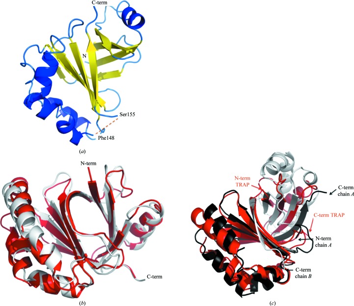
(a) The overall structure of TRAP. (b) A superposition of TRAP (in red) with the model obtained using SWISS-MODEL based on PDB entry 3fez (white); the r.m.s.d. is 1.85 Å over 136 Cα atoms. All figures were produced using PyMOL (Schrödinger LLC) and all superpositions were carried out with SSM (Krissinel & Henrick, 2004 ▶). (c) Superposition of TRAP (red) with the homodimeric monooxygenase ACTVA-ORF6 from S. coelicolor (PDB enry 1n5v ); the two identical chains are shown in white and black and the r.m.s.d. is 2.36 Å over 139 Cα atoms.
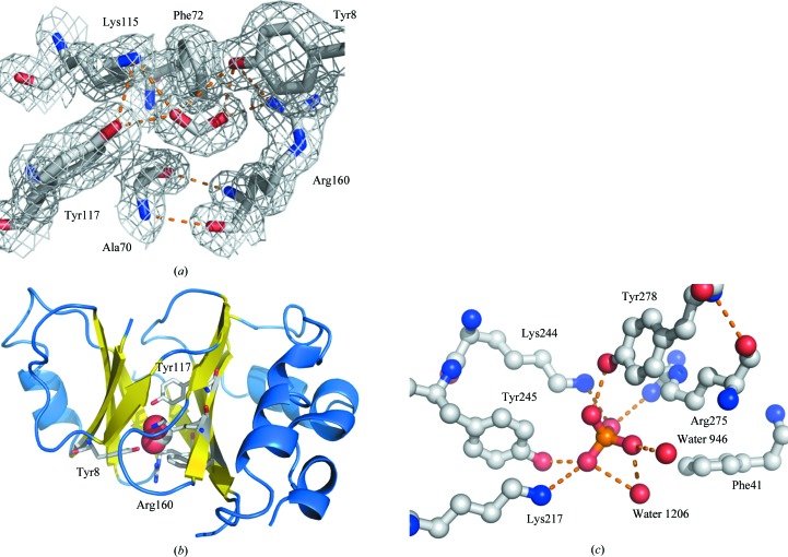
(a, b) The formate ion-binding site of TRAP. The site consists of hydrogen-bonding side chains from Tyr8, Lys115, Tyr117 and Arg160, with van der Waals contacts with Phe72 and Ala70. The main-chain atoms of Ala70 hold the Arg160 side chain in place. (a) 3F
o − 2F
c electron-density map at the 1.0σ level of the binding site. (b) The location of the site in the overall TRAP fold. The formate is shown as a stick model in (a) and as a CPK model in (b); hydrogen bonds are indicated by dotted orange lines. (c) The phosphate-binding site of amidohydrolase from M. synoviae (PDB entry 3ovg ; New York SGX Research Center for Structural Genomics, unpublished work). Colour scheme: phosphate ion, orange; amino-acid C atoms, grey; amino-acid O atoms, red; amino-acid N atoms, blue. Water molecules are shown as red spheres and hydrogen bonds as orange broken lines.
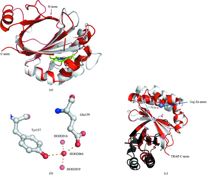
Predicted ligand-binding sites. (a) Superposition of TRAP (red) with a single chain of the S. aureus dimeric haem-degrading enzyme IsdG (PDB entry 2zdo ; white; r.m.s.d. of 1.87 Å over 81 Cα atoms). The 2zdo haem group is shown as a stick model. (b) A predicted zinc-binding site in TRAP. Water molecules occupying the site in the present crystal structure are shown as red balls, hydrogen bonds are shown as broken orange lines and amino acids are shown as ball-and-stick models. (c) Superposition of TRAP (red) with the homodimeric structure 1iuj (black and white chains; r.m.s.d. of 2.04 Å over 136 Cα atoms) indicating TRAP residues Tyr157 and Glu159 and the position of the zinc ion in TT1380.
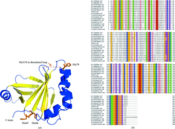
Histidines in TRAP. (a) Histidines 65, 66 and 79 are denoted as orange stick models. Note that His154 is unobserved in the present crystal structure. His79 and His154 are located near the disordered loop 149–154, while His65 and His66 are positioned on the C-terminal face of the TRAP β-barrel. (b) JalView (Waterhouse et al., 2009 ▶) alignment of TRAP with other Staphylococcus strains. The alignment is coloured at 75% identity using the Zappo colouring scheme. The strains of the sequences presented are TRAP (S. aureus RN6390B ATCC 55620), CAD33701 (S. aureus), NP_372359 (S. aureus subsp. aureus Mu50), YP_041300 (S. aureus subsp. aureus MRSA2520), ZP_06316969 (S. aureus subsp. aureus WW2703/97), ZP_05602371 (S. aureus subsp. aureus 55/2053), YP_001247249 (S. aureus subsp. aureus JH9), YP_003471354 (S. lugdunensis HKU09-01), NP_765069 (S. epidermidis ATCC 12228), YP_188938 (S. epidermidis RP62A), ZP_04797500 (S. epidermidis W23144), ZP_04819658 (S. epidermidis M23864:W1), ZP_03613577 (S. capitis SK14), ZP_04678846 (S. warneri L37603), ZP_04060607 (S. hominis SK119), ZP_07843459 (S. hominis subsp. hominis C80) and YP_253042 (S. haemolyticus JCSC1435).
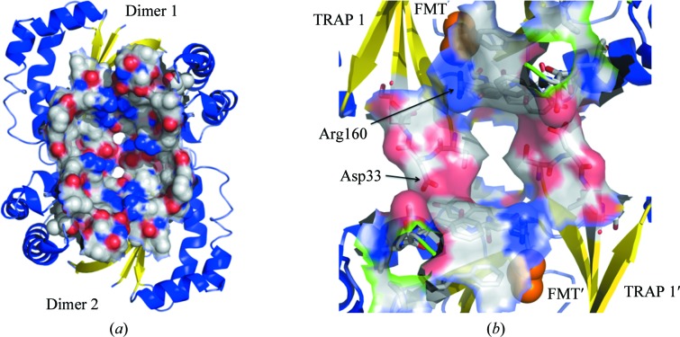
Potential TRAP dimer. (a) The structure of the 1iuj dimer of dimers as recorded in its PDB entry, showing the cleft formed at the dimer–dimer interface and represented as a surface. (b) TRAP molecules superposed on the 1iuj dimer of dimers, showing the formate as van der Waals spheres (orange) and the surface of the TRAP residues that correspond to the 1iuj interface residues; the residues that flank the unobserved loop, 148 and 155, are shown in green. Arg160 and Asp33 are discussed in the text.
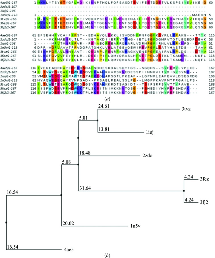
(a) JalView alignment of the TRAP-related proteins mentioned in this paper. It is apparent that although the structures discussed here have a close structural homology to TRAP, there is little sequence homology. Species: 4ae5 and 2zdo , Staphylococcus aureus; 1iuj , Thermus thermophilus; 1n5v , Streptomyces coelicolor; 3fez , Listeria monocytogenes; 3fj2 , L. innocua; 3tvz , Bacillus subtilis. (b) A JalView-generated dendrogram created using the neighbour-joining algorithm option also suggests that TRAP is potentially close to an unknown parent gene rather any other sequence given here.
References
-
- Adams, P. D. et al. (2010). Acta Cryst. D66, 213–221.
-
- Arnold, K., Bordoli, L., Kopp, J. & Schwede, T. (2006). Bioinformatics, 22, 195–201. - PubMed
-
- Balaban, N., Goldkorn, T., Gov, Y., Hirshberg, M., Koyfman, N., Matthews, H. R., Nhan, R. T., Singh, B. & Uziel, O. (2001). J. Biol. Chem. 276, 2658–2667. - PubMed
-
- Dauter, Z., Dauter, M. & Rajashankar, K. R. (2000). Acta Cryst. D56, 232–237. - PubMed
MeSH terms
Substances
Associated data
- Actions
LinkOut - more resources
Full Text Sources
Other Literature Sources
