. 2012 Jul 1;30(7):715-20.
doi: 10.1038/nbt.2249.

Combined small-molecule inhibition accelerates developmental timing and converts human pluripotent stem cells into nociceptors

Affiliations

Combined small-molecule inhibition accelerates developmental timing and converts human pluripotent stem cells into nociceptors

Stuart M Chambers et al. Nat Biotechnol. .

Abstract

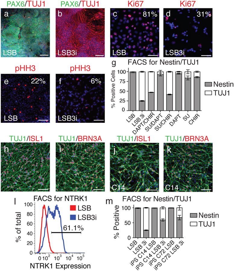
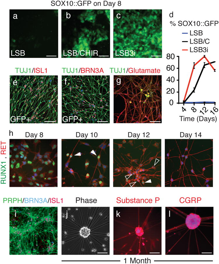
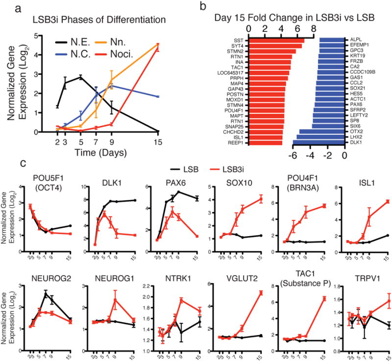
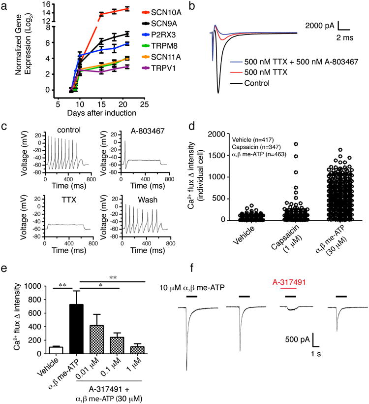
Considerable progress has been made in identifying signaling pathways that direct the differentiation of human pluripotent stem cells (hPSCs) into specialized cell types, including neurons. However, differentiation of hPSCs with extrinsic factors is a slow, step-wise process, mimicking the protracted timing of human development. Using a small-molecule screen, we identified a combination of five small-molecule pathway inhibitors that yield hPSC-derived neurons at >75% efficiency within 10 d of differentiation. The resulting neurons express canonical markers and functional properties of human nociceptors, including tetrodotoxin (TTX)-resistant, SCN10A-dependent sodium currents and response to nociceptive stimuli such as ATP and capsaicin. Neuronal fate acquisition occurs about threefold faster than during in vivo development, suggesting that use of small-molecule pathway inhibitors could become a general strategy for accelerating developmental timing in vitro. The quick and high-efficiency derivation of nociceptors offers unprecedented access to this medically relevant cell type for studies of human pain.


Figures

Figure 1
Figure 1. LSB3i treated hPSCs rapidly acquire a nociceptor phenotype within 12 days
Upon staining for TUJ1, a neuron marker, (a) compared to LSB alone, (b) far greater numbers of positive cells are observed when three inhibitors (CHIR99021, DAPT, SU5402; termed 3i) are applied 48 hours after treatment of hPSCs with LSB. Comparing expression of Ki67 and phospho-histone H3 in LSB (c,e) and LSB3i (d,f) treated hPSCs indicated a stark decline in proliferation by day 12. (g) Intracellular antibody FACS staining for Nestin and TUJ1 indicated a stark contrast in the number of neurons generated by LSB (2% TUJ1+) compared to LSB3i (75% TUJ1+). When one or two of the three inhibitors used in 3i are added, the same level of TUJ1 cells is not achieved, however CHIR with either SU5402 or DAPT can achieve greater than 53% neurons, indicating a requirement for CHIR in the formation of TUJ1+ neurons. TUJ1 positive neurons from LSB3i treated hPSCs express (h) ISL1, (i) BRN3A. (j,k) Similar neurons are observed when hiPSC lines are treated with LSB3i (C14 shown). (l) Greater than 61% of all cells express NTRK1 measured by FACS. (m) LSB3i treated hiPSCs form neurons at a moderate efficiency. Scale bars for (a,b) are 200 μm and (c-f,h-k) are 100 μm.
Figure 2
Figure 2. LSB3i treated hPSCs accelerate via a neural crest intermediate into mature bipolar nociceptors with an action potential
To monitor the emergence of neural crest stem cells, a transgenic SOX10∷GFP BAC hESC cell line was treated with (a) LSB, (b) LSB and CHIR99021, or (c) LSB3i. (d) SOX10∷GFP+ expression was accelerated and maximal expression (80% GFP+ by day 12) occurred earlier compared to LSB and CHIR99021 or LSB treatment alone. Sorted SOX10∷GFP+ cells gave rise to (e) ISL1 and (f) BRN3A positive neurons. (g) LSB3i neurons stain for glutamate. (h) Between days 8 and 14 RUNX1 expression is extinguished in some of the cells (higher expression in filled arrowhead, lower in empty arrowhead) and RET upregulates. (i) By day 15, mature neurons express peripherin, and after 1 month, (j) neuronal cell bodies arrange as clusters positive for (k) Substance P and (l) CGRP. Scale bars for (a-g, i-m) are 100 μm and 50 μm for (h).
Figure 3
Figure 3. Gene expression of LSB3i nociceptors
Gene expression analysis was performed on days 2, 3, 5, 7, 9, and 15 for both LSB and LSB3i treated cells. (a) Distinct phases of differentiation are observed when examining markers for neuroectoderm, neural crest, neurons, and nociceptors (N.E., N.C., Nn., and Noci.). (b) Top twenty significant up- (red) and downregulated (blue) genes by fold change at day 15 for LSB3i compared to LSB. (c) Expression of OCT4, DLK1, PAX6, SOX10, POU4F1 (BRN3A), ISL1, NEUROG2, NEUROG1, NTRK1, VGLUT2, TAC1, and TRPV1 are consistent with emergence of a nociceptor.
Fig. 4
Fig. 4. Functional characterization of mature LSB3i nociceptors
(a) Time course for expression of nociceptor-specific channels and receptors during LSB3i induction and maintenance of induced neurons. (b) Example voltage-clamp recording demonstrating both TTX-S and TTX-R Na+ currents, where the TTX-R component is further blocked by A-803467. Currents were evoked by a test pulse to 0 mV (see materials and methods). (c) A train of action potentials following current injection (75 pA). Application of 500 nM A-803467 to the same cell abolished repetitive firing with only the first action potential remaining. Additional application of 500 nM TTX blocked all action potential generation. Action potential activity recovered completely following wash-off of compounds. (d) Calcium flux induced by application of 30μM α,β Methylene ATP and 1μM capsaicin. Capsaicin induced a calcium response in 1-2% of cells, whereas a majority of LSB3i induced neurons responded to α,β Methylene ATP. To confirm that α,β Methylene ATP is acting through the P2RX3 receptor, the selective P2RX3 antagonist A-317491 was added (e). A-317491 significantly (*p<0.05, **p<0.01; one-way ANOVA, Dunnett's test) antagonized the response to α,β Methylene ATP. Electrophysiological recordings were used to analyze the current induced by α,β Methylene ATP. (f) Example of a current evoked by 10 μM α,β Methylene ATP, demonstrating typical fast activation and desensitization. The current was blocked by 1 μM A-317491 with partial recovery after wash.

References

    1. Bystron I, Rakic P, Molnar Z, Blakemore C. The first neurons of the human cerebral cortex. Nat Neurosci. 2006;9:880–886. - PubMed
    1. Zhang XQ, Zhang SC. Differentiation of neural precursors and dopaminergic neurons from human embryonic stem cells. Methods Mol Biol. 2009;584:355–366. - PMC - PubMed
    1. Elkabetz Y, et al. Human ES cell-derived neural rosettes reveal a functionally distinct early neural stem cell stage. Genes Dev. 2008;22:152–165. - PMC - PubMed
    1. Perrier AL, et al. Derivation of midbrain dopamine neurons from human embryonic stem cells. Proc Natl Acad Sci U S A. 2004;101:12543–12548. - PMC - PubMed
    1. Saha K, Jaenisch R. Technical challenges in using human induced pluripotent stem cells to model disease. Cell Stem Cell. 2009;5:584–595. - PMC - PubMed

Publication types

MeSH terms

Associated data