. 2012 Aug;19(8):788-96.
doi: 10.1038/nsmb.2334. Epub 2012 Jul 1.

Architecture of the RNA polymerase II preinitiation complex and mechanism of ATP-dependent promoter opening

Affiliations

Architecture of the RNA polymerase II preinitiation complex and mechanism of ATP-dependent promoter opening

Sebastian Grünberg et al. Nat Struct Mol Biol. 2012 Aug.

Abstract

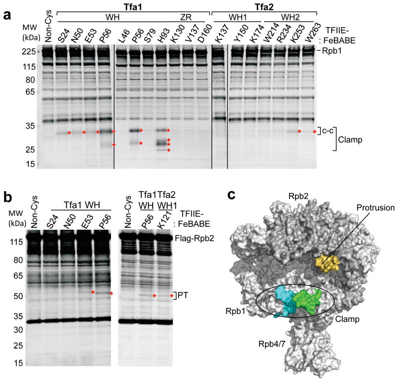
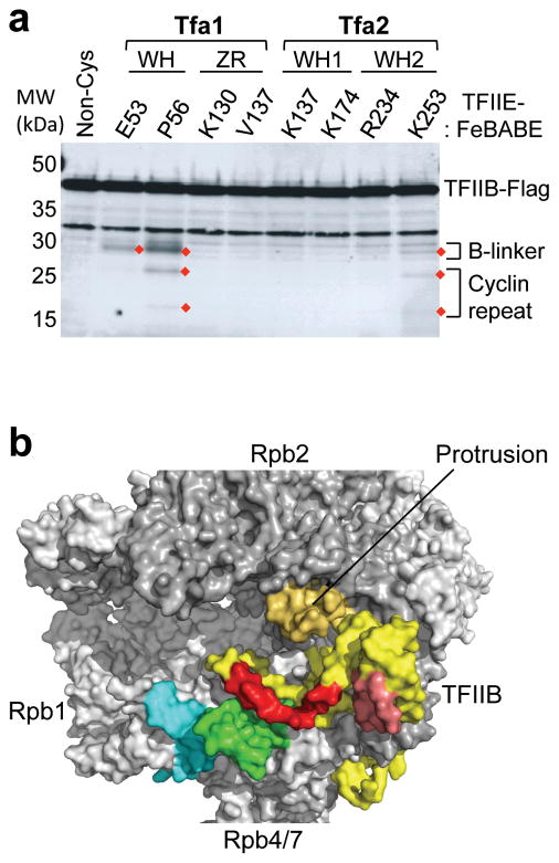
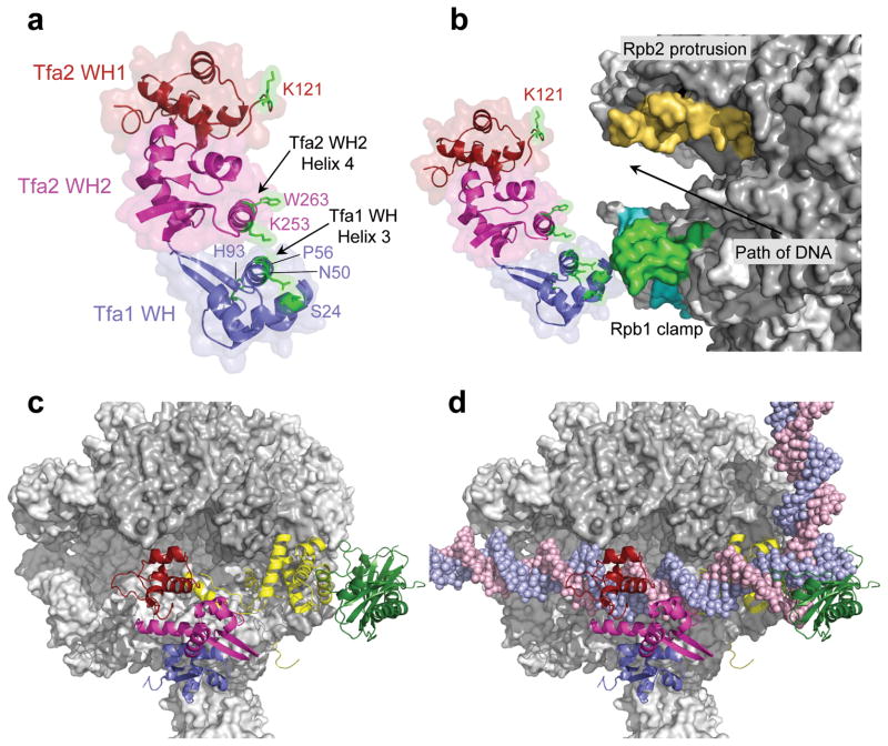
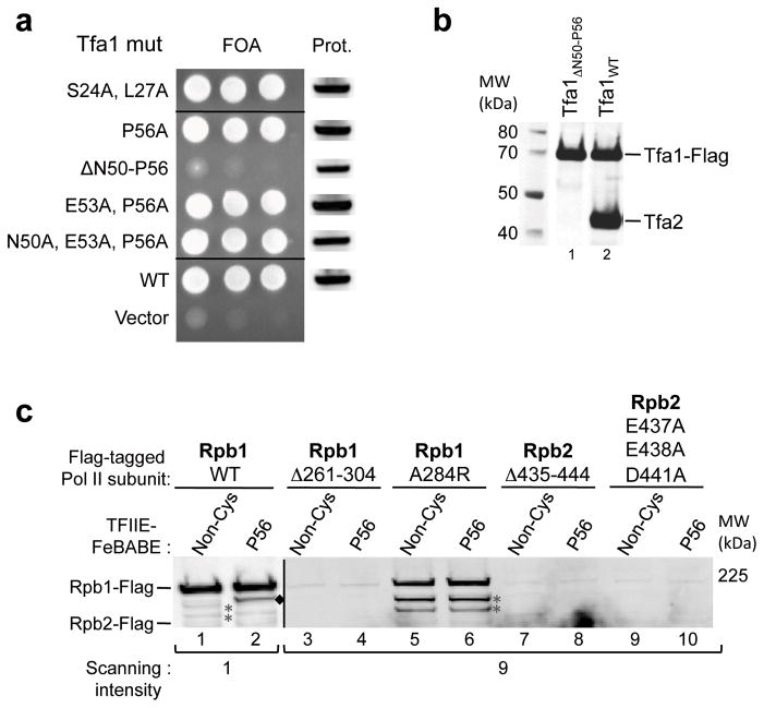
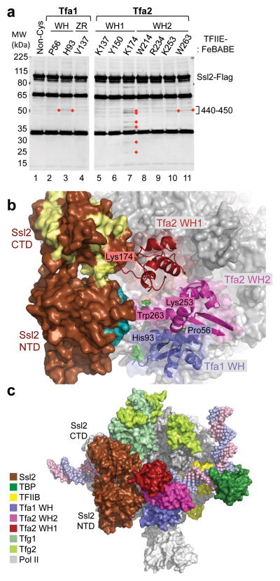
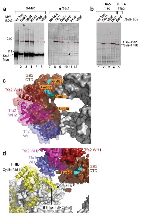
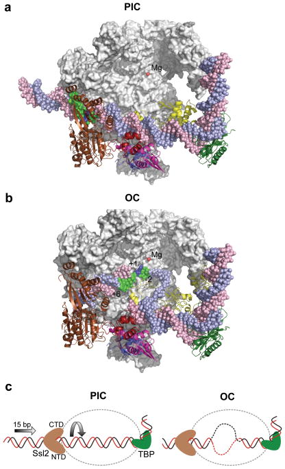
Yeast RNA polymerase II (Pol II) general transcription factor TFIIE and the TFIIH subunit Ssl2 (yeast ortholog of mammalian XPB) function in the transition of the preinitiation complex (PIC) to the open complex. We show that the three TFIIE winged-helix (WH) domains form a heterodimer, with the Tfa1 (TFIIEα) WH binding the Pol II clamp and the Tfa2 (TFIIEβ) tandem WH domain encircling promoter DNA that becomes single-stranded in the open complex. Ssl2 lies adjacent to TFIIE, enclosing downstream promoter DNA. Unlike previous proposals, comparison of the PIC and open-complex models strongly suggests that Ssl2 promotes DNA opening by functioning as a double-stranded-DNA translocase, feeding 15 base pairs into the Pol II cleft. Right-handed threading of DNA through the Ssl2 binding groove, combined with the fixed position of upstream promoter DNA, leads to DNA unwinding and the open state.


Figures

Figure 1
Figure 1
Functionally important regions in TFIIE. Summary of TFIIE subunit domains showing intramolecular FeBABE cleavage and in vivo function. Structured domains in either subunit are labeled (WH = winged helix, ZR = zinc ribbon), as are positions that were linked to FeBABE in the course of this work. Shaded bars summarize functional assays shown in Supplementary Fig. 1a, b (black = essential for yeast growth, grey = partially redundant WH domains (lethal when both deleted), white = no phenotype upon mutation). Lines connecting Tfa1 and Tfa2 represent intramolecular FeBABE cleavages when FeBABE was conjugated to the indicated positions in Tfa1 (Supplementary Fig. 1c; Supplementary Table 1).
Figure 2
Figure 2
Mapping of TFIIE-FeBABE cleavage in Pol II. (a) Hydroxyl radical-mediated cleavage in Pol II subunit Rpb1 by the indicated TFIIE-FeBABE variants. PICs were formed at the HIS4 promoter using yeast nuclear extracts that were supplemented with recombinant TFIIE-FeBABE. Rpb1 cleavage products were visualized by Western blot using an antibody against the N-terminus of Rpb1 . Red diamonds indicate reproducibly observed cleavage products compared to a reaction with a non cysteine-containing TFIIE (Non-Cys; lane 1). Brackets on the right indicate cleavage in the clamp and the coiled-coil domain (c-c). Bands in the Non-Cys control (lane 1) correspond to full length Rpb1 and non-specific reactivity of the primary/secondary antibodies with other proteins in the reaction. (b) Rpb2 cleavage using the indicated TFIIE-FeBABE derivatives. The right bracket indicates cleavage in the protrusion domain (PT). (c) Summary of the TFIIE-FeBABE mediated cleavages on Pol II. All cleavage events that are summarized in Table S1 were mapped on the Pol II surface and 10 residues on either side of the calculated cleavage sites are colored (green = cleavage in the Rpb1 clamp coiled-coil domain generated by FeBABE linked to residues in the TFIIE dimerization domain; cyan = cleavage in the Rpb1 clamp head by FeBABE conjugated to residue Tfa1 His93; orange = cleavage in the Rpb2 protrusion domain by FeBABE at Tfa1 Glu53, Pro56, and Tfa2 Lys121).
Figure 3
Figure 3
Cleavage of TFIIB by TFIIE-FeBABE in the PIC. (a) TFIIB cleavage products generated by TFIIE-FeBABE. PICs containing the indicated TFIIE-FeBABE derivatives and TFIIB-Flag were treated with H2O2 and analyzed by Western blot. Cleavage products are highlighted with red diamonds, and the brackets on the right indicate cleavage in either the B-linker or the first TFIIB cyclin repeat domain. (b) Cleavage results from a were mapped to TFIIB (yellow) where 10 residues on either side of the calculated cleavage site are highlighted (red = cleavage in the TFIIB linker region; salmon = cleavage in the first TFIIB cyclin repeat). Cleavages in Pol II are colored as described in Figure 2c.
Figure 4
Figure 4
Position of TFIIE in the PIC. (a) The TFIIE dimerization domain is formed in part by helix 3 in the Tfa1 WH domain and helix 4 in the Tfa2 WH2 domain. FeBABE-positions that cleave Pol II are highlighted in green. The Tfa1 WH domain is colored blue; the Tfa2 WH1 and WH2 domains are colored red and pink, respectively. (b) Position of the three TFIIE WH domains in the PIC based on the combined FeBABE cleavage data. Residues in TFIIE that cleave Pol II when linked to FeBABE are highlighted in green, as is the corresponding cleavage site in the coiled-coil region of the Pol II clamp. The cleavage site in the clamp head by Tfa1 residue His93 is colored in cyan, and cleavage in the Rpb2 protrusion domain is colored orange. An arrow indicates the path of the double stranded DNA in the PIC. (c, d) Top view of the Pol II PIC model containing Pol II (grey), TFIIB (yellow), TBP (dark green), and TFIIE without (c) and with (d) promoter DNA.
Figure 5
Figure 5
Effect of mutations in the TFIIE dimerization, Pol II clamp and protrusion domains. (a) Wild type TFIIE was selected against by plasmid shuffle assay and growth of TFA1 mutants measured on 5-fluoroorotic acid plates. On the left is growth of serial yeast cell dilutions and the right shows Western blot analysis of Flag- Tfa1 expression level in cells before plasmid shuffle. (b) A small deletion in helix 3 of the Tfa1 WH domain abolishes TFIIE dimerization. Tfa1 in which residues 50–56 of helix 3 were deleted (lane 1), and wild type Tfa1 (lane 2) were purified via anti-Flag affinity beads from yeast whole cell extracts and analyzed for CoIP of Tfa2. (c) The effect of Pol II mutations on PIC formation and TFIIE binding. PIC formation was attempted with yeast nuclear extracts containing Flag-tagged Pol II mutants (Rpb1 Δ261-304: deletion of the clamp coiled-coil domain; Rpb1 A284R: substitution of conserved A284 at the clamp coiled-coil; Rpb2 Δ435-444: deletion of the unstructured Rpb2 protrusion; Rpb2 E437A E438A D441A: substitution of acidic residues in the Rpb2 protrusion) supplemented with TFIIE −/+ FeBABE linked to residue Pro56. Purified PICs and cleavage products were analyzed by Western Blot, probing for Flag-tagged Rpb1 or Rpb2. Cleavage of wild type Rpb1 (WT) is labeled with black diamonds. Due to different Pol II expression levels, lanes containing wild type Pol II were scanned at intensity 1 on the Odyssey LI-COR system, reactions containing mutant Pol II were scanned at intensity 9. Asterisks indicate non specific Western blot signals, darker in lanes 5,6 because of the higher scanning intensity.
Figure 6
Figure 6
Orientation of the TFIIH subunit Ssl2/XPB in the PIC. (a) Cleavage in Ssl2 by TFIIE-FeBABE variants. PICs were formed with nuclear extracts containing Ssl2-Flag and supplemented with TFIIE-FeBABE. Cleavage was induced by addition of H2O2 and products were visualized by Western blot using an anti-Flag antibody. Reproducibly observed cleavage products are highlighted with red diamonds. The majority of TFIIE-FeBABE variants that cut Ssl2 cleaved in a region spanning residues 440–450 on the Ssl2 N-terminal domain. (b) PIC model containing TFIIE and Ssl2. Ssl2 was positioned in the PIC based on hydroxyl-radical cleavage by FeBABE linked to various positions in TFIIE (shown as green, or yellow for Lys174). Ssl2 cleavage by residues in the TFIIE dimerization domain is colored teal, cleavage by Tfa2 residue Lys174 is colored pale yellow. (c) Model of the PIC containing Ssl2. Different components of the PIC are colored according to the color-legend.
Figure 7
Figure 7
Protein crosslinking reveals Ssl2 interactions with TFIIE and TFIIB. (a) Nuclear extracts from strains with the crosslinker Bpa inserted at the indicated positions of Ssl2-13xMyc were used to form PICs and UV crosslinked. Proteins were separated on a 3–8% tris-acetate gel for 3 hrs, then visualized by probing with anti-Myc or anti-Tfa2. Anti-Myc blots contain 1X PIC per lane and anti-Tfa2 blots contain 4X PIC per lane. Crosslinking products are indicated by a red diamond. (b) Same as a except that strains used for the nuclear extract also contained either a 3x Flag epitope tagged Tfa2 or TFIIB as indicated. Anti-Flag blots contain 3X PIC per lane. (c) Residues in Ssl2 that crosslink to Tfa2 and TFIIB are shown on the Ssl2 surface in the PIC model. Residues that strongly and exclusively crosslinked to Tfa2 are colored in cyan. Colored in red is residue Ser440 which crosslinks strongest to TFIIB. (d) Same as c except that the model has been rotated and Rpb2 subunit removed to reveal TFIIB. Ssl2 Ser440 is colored red and is pointed toward the TFIIB linker helix, which is ~31Å distant.
Figure 8
Figure 8
Comparison of the PIC and open complex (OC) models suggests the role of Ssl2 in DNA strand opening. (a) The PIC model containing Pol II (grey), TBP (dark green), TFIIB (yellow), TFIIE (Tfa1 WH (blue), Tfa2 WH1 (red), and Tfa2 WH2 (pink)), Ssl2 (brown) and the active site Mg2+ (red). Rpb2 is removed to allow viewing into the Pol cleft. The blue template strand DNA is colored green from position −2 to +6 relative to the mammalian transcription start site (blue). (b) The OC model where double stranded DNA in the PIC was replaced with the DNA from the OC model . Compared to the PIC, 15 bases of upstream DNA is moved into the Pol II cleft and template strand DNA spanning positions −2 to +6 (green) relative to the transcription start site +1 (blue) is positioned close to the active center. (c) Diagram of Ssl2 function in OC formation. In the left panel, upstream promoter DNA is in a fixed position due to binding by TBP/TFIIB/Pol II. Ssl2 lies on downstream DNA at a fixed position due to interaction with TFIIE. Ssl2 feeds DNA into the cleft by a right handed threading mechanism, rotating and unwinding DNA.

Comment in

  • PICking apart Pol II initiation.
    Carey M. Carey M. Nat Struct Mol Biol. 2012 Aug;19(8):737-8. doi: 10.1038/nsmb.2349. Nat Struct Mol Biol. 2012. PMID: 22864359 No abstract available.

References

    1. Hahn S, Young ET. Transcriptional regulation in Saccharomyces cerevisiae: transcription factor regulation and function, mechanisms of initiation, and roles of activators and coactivators. Genetics. 2011;189:705–36. - PMC - PubMed
    1. Thomas MC, Chiang CM. The general transcription machinery and general cofactors. Crit Rev Biochem Mol Biol. 2006;41:105–78. - PubMed
    1. Wang W, Carey M, Gralla JD. Polymerase II promoter activation: closed complex formation and ATP-driven start site opening. Science. 1992;255:450–3. - PubMed
    1. Holstege FCP, Fiedler U, Timmers HTM. Three transitions in the RNA polymerase II transcription complex during initiation. EMBO J. 1997;16:7468–7480. - PMC - PubMed
    1. Chen HT, Warfield L, Hahn S. The positions of TFIIF and TFIIE in the RNA polymerase II transcription preinitiation complex. Nat Struct Mol Biol. 2007;14:696–703. - PMC - PubMed

Publication types

MeSH terms