. 2012 Aug;122(8):2793-806.
doi: 10.1172/JCI63051. Epub 2012 Jul 2.

Multiple myeloma-associated chromosomal translocation activates orphan snoRNA ACA11 to suppress oxidative stress

Affiliations

Multiple myeloma-associated chromosomal translocation activates orphan snoRNA ACA11 to suppress oxidative stress

Liang Chu et al. J Clin Invest. 2012 Aug.

Abstract

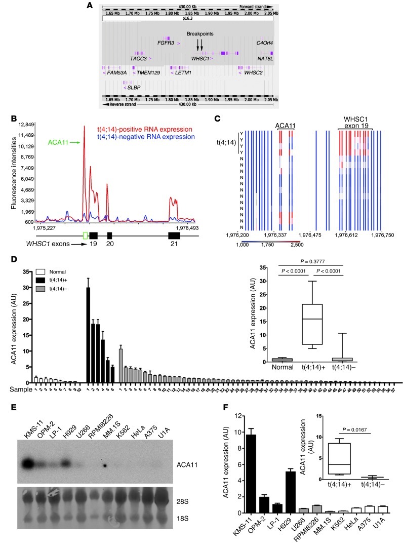
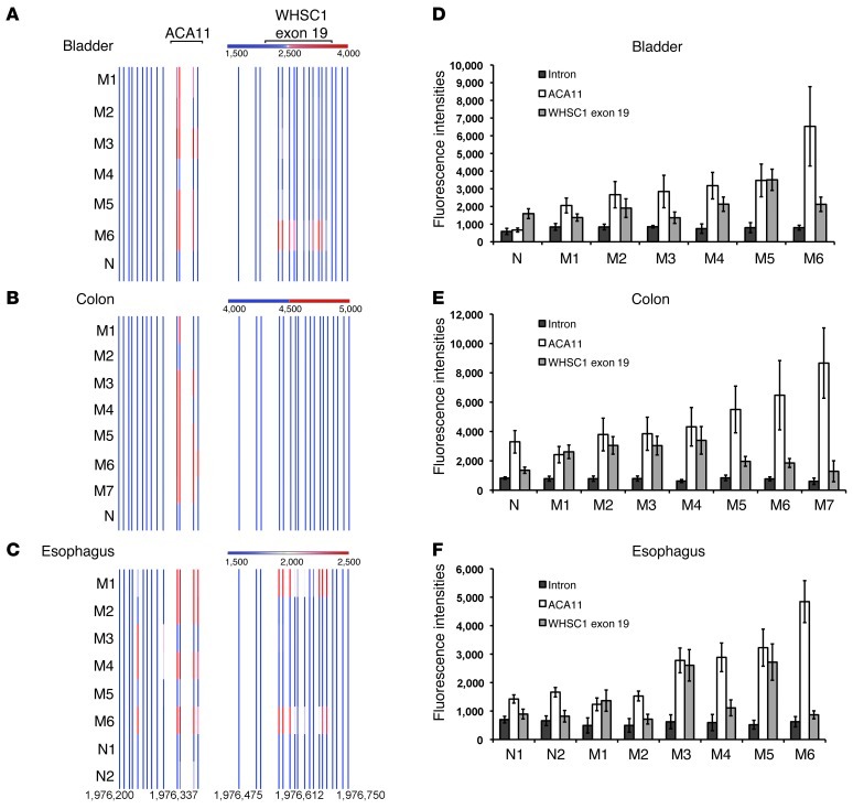
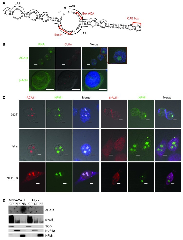
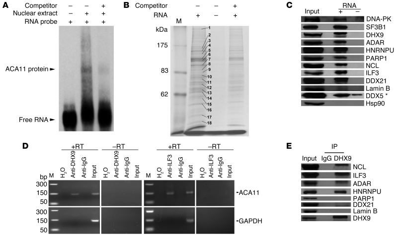
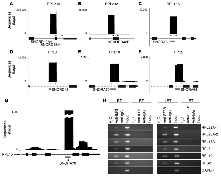
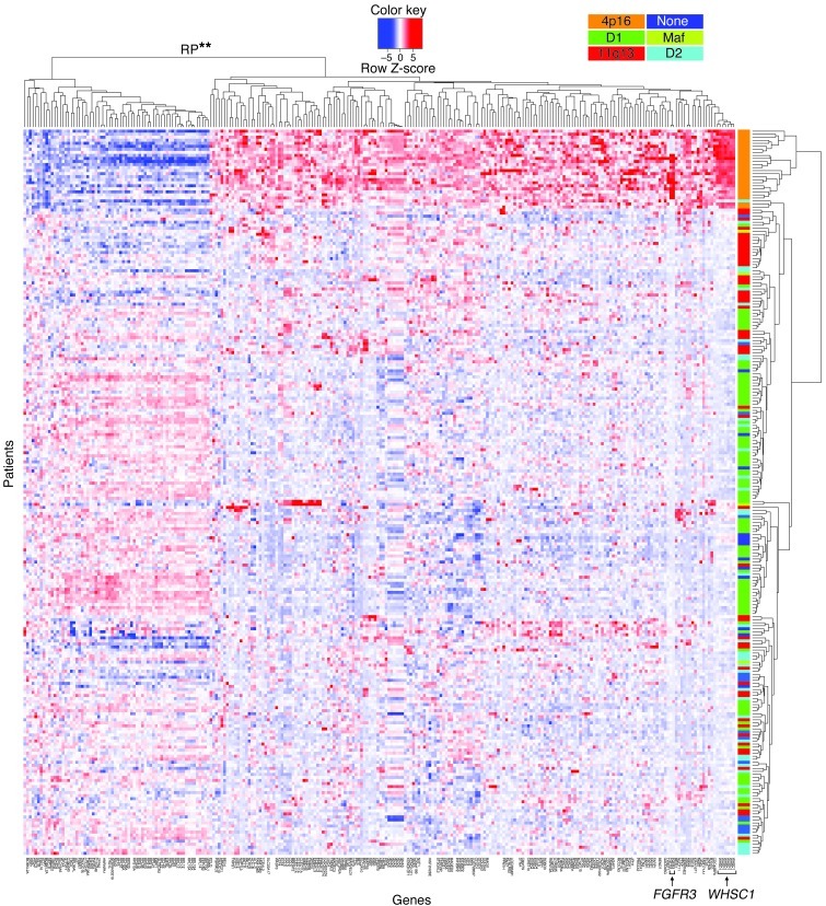
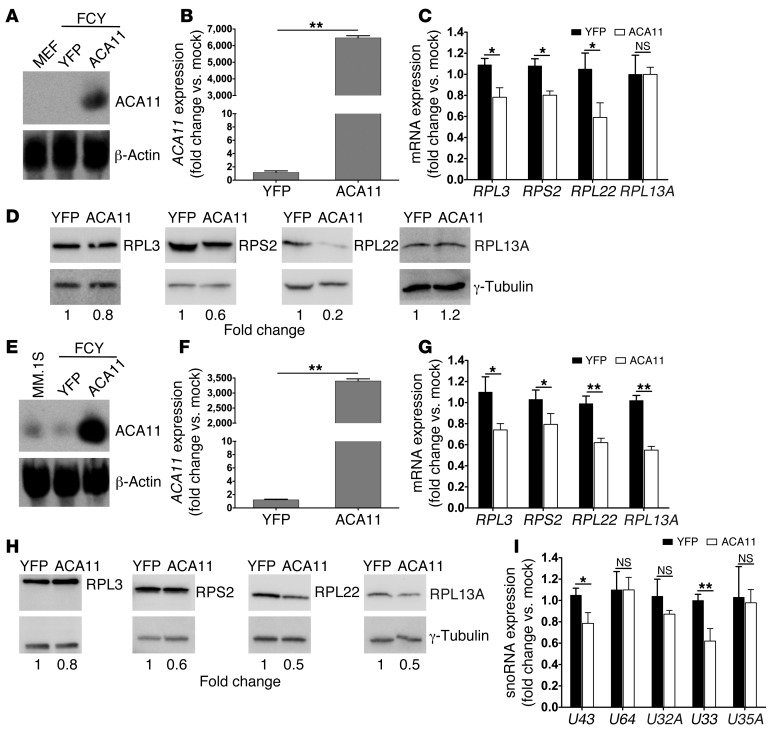
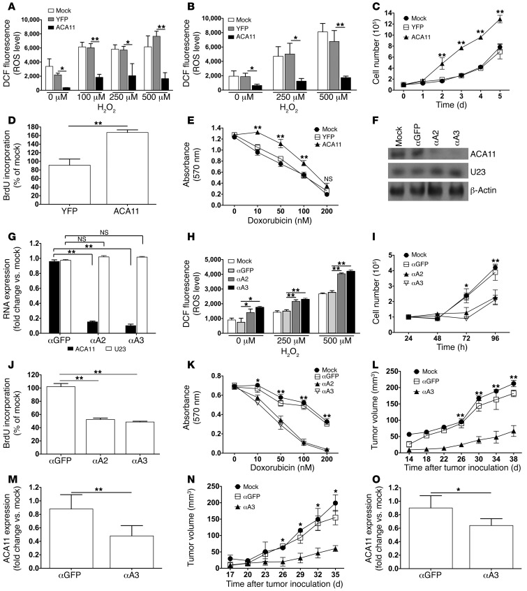
The histone methyltransferase WHSC1 (also known as MMSET) is overexpressed in multiple myeloma (MM) as a result of the t(4;14) chromosomal translocation and in a broad variety of other cancers by unclear mechanisms. Overexpression of WHSC1 did not transform wild-type or tumor-prone primary hematopoietic cells. We found that ACA11, an orphan box H/ACA class small nucleolar RNA (snoRNA) encoded within an intron of WHSC1, was highly expressed in t(4;14)-positive MM and other cancers. ACA11 localized to nucleoli and bound what we believe to be a novel small nuclear ribonucleoprotein (snRNP) complex composed of several proteins involved in postsplicing intron complexes. RNA targets of this uncharacterized snRNP included snoRNA intermediates hosted within ribosomal protein (RP) genes, and an RP gene signature was strongly associated with t(4;14) in patients with MM. Expression of ACA11 was sufficient to downregulate RP genes and other snoRNAs implicated in the control of oxidative stress. ACA11 suppressed oxidative stress, afforded resistance to chemotherapy, and increased the proliferation of MM cells, demonstrating that ACA11 is a critical target of the t(4;14) translocation in MM and suggesting an oncogenic role in other cancers as well.


Figures

Figure 1
Figure 1. ACA11 is activated by t(4;14) in MM.
(A) Schematic diagram of the chromosome 4p16.3 tiling oligonucleotide microarray, spanning 430 Kb across the t(4;14) breakpoint region (breakpoints are indicated by arrows). Annotated genes are indicated with exon structures. (B) Tiling microarray expression data of representative t(4;14)-positive and -negative patients with MM. (C) Heat map of t(4;14)-positive (Y; n = 4) and -negative (N; n = 12) patient samples. (B and C) Numbers indicate base pairs on chromosome 4 (human genome assembly GRCh37). (D) Real-time PCR of ACA11 in samples from patients with MM. The inset shows that ACA11 is significantly associated with t(4;14) status. Normal, normal plasma cells (n = 10); t(4;14)+, t(4;14)-positive patient samples (n = 6); t(4;14), t(4;14)-negative patient samples (n = 57). (E) Northern blot of ACA11 expression in human MM cell lines. t(4;14)-positive cell lines included KMS-11, OPM-2, LP-1, and H929 cells; t(4;14)-negative cell lines included U266, RPMI8226, and MM.1S cells; and non-MM cell lines included K562, HeLa, A375, and U1A cells. Loading controls were 28S and 18S rRNA. (F) ACA11 expression by real-time PCR is significantly associated with t(4;14) in MM cell lines. qRT-PCR data in D and F were normalized to the mean of 3 reference genes (GAPDH, UBC, YWHAZ). Data are presented as mean ± SD, with a box-whisker plot of minimum-to-maximum values (inset) (n = 3). The line within the box is the median value. The box represents the first and third quartiles. The whiskers show the largest and smallest events within at least 1.5 times the size of the box from the nearest edge.
Figure 2
Figure 2. ACA11 is overexpressed in bladder, colon, and esophageal cancers.
(AC) ACA11 is overexpressed in samples from patients with (A) bladder, (B) colon, and (C) esophageal cancer. Heat maps of RNA expression at the ACA11 locus were measured using our custom chromosome 4 tiling arrays. Each bar represents the normalized mean expression value obtained from quadruplicate probes representing each physical location. Numbers indicate base pairs on chromosome 4 (human genome assembly GRCh37). (DF) Graphs of data shown in AC. ACA11 levels in (D) bladder, (E) colon, and (F) esophagus samples were plotted using raw expression values of 4 probes. Intron, intron 18–19 of WHSC1 upstream of ACA11. Data in DF are presented as mean ± SD (n = 4). M, malignant tumor samples. N, normal control samples.
Figure 3
Figure 3. ACA11 localizes to nucleoli.
(A) Predicted secondary structure of ACA11. Red brackets indicate H box, ACA box, and CAB motifs. Black lines indicate ASOs. (B) Endogenous ACA11 is not significantly localized to Cajal bodies (coilin, red) in MM cells. ACA11 or β-actin is in green; DNA (DAPI) is in blue. Scale bar: 5 μm. (C) Localization of ACA11 to nucleoli. ACA11 or β-actin is shown in red; NPM1 is shown in green; DNA (DAPI) is in blue. Scale bar: 5 μm. (D) Northern blot (top 2 panels) for ACA11 in cytoplasmic (CP), nucleoplasmic (NP), and nucleolar (NS) fractions. Western blot (bottom 3 panels) of cytoplasmic (SOD), nuclear (NUP62), and nucleolar (NPM1) markers.
Figure 4
Figure 4. ACA11 binds a noncanonical ribonucleoprotein complex consisting of a set of proteins implicated in RNA processing.
(A) RNA EMSA confirming ACA11 binding to proteins from H929 cell nuclear extracts. Competitor, unlabeled ACA11 RNA. (B) Coomassie-stained SDS-PAGE gel of ACA11 pull-downs used for identification of proteins binding to ACA11 by MS. Representative proteins with the most abundant, unique peptides in each indicated band are marked with numbers that correspond to those in Table 2. M, protein marker. (C) Confirmation of ACA11-binding proteins by Western blot. The ACA11-protein complex was purified as in B and subjected to Western blot with antibodies to proteins identified by MS. The asterisk indicates nonspecific binding. Hsp90 was used as a negative control. (D) Confirmation of ACA11 presence in a DHX9/ILF3 ribonucleoprotein complex by CLIP followed by RT-PCR using H929 cells. H2O was used as a water template for RT-PCR. GAPDH was used as an internal control. M, DNA marker. (E) Physical interaction of ACA11 ribonucleoprotein complex proteins NCL, ILF3, ADAR, and HNRNPU with DHX9 in H929 cells was confirmed by immunoprecipitation.
Figure 5
Figure 5. RNA targets of the ACA11 ribonucleoprotein complex identified by RNP immunoprecipitation and deep sequencing.
(AF) Graphical representation of sequencing peaks mapping to the 6 targets of ACA11 snRNP. The horizontal gray bars indicate the locations of snoRNAs visualized in the UCSC genome browser. Arrows represent primers used for RT-PCR in H. (G) The same data as in E shown with lower vertical scale demonstrate smaller peaks of RPL10 exons. (H) RT-PCR confirms the presence of the indicated snoRNA intermediates resident within RP genes in the ILF3-, SF3B1-, or control IgG-precipitated complexes from H929 cells. H2O was used as a water template. GAPDH was used as control.
Figure 6
Figure 6. A t(4;14) gene expression signature defined by mRNA expression microarray data from 239 samples from patients with myeloma.
WHSC1 is upregulated in all 4p16 patients (arrow), and the nearby FGFR3 gene (arrow) is upregulated in most but not all 4p16 patients. Previously defined patient subgroup clusters (see Methods) are indicated by orange, dark green, red, dark blue, light green, and light blue. Highly significant downregulation (**P = 4.8 × 10⁻²⁰) of RP genes was found in the 4p16 (i.e., WHSC1, orange) subgroup.
Figure 7
Figure 7. ACA11 provides evidence for the t(4;14) gene signature in part — ACA11 is sufficient to downregulate RP genes.
(A) Overexpression of ACA11 confirmed by Northern blot analysis in Arf–/– MEFs stably transduced with ACA11 lentiviruses. β-Actin was used as loading control. (B) Overexpression of ACA11 in Arf–/– MEFs as measured by qRT-PCR. (C) RP mRNA expression in Arf–/– MEFs overexpressing ACA11 was analyzed by qRT-PCR. (D) Expression of RPs in Arf–/– MEFs overexpressing ACA11 demonstrated by Western blot. γ-Tubulin was used as loading control. (E) Overexpression of ACA11 confirmed by Northern blot analysis in MM.1S cells stably transduced with ACA11 lentiviruses. β-Actin was used as loading control. (F) qRT-PCR showing the expression of ACA11 in MM.1S cells in E. (G) RP mRNA expression in MM.1S cells overexpressing ACA11 was analyzed by qRT-PCR. (H) Downregulation of RPs in MM.1S cells overexpressing ACA11 demonstrated by Western blot. γ-Tubulin was used as loading control. (I) Small RNAs were isolated from MM.1S cells overexpressing ACA11. snoRNA expression was analyzed by qRT-PCR. U43, snoRNA located in RPL3; U64, snoRNA located in RPS2; U32A, U33, and U35A, snoRNAs located in RPL13A. qRT-PCR data were normalized to the average of 3 reference genes, (B, C, F, and G) GAPDH, UBC, and YWHAZ or (I) U6, U44, and U48, and are shown as fold change relative to that of mock-treated cells. Data represent mean ± SD (n = 3). *P < 0.05, **P < 0.01.
Figure 8
Figure 8. ACA11 functions to modulate oxidative stress, cell proliferation, and resistance to chemotherapy.
(A and B) ROS levels, quantified by DCFH-DA (DCF) labeling, are suppressed by ACA11 expression in (A) Arf–/– MEFs or (B) MM.1S cells at baseline and upon challenge with hydrogen peroxide (H2O2). (C and D) ACA11 expression increased growth and proliferation of MM.1S cells as measured by (C) cell counting and (D) BrdU-ELISA relative to mock-treated cells. (E) ACA11 increased resistance of MM.1S cells to cytotoxic chemotherapy as measured by MTT viability assay. (F and G) Northern blot and qRT-PCR analyses of ACA11 knockdown in H929 cells 4 days after transfection with ASOs. β-Actin was used as loading control. U23, unrelated snoRNA. αA2 and αA3 are ASOs that target ACA11. (HK) Knockdown of ACA11 in H929 cells (H) increased ROS levels, (I) decreased cell growth, (J) decreased cell proliferation, and (K) decreased resistance to cytotoxic chemotherapy. (L and N) Growth of (L) KMS-11 and (N) RPMI8226 cells in immunocompromised mice is reduced by ACA11 knockdown. (M and O) qRT-PCR showing the expression of ACA11 in (M) KMS-11 and (O) RPMI8226 xenograft tumors. Cells were nucleofected with 600 pmol ASOs per 106 cells and cultured for (I) 24 hours or (H, JL, and N) 48 hours before further analysis. qRT-PCR data in G, M, and O were normalized to the average of 3 reference genes (GAPDH, UBC, YWHAZ) and are shown as fold change relative to that of mock-treated cells. All data shown represent mean ± SD (n = 3 [AE and GK], n = 5 [LO]). *P < 0.05, **P < 0.01.

Comment in

Similar articles

Cited by

References

    1. Palumbo A, Anderson K. Multiple myeloma. N Engl J Med. 2011;364(11):1046–1060. doi: 10.1056/NEJMra1011442. - DOI - PubMed
    1. Richelda R, et al. A novel chromosomal translocation t(4; 14)(p16.3; q32) in multiple myeloma involves the fibroblast growth-factor receptor 3 gene. Blood. 1997;90(10):4062–4070. - PubMed
    1. Chesi M, et al. Frequent translocation t(4;14)(p16.3;q32.3) in multiple myeloma is associated with increased expression and activating mutations of fibroblast growth factor receptor 3. Nat Genet. 1997;16(3):260–264. doi: 10.1038/ng0797-260. - DOI - PMC - PubMed
    1. Winkler JM, Greipp P, Fonseca R. t(4;14)(p16.3;q32) is strongly associated with a shorter survival in myeloma patients. Br J Haematol. 2003;120(1):170–171. doi: 10.1046/j.1365-2141.2003.03983_5.x. - DOI - PubMed
    1. Karlin L, et al. Clinical and biological features of t(4;14) multiple myeloma: a prospective study. Leuk Lymphoma. 2011;52(2):238–246. doi: 10.3109/10428194.2010.537795. - DOI - PubMed

Publication types

MeSH terms