. 2012 Aug;122(8):2940-54.
doi: 10.1172/JCI61884. Epub 2012 Jul 2.

Brain indoleamine 2,3-dioxygenase contributes to the comorbidity of pain and depression

Affiliations

Brain indoleamine 2,3-dioxygenase contributes to the comorbidity of pain and depression

Hyangin Kim et al. J Clin Invest. 2012 Aug.

Abstract

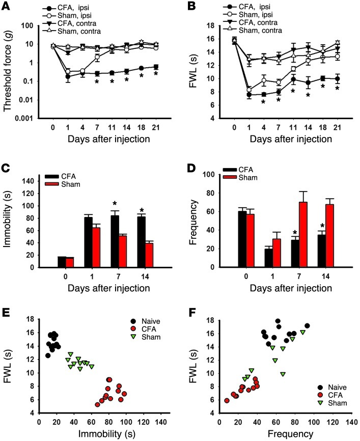
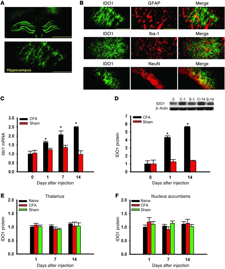
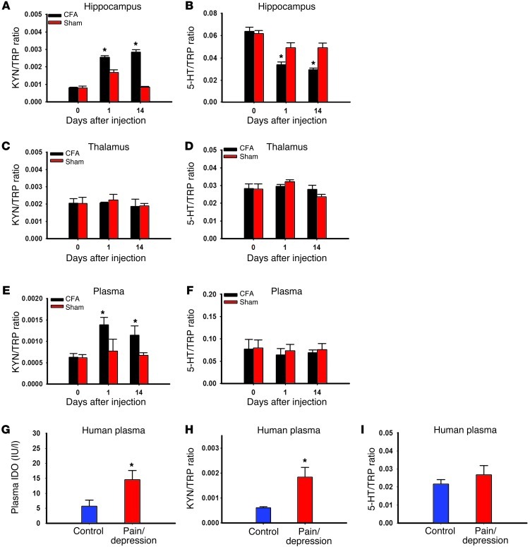
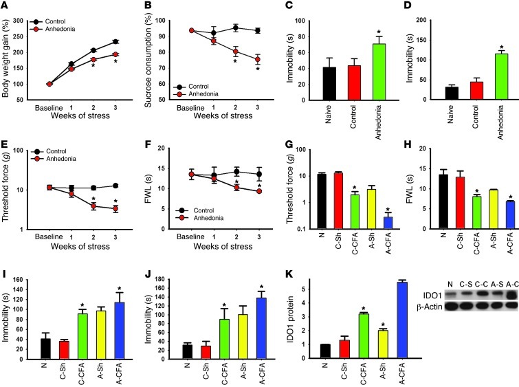
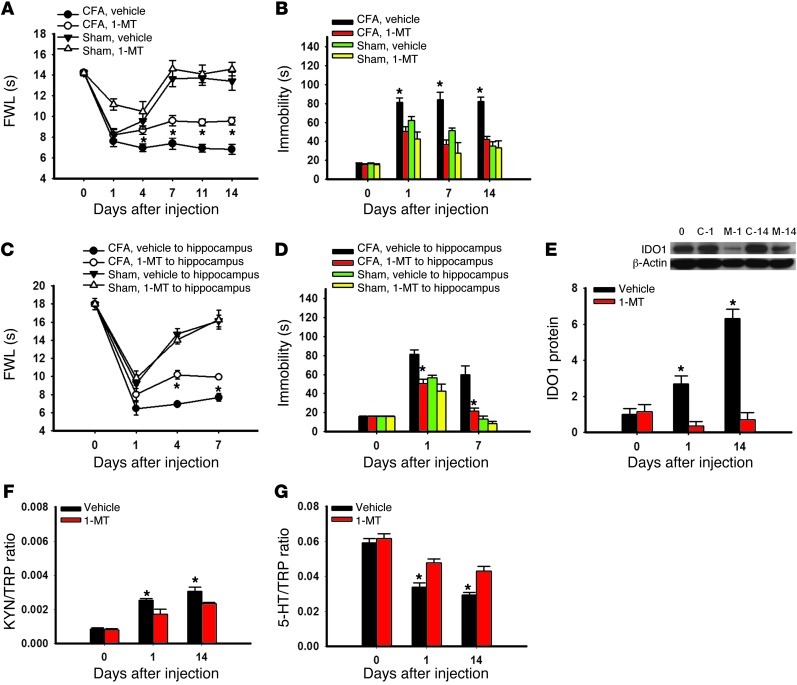
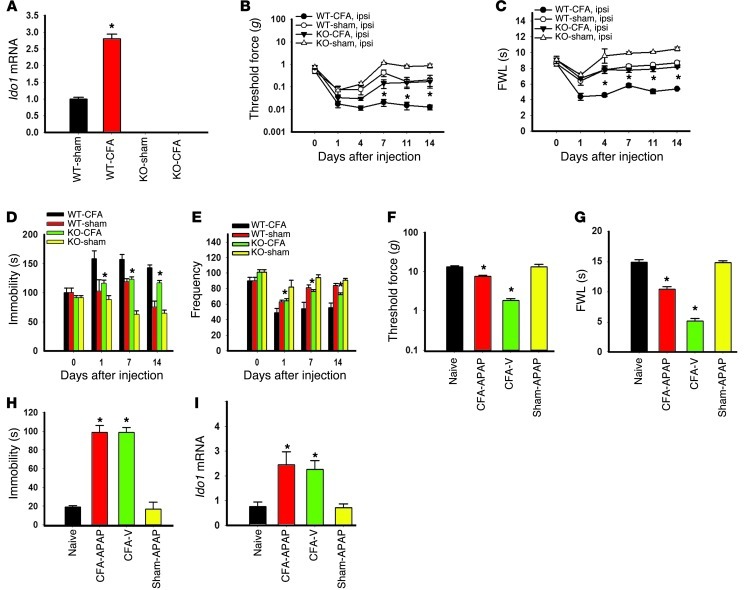
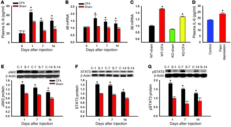
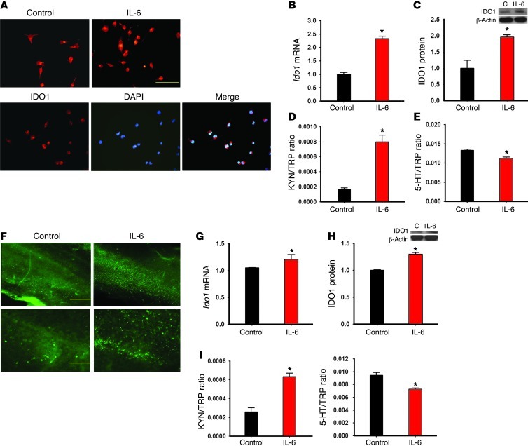
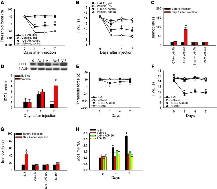
Pain and depression are frequently comorbid disorders, but the mechanism underlying this association is unknown. Here, we report that brain indoleamine 2,3-dioxygenase 1 (IDO1), a rate-limiting enzyme in tryptophan metabolism, plays a key role in this comorbidity. We found that chronic pain in rats induced depressive behavior and IDO1 upregulation in the bilateral hippocampus. Upregulation of IDO1 resulted in the increased kynurenine/tryptophan ratio and decreased serotonin/tryptophan ratio in the bilateral hippocampus. We observed elevated plasma IDO activity in patients with both pain and depression, as well as in rats with anhedonia induced by chronic social stress. Intra-hippocampal administration of IL-6 in rats, in addition to in vitro experiments, demonstrated that IL-6 induces IDO1 expression through the JAK/STAT pathway. Further, either Ido1 gene knockout or pharmacological inhibition of hippocampal IDO1 activity attenuated both nociceptive and depressive behavior. These results reveal an IDO1-mediated regulatory mechanism underlying the comorbidity of pain and depression and suggest a new strategy for the concurrent treatment of both conditions via modulation of brain IDO1 activity.


Figures

Figure 1
Figure 1. Correlation of nociceptive and depressive behaviors.
(AD) The injection of CFA into the right tibiotarsal joint of Wistar rats produced mechanical allodynia (A) and thermal hyperalgesia (B) on the ipsilateral hind paw, as well as prolonging the immobility time in FST (C) and reducing the frequency (number of squares crossed) in OFT (D). Mean ± SEM, n = 10–11, *P < 0.05, compared with sham controls. FWL, foot withdrawal latency; ipsi, ipsilateral; contra, contralateral. (E) The prolonged immobility time in FST was inversely correlated with the reduced hind paw withdrawal latency in the thermal hyperalgesia test. The data were obtained on day 14. r = –0.913, n = 10–11, *P < 0.01. (F) The reduced frequency in OFT correlated with the reduced hind paw withdrawal latency in the thermal hyperalgesia test. r = 0.840, n = 10–11, P < 0.01.
Figure 2
Figure 2. IDO1 expression in the hippocampus.
(A) IDO1 immunoreactivity was detected in the hippocampus. Scale bars: 1.0 mm (top), 50 μm (bottom). (B) Photomicrographs of colocalization between IDO1 and GFAP, Iba-1, or NeuN in the hippocampus. Scale bars: 50 μm. (C and D) Ido1 mRNA (C) and protein (D) expression was increased in the contralateral hippocampus of Wistar rats injected with CFA as detected by real-time PCR (C) and Western blot analysis (D). Day 0, baseline (naive rats); C-1 and C-14, samples taken on day 1 and day 14 from rats with CFA-induced arthritis; S-1 and S-14, samples taken on day 1 and day 14 from sham control rats. β-Actin was used as loading control. y axis shows fold change in IDO1 mRNA and protein expression. Mean ± SEM, n = 6–10, *P < 0.05 compared with sham control. (E and F) IDO1 expression (Western blot) was not upregulated in the thalamus (E) or nucleus accumbens (F) in Wistar rats with CFA-induced arthritis; n = 6, P > 0.05.
Figure 3
Figure 3. Altered tryptophan metabolites by IDO enzyme activity.
(A and B) The kynurenine (KYN)/tryptophan (TRP) ratio was increased (A), whereas the serotonin (5-HT)/TRP ratio was decreased (B), in the contralateral hippocampus of Wistar rats with CFA-induced arthritis as assayed by HPLC. Mean ± SEM, n = 6, *P < 0.05 compared with sham control. (C and D) The KYN/TRP ratio (C) and the 5-HT/TRP ratio (D) were not changed in the thalamus of Wistar rats with CFA-induced arthritis as assayed by HPLC. The plasma KYN/TRP ratio (E), but not the 5-HT/TRP ratio (F), was increased in Wistar rats with CFA-induced arthritis when both were examined on day 14. Mean ± SEM, n = 6, *P < 0.05 compared with sham control. The plasma IDO level (G; ELISA) and the KYN/TRP ratio (H; HPLC), but not the 5-HT/TRP ratio (I; HPLC), were elevated in patients with both chronic back pain and depression. Mean ± SEM, n = 13–20, *P < 0.05 compared with healthy control.
Figure 4
Figure 4. Relationship between anhedonic and nociceptive behavior.
(A) The body weight gain was calculated as the percentage of the initial (baseline) body weight. Anhedonic rats gained less weight than control rats. (B) Sucrose preference was calculated as the percentage of the total fluid intake over 24 hours. Sucrose preference was diminished in anhedonic rats. (CF) The prolonged immobility time in FST (C) and TST (D) was associated with mechanical allodynia (E) and thermal hyperalgesia (F) in anhedonic rats. Data in AF are mean ± SEM, n = 6 *P < 0.05 compared with control. (GJ) Mechanical allodynia (G) and thermal hyperalgesia (H) were exacerbated, and immobility time was prolonged, in FST (I) and TST (J) in anhedonic rats when examined at 7 days after the CFA-induced monoarthritis. (K) IDO1 protein expression was increased in anhedonic rats (A-Sh and A-CFA) as compared with control rats (C-Sh and C-CFA) with and without CFA injection. Data in GK are mean ± SEM, n = 6, *P < 0.05 compared with sham control. N, naive.
Figure 5
Figure 5. Effect of IDO1 inhibition on behavioral changes.
(A) Intraperitoneal injection of the IDO1 inhibitor 1-MT (10 mg/d), given twice daily for 14 consecutive days beginning immediately after the CFA injection, attenuated mechanical allodynia on the ipsilateral hind paw of rats. (B) The same 1-MT treatment regimen concurrently improved the immobility time in FST in the same rats. Mean ± SEM, n = 6, *P < 0.05 compared with vehicle control. (C and D) Intra-hippocampal microinjection of 1-MT (5 μg in 0.5 μl), once daily for 7 days beginning immediately after the CFA injection, also attenuated thermal hyperalgesia (C) on the ipsilateral hind paw of rats as well as reducing the immobility time in FST (D) in the same rats. Mean ± SEM, n = 6, *P < 0.05 compared with vehicle control. (E) IDO1 expression in the hippocampus was decreased in rats receiving a 14-day intraperitoneal administration of 1-MT (10 mg/d). Day 0, baseline (naive rats); C-1 and C-14, samples taken on day 1 and day 14 from rats with CFA-induced arthritis; S-1 and S-14, samples taken on day 1 and day 14 from control rats. β-Actin was used as loading control. Mean ± SEM, n = 6, *P < 0.05 compared with sham control. The same systemic 1-MT treatment regimen reduced the KYN/TRP ratio (F) and increased the 5-HT/TRP ratio (G) in the hippocampus. Mean ± SEM, n = 6, *P < 0.05 compared with vehicle control.
Figure 6
Figure 6. Effect of IDO-knockout on behavioral changes.
(A) IDO-knockout mice had no Ido1 mRNA expression (real-time PCR) in the hippocampus. Ido1 mRNA expression in wild-type mice was increased in the hippocampus after the CFA injection. Mean ± SEM, n = 6, *P < 0.05 compared with sham control. (B and C) Mechanical allodynia (B) and thermal hyperalgesia (C) on the ipsilateral hind paw were attenuated in IDO-knockout mice. (D and E) IDO-knockout also reduced the immobility time in FST (D) and the decreased frequency in OFT (E) in the same mice with CFA-induced arthritis. Mean ± SEM, n = 6, *P < 0.05 compared with wild-type mice. (F and G) Intraperitoneal injection of acetaminophen (APAP; 100 mg/kg), given once on day 14, attenuated ipsilateral mechanical allodynia (F) and thermal hyperalgesia (G) when mice were examined at 1 hour after the injection. Mean ± SEM, n = 6, *P < 0.05 compared with vehicle control. (H) The same APAP treatment did not change the immobility time in FST in the same rats. (I) Contralateral hippocampal Ido1 mRNA expression was increased after the CFA injection, which was not reversed by a single APAP treatment. Data in H and I are mean ± SEM, n = 6, *P < 0.05 compared with vehicle control.
Figure 7
Figure 7. Role of IL-6 in the hippocampal JAK/STAT pathway.
(A and B) The plasma IL-6 concentration was increased (A, ELISA), as was Il6 mRNA expression in the hippocampus (B, real-time PCR), in rats with CFA-induced arthritis. (C) Il6 mRNA expression was also increased in the hippocampus of both IDO-knockout and wild-type mice with CFA-induced arthritis. Mean ± SEM, n = 6, *P < 0.05 compared with sham control. (D) The plasma IL-6 level was elevated in patients with both chronic back pain and depression (ELISA). Mean ± SEM, n = 13–20, *P < 0.05 compared with healthy control. (EG) The expression of JAK2 (E), STAT3 (F), and p-STAT3 (G) in the hippocampus was increased in rats with CFA-induced arthritis (Western blot analysis). C-1, C-7, and C-14, samples taken on days 1, 7, and 14 from rats with CFA-induced arthritis; S-1, S-7, and S-14, samples taken on days 1, 7, and 14 from sham control rats. β-Actin was used as loading control. Mean ± SEM, n = 4–5, *P < 0.05 compared with sham control.
Figure 8
Figure 8. Effects of IL-6 on IDO1 expression in Neuro2a cells and organotypic hippocampal culture.
(A) IDO1 immunoreactivity was increased in IL-6–treated Neuro2a cells, as expressed in perinuclear cytoplasm when samples were costained with DAPI. Scale bar: 50 μm. (BE) IDO1 mRNA (B) and protein (C) expression, as well as IDO activity (KYN/TRP ratio [D] and 5-HT/TRP ratio [E]), was increased in cultured Neuro2a cells after addition of IL-6 (0.5 ng/ml) for 24 hours. (FH) Exposure of exogenous IL-6 (100 ng/ml) for 24 hours increased the expression of IDO1 (F: immunoreactivity; G: mRNA; H: Western blot) in hippocampal organotypic slice culture. Scale bars: 500 μm (top row) and 50 μm (bottom row). C, control. In addition, adding IL-6 for 24 hours also increased the KYN/TRP ratio and decreased the 5-HT/TRP ratio (HPLC) in the culture medium (I). *P < 0.05 compared with vehicle control.
Figure 9
Figure 9. Role of IL-6 signaling in hippocampal IDO expression and behavioral changes.
(AD) Microinjection of an IL-6 antiserum (IL-6 Ab; 0.5 μg/d; once daily for 7 days) into the hippocampus contralateral to the hind paw with CFA-induced arthritis attenuated mechanical allodynia (A) and thermal hyperalgesia (B) on the ipsilateral hind paw, without a change in nociceptive threshold in sham rats (data not shown). The same IL-6 antiserum microinjection regimen concurrently improved depressive behavior in FST (C) and prevented the hippocampal IDO1 upregulation (D) in these same rats. Day 0, baseline (naive rats); Ab-1 and Ab-7, samples taken on day 1 and day 7 from Wistar rats treated with IL-6 antiserum; V-1 and V-7, samples taken on day 1 and day 7 from Wistar rats treated with control serum. β-Actin was used as loading control. Mean ± SEM, n = 6, *P < 0.05 compared with vehicle (control serum). (EH) Microinjection of exogenous IL-6 (0.1 μg/0.5 μl; once daily for 7 days) into the left hippocampus of naive rats (without CFA injection) induced mechanical allodynia (E) and thermal hyperalgesia (F) on the right hind paw. The same IL-6 microinjection regimen concurrently induced depressive behavior in FST (G) and upregulated hippocampal Ido1 mRNA expression (H) in these same rats. The effects from the intra-hippocampal microinjection of IL-6 were blocked when IL-6 was co-administered with AG490 (a JAK/STAT inhibitor; 5 μg/0.5 μl) for 7 days. Mean ± SEM, n = 4–5, *P < 0.05 compared with vehicle control.

References

    1. Sullivan MJ, Reesor K, Mikail S, Fisher R. The treatment of depression in chronic low back pain: review and recommendations. Pain. 1992;50(1):5–13. doi: 10.1016/0304-3959(92)90107-M. - DOI - PubMed
    1. Von KM, Simon G. The relationship between pain and depression. Br J Psychiatry Suppl. 1996;(30):101–108. - PubMed
    1. Mao J, Gold MS, Backonja MM. Combination drug therapy for chronic pain: a call for more clinical studies. J Pain. 2011;12(2):157–166. - PMC - PubMed
    1. Ganong AH, Cotman CW. Kynurenic acid and quinolinic acid act at N-methyl-D-aspartate receptors in the rat hippocampus. J Pharmacol Exp Ther. 1986;236(1):293–299. - PubMed
    1. Dantzer R, O’Connor JC, Freund GG, Johnson RW, Kelley KW. From inflammation to sickness and depression: when the immune system subjugates the brain. Nat Rev Neurosci. 2008;9(1):46–56. doi: 10.1038/nrn2297. - DOI - PMC - PubMed

Publication types

MeSH terms

Substances