. 2013 May 2;32(18):2356-64.
doi: 10.1038/onc.2012.270. Epub 2012 Jul 2.

p16(INK4A) represses the paracrine tumor-promoting effects of breast stromal fibroblasts

Affiliations
Free PMC article

p16(INK4A) represses the paracrine tumor-promoting effects of breast stromal fibroblasts

M M Al-Ansari et al. Oncogene. .
Free PMC article

Erratum in

  • Oncogene. 2013 May 2;32(18):2372

Abstract

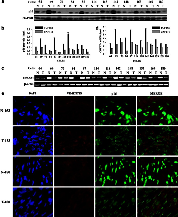
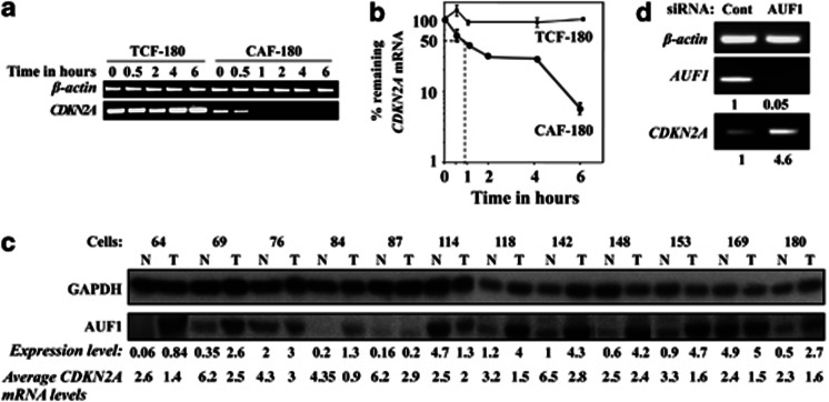
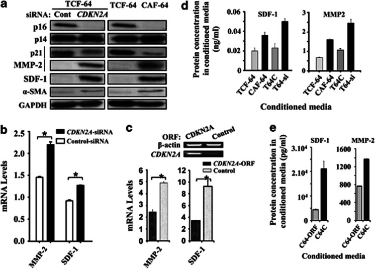
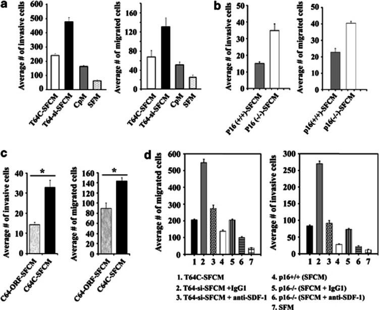
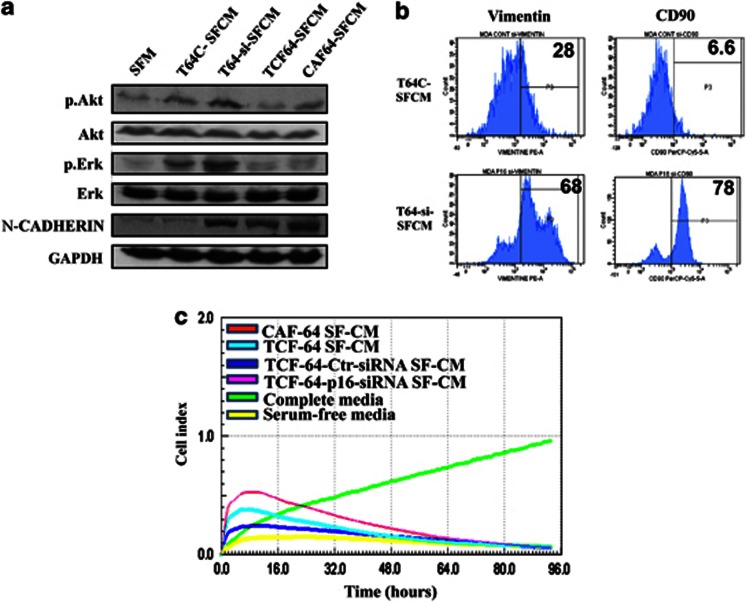
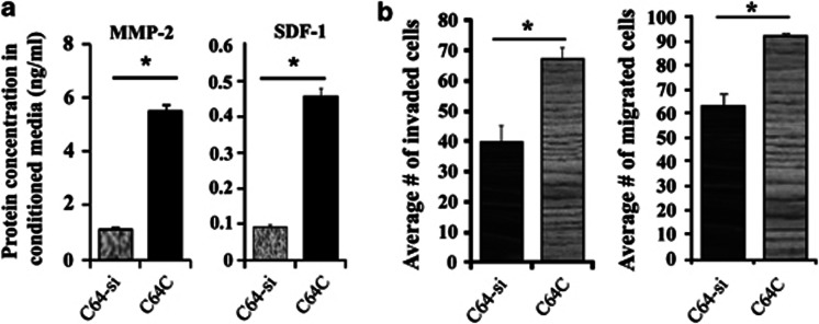
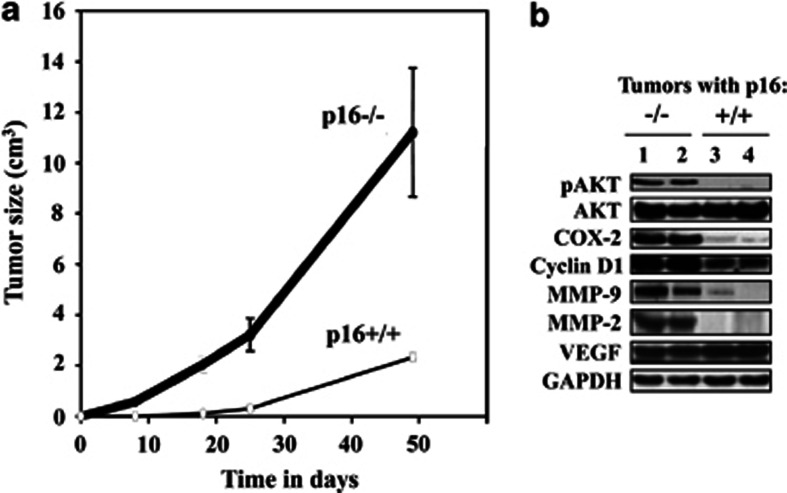
Cancer-associated fibroblasts (CAFs), the most abundant and probably the most active cellular component of breast cancer-associated stroma, promote carcinogenesis through paracrine effects; however, the molecular basis remains elusive. We have shown here that p16(INK4A) expression is reduced in 83% CAFs as compared with their normal adjacent counterparts cancer-free tissues isolated from the same patients. This decrease is mainly due to AUF1-dependent higher turnover of the CDKN2A mRNA in CAFs. Importantly, p16(INK4A) downregulation using specific siRNA activated breast fibroblasts and increased the expression/secretion levels of stromal cell-derived factor 1 (SDF-1) and matrix metalloproteinase (MMP)-2. Consequently, media conditioned with these cells stimulated the proliferation of epithelial cells. Furthermore, the migration/invasion of breast cancer cells was also enhanced in an SDF-1-dependent manner. This effect was mediated through inducing an epithelial-mesenchymal transition state. By contrast, increase in p16(INK4A) level through ectopic expression or AUF1 downregulation, reduced the secreted levels of SDF-1 and MMP-2 and suppressed the pro-carcinogenic effects of CAFs. In addition, p16(INK4A)-defective fibroblasts accelerated breast tumor xenograft formation and growth rate in mice. Importantly, tumors formed in the presence of p16(INK4A)-defective fibroblasts exhibited higher levels of active Akt, Cox-2, MMP-2 and MMP-9, showing their greater aggressiveness as compared with xenografts formed in the presence of p16(INK4A)-proficient fibroblasts. These results provide the first indication that p16(INK4A) downregulation in breast stromal fibroblasts is an important step toward their activation.


Figures

Figure 1
Figure 1
p16 Expression level is downregulated in CAFs. (a) Whole-cell lysates were prepared from the indicated cells and 50 μg of proteins were used for immunoblot analysis using the indicated antibodies. (b, d) Histograms, the values were determined by densitometry and normalized against the internal controls glyceraldehyde-3-phosphate dehydrogenase (GAPDH) and β-actin. (c) Total RNA was extracted and the amount of CDKN2A mRNA was assessed by RT–PCR. The amplified fragments were electrophoresed on ethidium bromide stained agarose gel. Error bars represent means±s.d. (e) Immunofluorescence analysis using the indicated frozen sections N, T and antibodies. N, normal; T, tumor.
Figure 2
Figure 2
CAFs exhibit unstable CDKN2A mRNA. (a) CAF/TCF-180 cells were treated with 5 μg/ml actinomycin D for various times, and total RNA was extracted and the level of the CDKN2A mRNA was assessed using RT–PCR. The amplified fragments were electrophoresed on ethidium bromide stained agarose gel. (b) Graph showing the proportion of CDKN2A mRNA remaining post treatment, and the dotted lines indicate the CDKN2A mRNA half-life. The values were determined by densitometry and normalized against β-actin. Error bars represent means±s.d. (c) Whole-cell lysates were prepared from the indicated cells, N and T correspond to TCF and CAF, respectively. The relative expression levels were determined relative to GAPDH. (d) Total RNA was extracted from CAF-64 cells expressing either AUF1-siRNA or control-siRNA, and the levels of AUF1 and CDKN2A mRNAs were assessed by RT–PCR and normalized against β-actin. The numbers below the bands indicate the corresponding expression levels.
Figure 3
Figure 3
p16 Suppresses the expression of α-SMA, SDF1 and MMP 2. (a) Whole-cell lysates were prepared from TCF/CAF-64 and TCF-64 expressing either CDKN2A-siRNA or control-siRNA, and were used for immunoblot analysis. (b) Total RNA was extracted and the amount of MMP-2 and SDF-1 mRNAs were assessed using real-time RT–PCR. Error bars represent means±s.d. *P-value<0.05. (c) Total RNA was extracted from CAF-64 cells expressing either CDKN2A ORF or control. Upper panel: the CDKN2A mRNA was amplified by RT–PCR, and the obtained fragments were electrophoresed on ethidium bromide stained agarose gel. Lower panel: the amount of MMP-2 and SDF-1 mRNAs were assessed using real-time RT–PCR. Error bars represent means±s.d. *P-value<0.05. (d, e) CM from the indicated cells were collected after 24 h and the levels of the indicated proteins were determined by ELISA and were presented in the respective histograms. Error bars represent means±s.d.
Figure 4
Figure 4
p16-Deficient fibroblast secretions stimulate breast cancer cell invasion/migration in an SDF1-dependent manner. SF-CM were collected after 24 h of incubation from the indicated cells, and were added independently into the lower compartments of 24-well BD BioCoat plates. A total of 105 MDA-MB-231 cells were seeded onto the upper compartment of the migration and invasion plates and incubated for 24 h in the presence of SF-CM. The number of migrated/invaded cells was represented in histograms. Error bars represent means±s.d. (a, c and d) Human breast fibroblast cells, *P<0.02. (b) Mice fibroblast cells.
Figure 5
Figure 5
p16-Deficient fibroblast secretions stimulate an epithelial–mesenchymal transition-like state in breast cancer cells. Whole-cell lysates were prepared from MDA-MB-231 cells that were treated for 24 h with SFM (used as control) or SF-CM collected from the indicated cells. (a) Immunoblotting analysis was performed using the indicated antibodies; (b) flow cytometry was used to assess the level of vimentin and CD90 in MDA-MB-231 cells treated with the indicated CM. (c) SF-CM collected after 24 h from the indicated cells were added separately to MCF-10 cells previously seeded into 96 wells, and cell proliferation was assessed by the real-time cell electronic sensing system.
Figure 6
Figure 6
AUF1 downregulation suppresses the effects of reduced levels of p16 in CAF cells. (a) CM from CAF-64 cells expressing either AUF1-siRNA (C64-si) or control-siRNA (C64C) were collected after 24 h and the levels of the indicated proteins were determined by ELISA and were presented in the respective histograms. (b) Figure legends are as in Figure 5. Error bars represent means±s.d., *P<0.003.
Figure 7
Figure 7
p16-Deficient fibroblasts enhances breast cancer xenografts formation and growth. Breast cancer xenografts were created by co-injecting MDA-MB-231 cells with p16−/− or p16+/+ MEFs subcutaneously into nude mice. (a) Graph showing tumor sizes. Error bars represent means±s.d. (b) Tumors were excised and whole-cell lysates were prepared and protein levels were assessed by immunoblotting using the indicated antibodies.

Similar articles

Cited by

References

    1. Jemal A, Bray F, Center MM, Ferlay J, Ward E, Forman D. Global cancer statistics. Ca Cancer J Clin. 2011;61:69–90. - PubMed
    1. Shimoda M, Mellody KT, Orimo A. Carcinoma-associated fibroblasts are a rate-limiting determinant for tumour progression. Semin Cell Dev Biol. 2009;21:19–25. - PMC - PubMed
    1. Franco OE, Shaw AK, Strand DW, Hayward SW. Cancer associated fibroblasts in cancer pathogenesis. Semin Cell Dev Biol. 2009;21:33–39. - PMC - PubMed
    1. Dong-Le Bourhis X, Berthois Y, Millot G, Degeorges A, Sylvi M, Martin PM, et al. Effect of stromal and epithelial cells derived from normal and tumorous breast tissue on the proliferation of human breast cancer cell lines in co-culture. Int J Cancer. 1997;71:42–48. - PubMed
    1. Kim JB, Stein R, O'Hare MJ. Tumour-stromal interactions in breast cancer: the role of stroma in tumourigenesis. Tumour Biol. 2005;26:173–185. - PubMed

Publication types

MeSH terms