. 2012 Sep;160(1):349-64.
doi: 10.1104/pp.112.201319. Epub 2012 Jun 29.

The subcellular localization of Tubby-like proteins and participation in stress signaling and root colonization by the mutualist Piriformospora indica

Affiliations

The subcellular localization of Tubby-like proteins and participation in stress signaling and root colonization by the mutualist Piriformospora indica

Marco Uwe Reitz et al. Plant Physiol. 2012 Sep.

Abstract

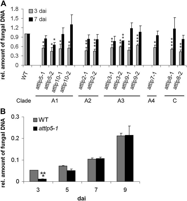
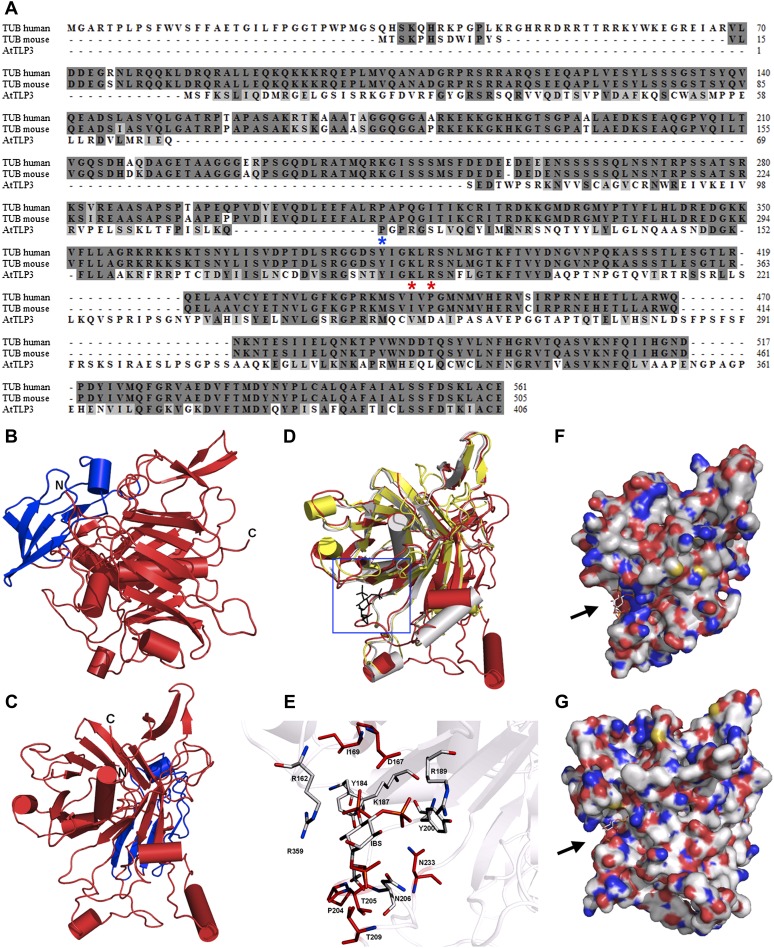
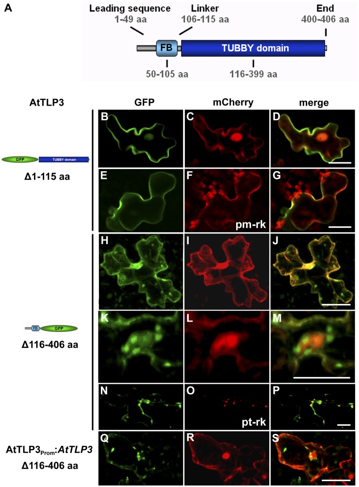
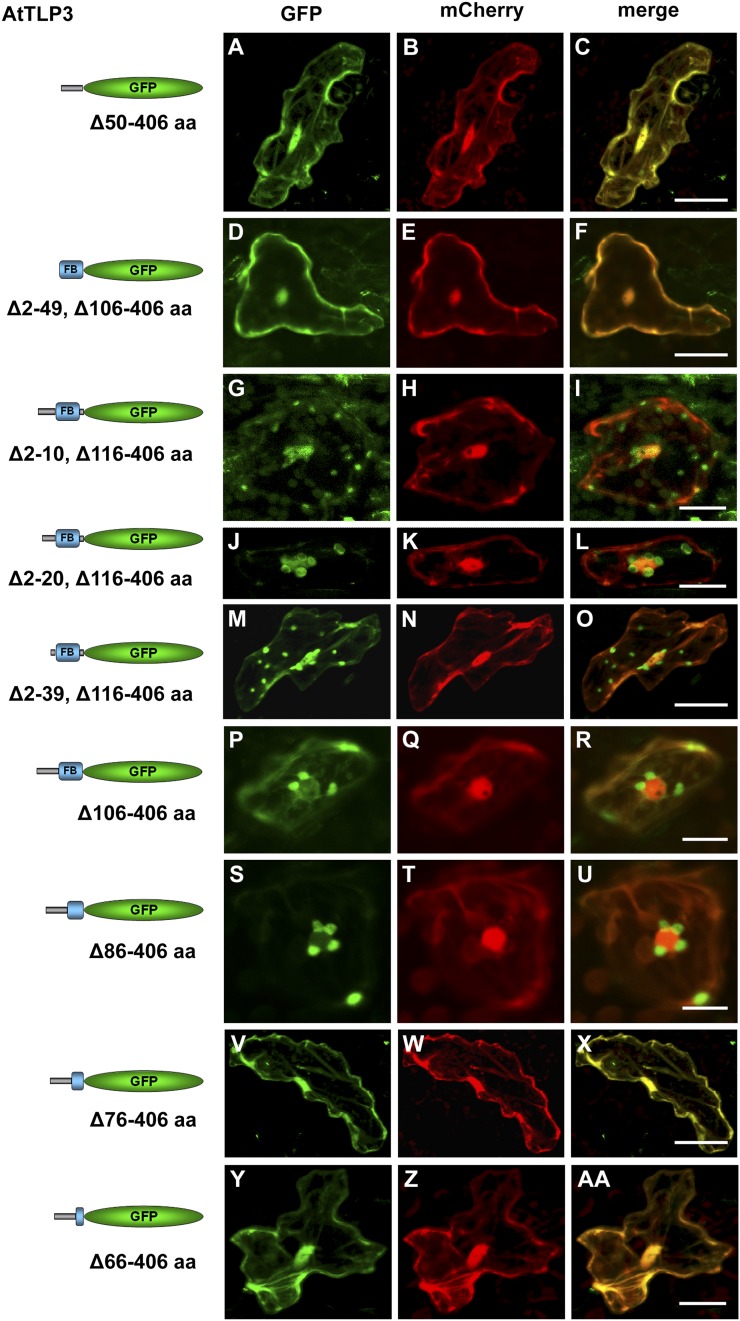
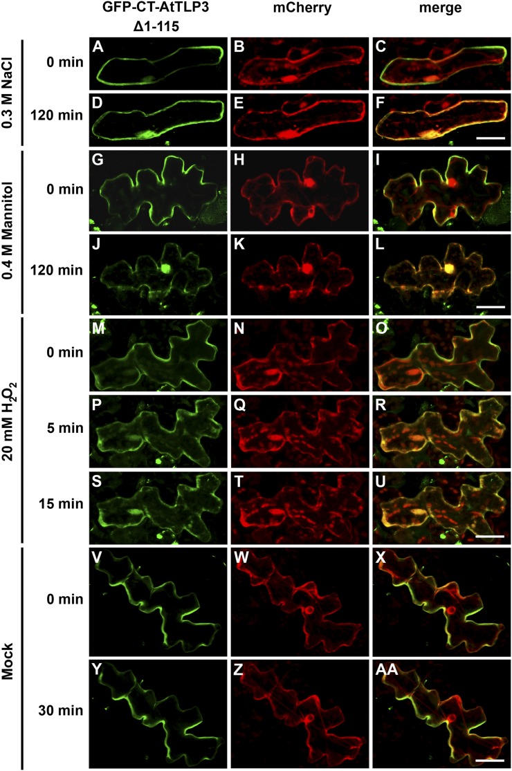
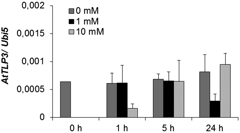
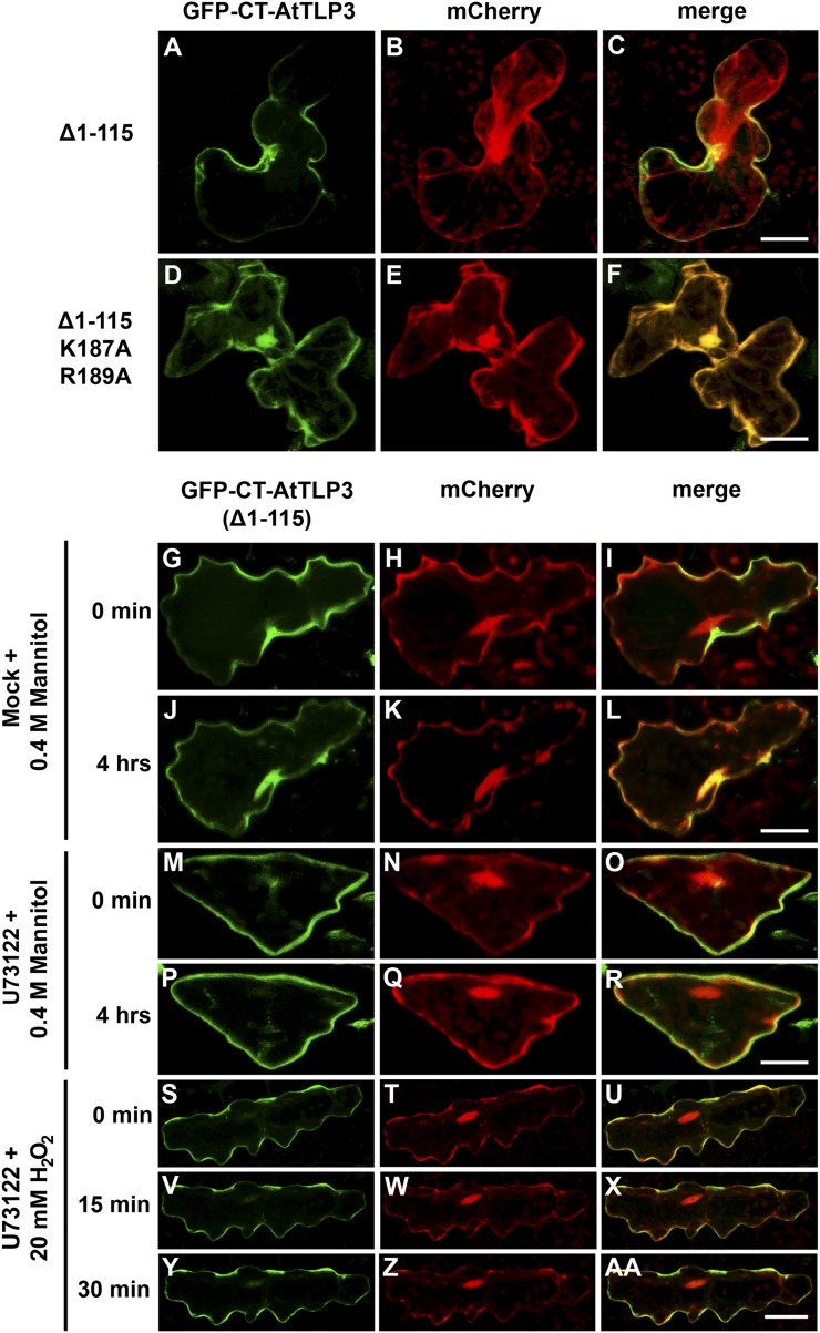
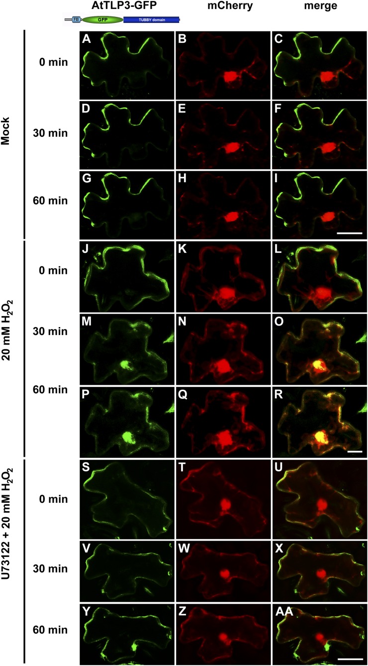
Tubby and Tubby-like proteins (TLPs) were first discovered in mammals, where they are involved in the development and function of neuronal cells. Due to their importance as plasma membrane (PM)-tethered transcription factors or mediators of vesicle trafficking, their lack causes obesity and other disease syndromes. Phosphatidylinositol 4,5-bisphosphate binding of the carboxyl-terminal Tubby domain attaches these proteins to the PM and vesicles and is essential for function. TLPs are conserved across eukaryotic kingdoms including plants, suggesting fundamental biological functions of TLPs. Plant TLPs possess an amino-terminal F-box domain that distinguishes them from other eukaryotic TLPs. Arabidopsis (Arabidopsis thaliana) encodes 11 AtTLPs that fall into six phylogenetic clades. We identified the significance of AtTLPs for root colonization of Arabidopsis by the mutualistic fungus Piriformospora indica. Our results further indicate conserved phosphatidylinositol 4,5-bisphosphate-binding sites in the Tubby domains that are required for PM anchoring of AtTLPs. More detailed studies revealed phospholipase C-triggered release of AtTLP3 from the PM, indicating a conserved mechanism as reported for mammalian Tubby and TLP3. We further show that hydrogen peroxide stimulates the release of AtTLP3 from the PM, presumably for activating downstream events. Different from mammalian homologs, the amino-terminal part of almost all AtTLPs has nucleocytosolic and plastidial localization patterns. Thus, it is tempting to assume that TLPs translate reactive oxygen species currents into signaling not only for transcriptional regulation in the nucleus but also affect plastid-associated functions after release from the PM.


Figures

Figure 1.
Figure 1.
Delayed colonization of attlp mutants by P. indica. Three-week-old plants were inoculated with P. indica, and fungal biomass was determined during biotrophic (3 dai) and cell death-dependent (7 dai) colonization (A) or at 3, 5, 7, and 9 dai (B) by qRT-PCR. A, Clades indicate the membership of tested mutants. The relative amount of fungal biomass in mutant roots was related to the wild type (WT; set to 1). Data are averages from at least three independent experiments, and error bars represent se. B, This experiment was performed twice with similar results. se values are from three technical replicates of one biological experiment. For both experiments, 200 plants were analyzed per mutant or wild type and per time point. Asterisks indicate significance at P < 0.05 (*), P < 0.01 (**), or P < 0.001 (***) as analyzed by Student’s t test.
Figure 2.
Figure 2.
Sequence and 3D structure of AtTLP3 and its Tubby domain. A, Sequence alignment for human (accession no. 19923167) and mouse (accession no. 11230782) Tubby (TUB) protein with AtTLP3 (accession no. 30690823). Identical and similar amino acids are shaded in dark gray and light gray, respectively. The two amino acids that are necessary for PIP2 binding (Santagata et al., 2001) are indicated (red asterisks) underneath those amino acids. The blue asterisk underneath one amino acid indicates the start of the Tubby domain. B and C, 3D homology model of AtTLP3 showing the FB domain (blue) and the Tubby domain (red). The N and C termini are highlighted. D, Superposition of the structural model of AtTLP3 (red) with the crystal structure of the human Tubby protein (PDB identifier 1S31; yellow) and the crystal structure of mouse brain Tubby protein (gray) bound to PIP2 (black, blue frame). E, Magnification of the frame in D shows the highly conserved inositol lipid-binding domain of AtTLP3. A comparison of AtTLP3, mouse, and human Tubby proteins was done. The PIP2 analog l-α-glycerophospho-d-myoinositol 4,5-bisphosphate (IBS) was used to show the conservation of binding sites. Amino acid residues that are similar in type and position (conformation) in all three proteins are colored in gray, and amino acids that are specific for the AtTLP3 protein are marked in red. F and G, Surface representation of the mouse Tubby protein with bound IBS (F) and structural model of AtTLP3 (G) with modeled IBS. The groove of highly positive charge (blue regions) is more significant in the mouse protein, and the dimension of the groove differs in the AtTLP3 structural model.
Figure 3.
Figure 3.
Organ- and tissue-specific expression of AtTLP3 in AtTLP3Prom:GUS plants. Four-day-old seedling (A), roots of 18-d-old plants (B–D), rosette leaf (E), anthotaxy (F), single flower with some sepals and petals removed (G), and mature silique (H) are shown. Bars = 1 mm in A and E to H and 0.1 mm in B to D.
Figure 4.
Figure 4.
Subcellular localization of the FB and Tubby domains of AtTLP3. A, Model of AtTLP3 with the N-terminal FB domain and the C-terminal Tubby domain. Numbers indicate the lengths and positions of domains within AtTLP3 in amino acids (aa). For all experiments below, leaf cells were cotransformed with the GFP-CT-AtTLP3 (Δ1115) (B and E) or NT-AtTLP3-GFP (Δ116406) (H, K, and N) fusions under the control of the 35S promoter (B–P) or the native promoter (Q) and cytosolic and nucleoplasmic marker mCherry (C, I, L, and R), PM marker pm-rk (F), or plastidial marker pt-rk (O) by biolistic transformation. D, G, J, M, P, and S are merged images indicating the subcellular localization of domains. Yellow color indicates the colocalization of green- and red-fluorescing proteins. Constructs used for transformation are indicated on the left. B to D, PM localization of GFP-CT-AtTLP3. E to G, GFP-CT-AtTLP3 colocalizes with the PM marker pm-rk at the PM. H to S, Plastidial and nucleocytosolic localization of NT-AtTLP3-GFP. K to M, Accumulation of plastids around the nucleus in a cell expressing NT-AtTLP3-GFP. N to P, Plastidial localization was confirmed by cotransformation of the NT-AtTLP3-GFP fusion with the plastidial marker pt-rk. Q to S, Cells transformed with NT-AtTLP3-GFP under the control of its endogenous promoter (AtTLP3PROM) also showed plastidial, cytosolic, and nucleoplasmic localization. Bars = 20 µm.
Figure 5.
Figure 5.
Parts of the leading sequence and FB domain determine plastidial localization of the N terminus of AtTLP3. A, D, G, J, M, P, S, V, and Y, Arabidopsis leaf cells were transiently transformed with truncated versions of the N terminus of AtTLP3 fused to GFP. Constructs used for transformation are indicated on the left. B, E, H, K, N, Q, T, W, and Z, Leaf cells were cotransformed with the cytosolic and nucleoplasmic marker mCherry. C, F, I, L, O, R, U, X, and AA, Merged images indicate the subcellular localization of truncated versions. Yellow color indicates the colocalization of green- and red-fluorescing proteins. All truncated versions lack amino acids (aa) of the C terminus (Tubby domain; amino acids 116–406) of AtTLP3, while the linker sequence (amino acids 106–115) is either missing (Δ116–406) or included (Δ106–406). In addition, various amino acids of the N terminus are missing as indicated. A to F, Cytosolic and nucleoplasmic localization of GFP fused to the leading sequence (Δ50–406; A–C) or the FB domain (Δ2–49, Δ106–406; D–F). G to O, Plastidial and nucleocytosolic localization of GFP fused to truncated N-terminal versions lacking amino acids 2 to 10 (Δ2–10, Δ106–406; G–I), 2 to 20 (Δ2–20, Δ106–406; J–l), or 2 to 39 (Δ2–39, Δ106–406; M–O). P to R, Plastidial localization of GFP fused to an N-terminal version lacking the linker sequence and Tubby domain (Δ106–406). S to U, Plastidial localization of GFP fused to an N-terminal version lacking the linker sequence, Tubby domain, and the last 20 amino acids of the FB (Δ86–406). V to AA, Cytosolic and nucleoplasmic localization of GFP fused to an N-terminal version lacking the linker sequence, Tubby domain, and the last 30 (Δ76–406; V–X) or 40 (Δ66–406; Y–AA) amino acids of the FB. Experiments were repeated three times with similar results. Bars = 20 µm.
Figure 6.
Figure 6.
Osmotic stress-induced translocation of the membrane-tethered AtTLP3 Tubby domain to plastids. A, D, G, J, M, P, S, V, Y, Arabidopsis leaf cells were transformed with GFP-CT-AtTLP3 (Δ1115). B, E, H, K, N, Q, T, W, Z, Arabidopsis leaf cells were transformed with the cytosolic and nucleoplasmic marker mCherry. C, F, I, L, O, R, U, X, AA, Merged images indicate the subcellular localization of truncated domain versions. Yellow color indicates the colocalization of green- and red-fluorescing proteins. A to F, Application of 0.3 m NaCl results in the relocalization of the C terminus of AtTLP3 [GFP-CT-AtTLP3 (Δ1–115)] from the PM (A–C) to the cytosol and nucleus within 120 min (D–F). G to L, Application of 0.4 m mannitol results in the relocalization of GFP-CT-AtTLP3 (Δ1–115) from the PM (G–I) to the cytosol and nucleus within 120 min (J–L). Exposure of cells to mannitol caused plasmolysis, thereby slightly changing the shape of cells (J–L). M to U, Application of 20 mm H2O2 results in the relocalization of GFP-CT-AtTLP3 (Δ1–115) from the PM to the cytosol and nucleus. M to O, PM localization of GFP-CT-AtTLP3 before treatment (0 min). P to R, Cytosolic and nucleoplasmic localization of GFP-CT-AtTLP3 (Δ1–115) at 5 min after application of 20 mm H2O2. S to U, Cytosolic and nucleoplasmic localization of GFP-CT-AtTLP3 (Δ1–115) at 15 min after application of 20 mm H2O2. V to AA, PM localization of GFP-CT-AtTLP3 (Δ1–115) at 0 and 30 min after mock treatment. Experiments were repeated three times with similar results. Bars = 20 µm.
Figure 7.
Figure 7.
Expression of AtTLP3 in Arabidopsis plants treated with H2O2. Arabidopsis plants were harvested at the indicated time points after mock treatment or treatment with 1 mm or 10 mm H2O2. Expression values were calculated by the 2−ΔCt method by relating cycle thresholds of AtTLP3 to those of the housekeeping gene AtUbi5. Values are means ± se of three independent experiments.
Figure 8.
Figure 8.
Lys-187 and Arg-189 represent PIP2-binding sites in the Tubby domain of AtTLP3, and phospholipase C releases CT-AtTLP3 from the PM. A, D, G, J, M, P, S, V, Y, Arabidopsis leaf cells were transformed with GFP-CT-AtTLP3 (Δ1115). B, E, H, K, N, Q, T, W, Z, Arabidopsis leaf cells were transformed with the cytosolic and nucleoplasmic marker mCherry. Amino acids eliminated in truncated versions used for transformation and treatments are indicated on the left. C, F, I, L, O, R, U, X, and AA, Merged images indicate the subcellular localization of truncated domain versions. Yellow color indicates the colocalization of green- and red-fluorescing proteins. A to C, A version of the C terminus of AtTLP3 fused to GFP [GFP-CT-AtTLP3 (Δ1–115)] displayed PM localization. D to F, GFP-CT-AtTLP3 (Δ1–115), in which Lys-187 and Arg-189 had been converted to Ala (A), has lost its PM-binding ability and shows cytosolic and nucleoplasmic localization. G to L, Application of 0.4 m mannitol results in the translocation of GFP-CT-AtTLP3 (Δ1–115) from the PM at 0 min (G–I) to the cytosol and nucleus within 4 h (J–L). M to R, Pretreatment with the phospholipase C inhibitor U73122 inhibits mannitol-triggered relocalization of GFP-TLP3 (Δ1–115) from the PM (M–O) to the cytosol and nucleus (P–R). S to AA, Pretreatment with the phospholipase C inhibitor U73122 inhibits H2O2-triggered relocalization of GFP-TLP3 (Δ1–115) from the PM to the cytosol and nucleus. Treated cells were observed at 0 min (S–U), 15 min (V–X), and 30 min (Y–AA) after H2O2 application. Experiments were repeated three times with similar results. Bars = 20 µm.
Figure 9.
Figure 9.
Phospholipase C releases full-length AtTLP3-GFP from the PM when heterologously expressed in N. benthamiana. A, D, G, J, M, P, S, V, Y, N. benthamiana leaf cells were transformed with full-length AtTLP3-GFP. B, E, H, K, N, Q, T, W, Z, N. benthamiana leaf cells were transformed the cytosolic and nucleoplasmic marker mCherry. C, F, I, L, O, R, U, X, AA, Merged images indicate the subcellular localization of full-length AtTLP3-GFP. Yellow color indicates the colocalization of green- and red-fluorescing proteins. A to I, Full-length AtTLP3 fused to GFP displayed PM localization over time. J to R, Application of 20 mm H2O2 results in the relocalization of full-length AtTLP3-GFP from the PM to the cytosol and nucleus. J to L, PM localization of full-length AtTLP3-GFP before treatment (0 min). M to O, Cytosolic and nucleoplasmic localization of full-length AtTLP3-GFP at 30 min after application of 20 mm H2O2. P to R, Cytosolic and nucleoplasmic localization of full-length AtTLP3-GFP at 60 min after application of 20 mm H2O2. S to AA, Pretreatment with the phospholipase C inhibitor U73122 inhibits H2O2-triggered relocalization of full-length AtTLP3-GFP from the PM to the cytosol and nucleus. Treated cells were observed at 0 min (S–U), 30 min (V–X), and 60 min (Y–AA) after H2O2 application. Experiments were repeated three times with similar results. Bars = 20 µm.

Similar articles

Cited by

References

    1. Abramoff MD, Magalhaes PJ, Ram SJ. (2004) Image processing with ImageJ. Biophotonics International 11: 36–42
    1. Alonso JM, Stepanova AN, Leisse TJ, Kim CJ, Chen H, Shinn P, Stevenson DK, Zimmerman J, Barajas P, Cheuk R, et al. (2003) Genome-wide insertional mutagenesis of Arabidopsis thaliana. Science 301: 653–657 - PubMed
    1. Arnold K, Bordoli L, Kopp J, Schwede T. (2006) The SWISS-MODEL workspace: a Web-based environment for protein structure homology modelling. Bioinformatics 22: 195–201 - PubMed
    1. Attard A, Gourgues M, Callemeyn-Torre N, Keller H. (2010) The immediate activation of defense responses in Arabidopsis roots is not sufficient to prevent Phytophthora parasitica infection. New Phytol 187: 449–460 - PubMed
    1. Bechtold N, Ellis J, Pelletier G. (1993) In planta Agrobacterium mediated gene transfer by infiltration of adult Arabidopsis thaliana plants. C R Acad Sci III 316: 1194–1199

Publication types

MeSH terms