Review
. 2013;19(2):213-24.
doi: 10.1177/1753425912451779. Epub 2012 Jun 29.

Particle disease: biologic mechanisms of periprosthetic osteolysis in total hip arthroplasty

Affiliations
Review

Particle disease: biologic mechanisms of periprosthetic osteolysis in total hip arthroplasty

Jiri Gallo et al. Innate Immun. 2013.

Abstract

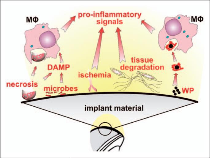
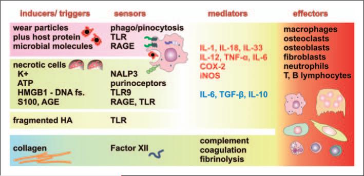
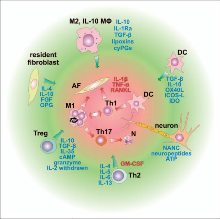
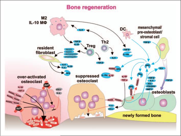
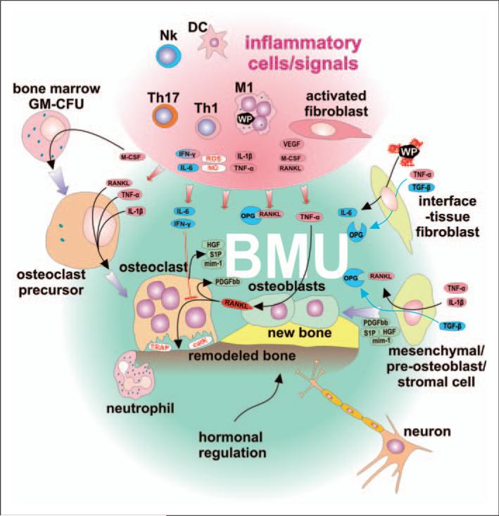
Numerous studies provide detailed insight into the triggering and amplification mechanisms of the inflammatory response associated with prosthetic wear particles, promoting final dominance of bone resorption over bone formation in multiple bone multicellular units around an implant. In fact, inflammation is a highly regulated process tightly linked to simultaneous stimulation of tissue protective and regenerative mechanisms in order to prevent collateral damage of periprosthetic tissues. A variety of cytokines, chemokines, hormones and specific cell populations, including macrophages, dendritic and stem cells, attempt to balance tissue architecture and minimize inflammation. Based on this fact, we postulate that the local tissue homeostatic mechanisms more effectively regulate the pro-inflammatory/pro-osteolytic cells/pathways in patients with none/mild periprosthetic osteolysis (PPOL) than in patients with severe PPOL. In this line of thinking, 'particle disease theory' can be understood, at least partially, in terms of the failure of local tissue homeostatic mechanisms. As a result, we envision focusing current research on homeostatic mechanisms in addition to traditional efforts to elucidate details of pro-inflammatory/pro-osteolytic pathways. We believe this approach could open new avenues for research and potential therapeutic strategies.


Figures

Figure 1
Figure 1
Initial triggers of inflammatory response to total hip arthroplasty (THA). Immediately after implantation of THA into the bone bed several factors can trigger an inflammatory response which can contribute to tissue damage, poor initial fixation and periprosthetic osteolysis. The main factors are: (i) tissue necrosis, ischemia and degradation of tissues sensed mainly by macrophages (MΦ), other cells and the complement system; (ii) microbial remnants (PAMPS/MAMPS) from direct contamination or from the blood; (iii) micromotion at the implant-host interface and formation of a synovial interface-like membrane around the implant leading to activation of fibroblasts and other mesenchymal cells, and to release of osteoclast-activating pro-inflammatory factors; (iv) prosthetic wear particles (WP), liberated from the articulating and/or non-articulating surfaces, together with adsorbed host proteins (shown as a red string around the wear particles) are sensed by MΦ, triggering inflammation.
Figure 2
Figure 2
Components of inflammatory response ‘inducers—sensors—mediators—effectors’ potentially associated with periprosthetic osteolysis and aseptic loosening. The host response is initiated by triggers (inducers), which are of both exogenous (wear particles, bacteria or microbial remnants) and endogenous (cells, extracellular matrix and bodily fluids) origin. Wear particles with adsorbed host proteins are phago-/pinocytosed (leading to a foreign body reaction) and sensed by pattern recognizing receptors, such as TLRs and receptor for advanced glycation end products (RAGE). Trauma-associated necrosis leads to the release of cellular components, such as ATP, K+ ions, fragmented DNA, members of the S100 calcium-binding protein family, advanced glycation end products (AGE) and others, effectively triggering inflammation after binding to, and activating, respective sensors, such as purinoreceptors, P2X7, NACHT, Leucine-Rich Repeat- and PYD-domains Containing Protein 3 (NALP3 or cryopyrin), high mobility group box 1 protein (HMGB1; the complex with DNA that can stimulate TLR9), RAGE and others, expressed mostly by macrophages. Trauma also leads to the breakdown of extracellular matrix components, such as hyaluronan (HA), fragments of which are sensed by TLRs. Receptors, or sensors, activate macrophages to release the pro-inflammatory factors IL-1, IL-12, IL-18, IL-33, TNF-α, cyclooxygenase 2 (COX-2) and inducible nitric oxide synthetase (iNOS), but also the anti-inflammatory factors IL-10 and TGF-β. IL-6 has multiple activities depending on differentiation and activation status, and receptor expression on target cells, such as osteoclasts; therefore, it could both suppress osteolysis but facilitate osteoclast formation. In addition to the above-mentioned cellular receptors, soluble factors, such as Factor XII, sense extracellular matrix components presented by collagen leading to activation of coagulation, fibrinolysis and complement, which could substantially contribute to the recruitment of inflammatory cells. The above-mentioned inflammatory mediators contribute to the activation or differentiation of several cell lines participating in periprosthetic osteolysis and aseptic loosening of THA. Anti-inflammatory factors associated with bone homeostasis are in blue, pro-inflammatory factors or those associated with bone resorption are in red.
Figure 3
Figure 3
Cell populations involved in the suppression of the inflammatory response. Cells involved in the inflammatory reaction, such as classically-activated macrophages (M1), Th1 cells, Th17 cells, activated fibroblasts, dendritic cells (DC) and neutrophils, contribute to bone resorption mostly through IL-1β, TNF-α and receptor activator of NF-κB ligand (RANKL) signaling. Activity of the above-mentioned cells is controlled and suppressed by several factors secreted predominantly from immune cells represented by macrophages stimulated through the IL-4, IL-13, α-tocopherol, IgG-containing immune complexes (IC), apoptotic cells or prostaglandins, leading to the ‘alternatively activated’ healing phenotype (M2), from regulatory IL-10-secreting macrophages (IL-10 MΦ), DCs, regulatory T cells (Treg) and Th2 cells. Depending on the modulating properties of signals they receive, DCs could play a pro-inflammatory role by activating pro-inflammatory Th1 or Th17 cells, or an anti-inflammatory role by activating regulatory T cells (Treg) that suppress immune reactions. Furthermore, inflammatory cells could be suppressed by resident fibroblasts and neurons. AF, activated fibroblast; cyPGs, cyclopentenone prostaglandins; GM-CSF, granulocyte-macrophage colony stimulating factor; OPG, soluble receptor for RANKL – osteoprotegerin; FGF, fibroblast growth factor; OX40L, tumor necrosis factor ligand superfamily member 4; ICOS-L, inducible T-cell co-stimulator ligand, IDO, indolamine 2,3 dioxygenase; NANC, non-adrenergic non-cholinergic neurotransmitters. Anti-inflammatory factors leading to bone remodeling are in blue, pro-inflammatory factors or those associated with bone resorption are in red.
Figure 4
Figure 4
Bone regeneration is orchestrated with a substantial contribution from immune cells. Within the bone multicellular unit (BMU), suppression of the inflammatory response is associated with a change from net bone resorption toward bone remodeling. Osteoclasts stimulated by RANKL, IL-1β, TNF-α and wear particles pump protons (H+), move toward the bone surface and secrete bone destructing cathepsin K (catK) and tartarate-resistant acid phosphatase (TRAP). A decrease in the level of the stimulating factors is a consequence of anti-inflammatory activity of several immune cell types (M2, IL-10-secreting macrophages, DC, Treg cells, Th2 cells), as well as non-immune cells, such as resident-tissue fibroblasts and mesenchymal/pre-osteoblast/stromal cells. Anti-inflammatory activity is mediated by soluble factors IL-4, IL-10, TGF-β, soluble receptor for IL-1 (IL-1Ra), and OPG secreted and acting within the area of BMU. Strong immunosuppressive Treg cells differentiate under the influence of TGF-β and IL-10 secreted from M2 and IL-10-secreting macrophages and specific populations of DC. Further, Treg cells secrete decorin, asporin, dermatopontin and amphiregullin contributing to a reparative bone remodeling. Bone remodeling is further supported by stimulation of osteoblasts by 17β-estradiol, differentiation factor Wnt and bone morphogenetic protein 2 (BMP-2)—some of them acting as autocrine factors. IFN-γ secreted by Th1 and NK cells or IL-6 secreted by T cells and macrophages contribute to bone remodeling by activation of osteoblasts to produce RANKL inhibitor OPG. The number of osteoblasts increases as osteoblast precursors differentiate under the influence of sphingosine-1-phosphate produced by mature osteoclasts. The mechanisms above and TGF-β, together with other factors, contribute to the recruitment and differentiation of pre-osteoblasts toward mature bone remodeling osteoblasts. Furthermore, IFN-γ and IL-6 suppress bone resorption by acting directly upon osteoclasts. Nevertheless, activated osteoclasts are not responsive to IFN-γ and IL-6 because intracellular response pathways are blocked by over-expressed p38.
Figure 5
Figure 5
The BMU is self regulatory and not affected by pro-inflammatory signals produced by activated immune cells and fibroblasts. Classically-activated M1 macrophages and subpopulations of DCs secrete Th1- and Th17-stimulating cytokines, as well as toxic (reactive) nitrogen and oxygen species, and COX-2. Stimulated Th1 lymphocytes secrete RANKL, one of the most important osteoclast-activating cytokines. M1 further secrete cytokines IL-1β and TNF-α, thus inducing pre-osteoclast differentiation and activation leading to increased bone resorption. IL-1β and TNF-α stimulate osteoblasts to secrete RANKL, thus creating positive feedback for bone resorption. Furthermore, inflammatory-activated fibroblasts support bone resorption by secretion of RANKL and stimulation of pre-osteoclast differentiation by M-CSF. Although the BMU is exposed to the above factors, it has its own regulatory mechanisms to help ensure homeostasis: the BMU is covered by a canopy of cells so that BMUs can undergo activation–resorption-formation cycles in bone remodeling compartments. Osteoclasts exposed to IL-6 and IFN-γ do not respond to RANKL-mediated activation. Furthermore, IL-6 is secreted by fibroblasts after exposure to wear particles. Osteoclasts, upon stimulation by RANKL, secrete platelet-derived growth factor bb (PDGFbb), which induces the proliferation of pre-osteoblasts leading to an increase in the number of osteoblast precursors. Furthermore, osteoclasts produce sphingosine 1-phosphate (S1P), myb-induced myeloid protein-1 (mim-1) and hepatocyte growth factor (HGF), collectively contributing to pre-osteoblasts migration and osteoblast survival. Attenuation of osteoclast activity leads to a decrease in the production of PDGFbb and S1P-induced differentiation of pre-osteoblasts toward active osteoblasts and bone remodeling. Physiologically-activated interface tissue fibroblasts/tissue-resident fibroblasts respond to TNF-α together with TGF-β by secretion of OPG. In addition, BMU neurons could sense local inflammation and modulate activity of both immune and BMU cells through ATP and neuropeptide mediators, and further activate the neuroendocrine system. All the above factors are most prominent early after total hip arthroplasty surgery or when the inflammatory response is more quiescent.

Similar articles

Cited by

References

    1. Kurtz S, Ong K, Lau E, Mowat F, Halpern M. Projections of primary and revision hip and knee arthroplasty in the United States from 2005 to 2030. J Bone Joint Surg Am 2007. 89:780–785. - PubMed
    1. Ulrich SD, Seyler TM, Bennett D, Delanois RE, Saleh KJ, Thongtrangan I, et al. Total hip arthroplasties: What are the reasons for revision? Int Orthop. 2008;32:597–604. - PMC - PubMed
    1. Gallo J, Kaminek P, Zapletalova J, Cechova I, Spicka J, Ditmar R. Is osteolysis associated with a stable total hip replacement asymptomatic? Acta Chir Orthop Traumatol Cech. 2004;71:20–25. - PubMed
    1. Maloney W, Rosenberg A. What is the outcome of treatment for osteolysis? J Am Acad Orthop Surg. 2008;16(Suppl. 1):S26–S32. - PubMed
    1. Deirmengian GK, Zmistowski B, O'Neil JT, Hozack WJ. Management of acetabular bone loss in revision total hip arthroplasty. J Bone Joint Surg Am. 2011;93:1842–1852. - PubMed

Publication types

MeSH terms

Substances