. 2012 Jul 15;11(14):2717-28.
doi: 10.4161/cc.21091. Epub 2012 Jul 15.

PIASy-mediated Tip60 sumoylation regulates p53-induced autophagy

Affiliations

PIASy-mediated Tip60 sumoylation regulates p53-induced autophagy

Samisubbu R Naidu et al. Cell Cycle. .

Abstract

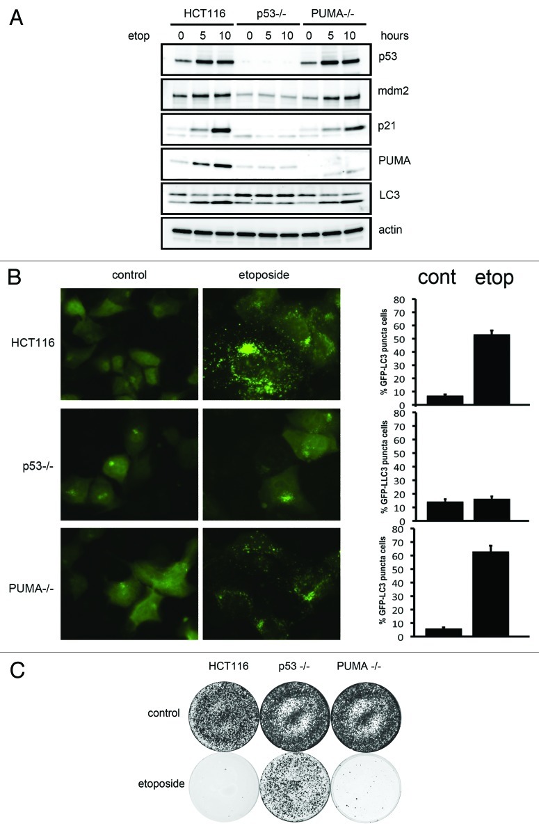
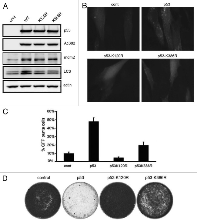
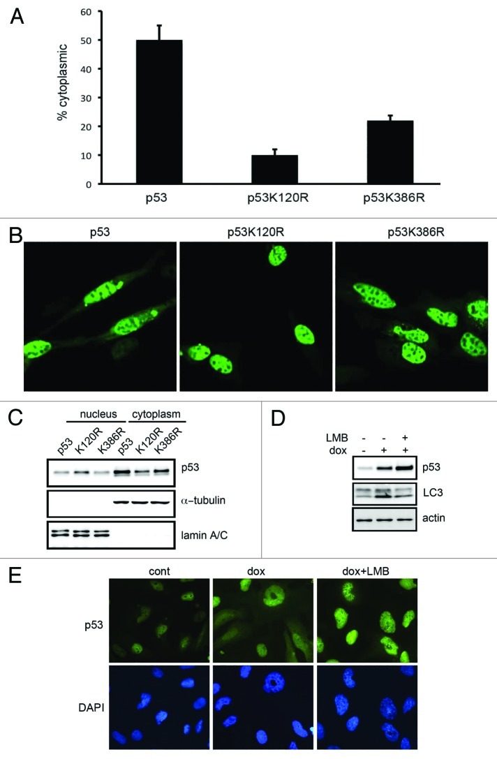
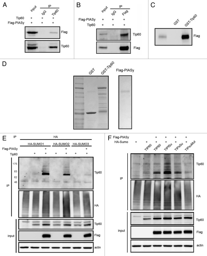
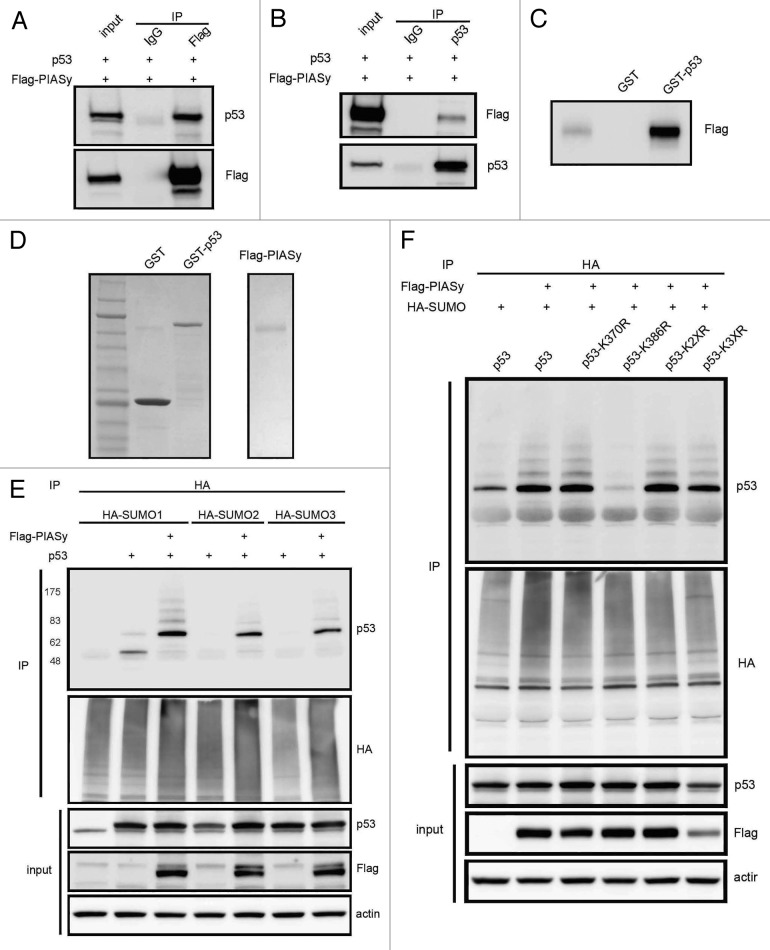
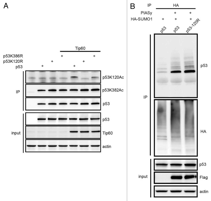
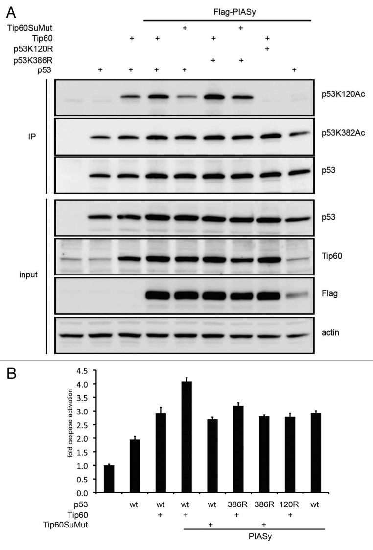
Posttranslational modifications of p53 integrate diverse stress signals and regulate its activity, but their combinatorial contribution to overall p53 function is not clear. We investigated the roles of lysine (K) acetylation and sumoylation on p53 and their relation to apoptosis and autophagy. Here we describe the collaborative role of the SUMO E3 ligase PIASy and the lysine acetyltransferase Tip60 in p53-mediated autophagy. PIASy binding to p53 and PIASy-activated Tip60 lead to K386 sumoylation and K120 acetylation of p53, respectively. Even though these two modifications are not dependent on each other, together they act as a "binary death signal" to promote cytoplasmic accumulation of p53 and execution of PUMA-independent autophagy. PIASy-induced Tip60 sumoylation augments p53 K120 acetylation and apoptosis. In addition to p14(ARF) inactivation, impairment in this intricate signaling may explain why p53 mutations are not found in nearly 50% of malignancies.


Figures

None
Figure 1. DNA damage induced autophagy is p53 dependent and PUMA independent. (A) Wild-type, p53-null or PUMA-null HCT116 cells were treated with etoposide for up to 10 h. Whole-cell lysates were blotted with the indicated antibodies (B) Control or etoposide treated wild-type, p53-null or PUMA-null HCT116 cells expressing GFP-LC3 were visualized for GFP puncta in three independent experiments. (C) HCT116 cells were transfected with a puromycin resistance gene and exposed to etoposide (bottom set). Proliferating puromycin resistant colonies were stained with crystal violet.
None
Figure 2. p53 K120R and K386R mutations fail to induce autophagy. (A) Saos2 cells were transfected with p53, K120R or K386R and whole cell extracts immunoblotted for indicated proteins. (B and C) Saos2 cells were transfected with indicated expression-plasmids along with GFP-LC3. LC3 puncta were counted in100 cells from three independent transfections. Error bars represent standard error. (D) Saos2 cells were co-transfected with control vector or wild-type, K120R or K386R p53 along with a puromycin-resistance encoding plasmid. Cells were selected with puromycin and the colonies stained with crystal violet.
None
Figure 3. p53 mutants K120R and K386R do not accumulate in the cytoplasm. (A and B) Saos2 cells were transfected, fixed with paraformaldehyde and p53 localization was observed. Cells showing cytoplasmic-p53 localization were counted and shown in the bar graph. Error bars represent standard error. (C) Cytoplasmic and nuclear fractions of Saos2 cells transfected and analyzed for p53 subcellular distribution. (D) SH-SY5Y cells were treated with doxorubicin in the presence or absence of leptomycin B (LMB) and the protein extracts were probed with indicated antibodies. (E) Cells treated as in (D) were examined for p53 localization.
None
Figure 4. PIASy interacts with and sumoylates Tip60. (A and B) RPE1 cells transfected with Flag-PIASy and Tip60 were immunoprecipitated with Tip60 or Flag antibodies and immunoblots were probed with reciprocal antibodies. (C) In vitro pull-down assay using the semi-purified proteins showing PIASy binds to GST-Tip60. (D) Coomassie blue staining of semi-purified proteins. (E) RPE1 cells transfected with HA tagged SUMO orthologs in the presence or absence of PIASy along with Tip60. After immunoprecipitation with HA antibody, proteins were blotted with Tip60 or HA antibodies. (F) Cells were transfected as in (D) with Tip60 or first (TIPfSu), second (TIPsSu) or double (TIPsuMut) sumo consensus site K-R mutants along with HA-SUMO1 and the cell extracts immunoblotted as indicated.
None
Figure 5. PIASy complexes with and sumoylates p53. (A and B) RPE1 cells transfected with Flag-PIASy and Tip60 were immunoprecipitated with Flag or p53 antibodies and western blotted with indicated antibodies. (C) In vitro binding assay using the proteins from (D) showing Flag-PIASy binds to GST-p53. (E) PIASy induces sumoylation of p53 as detected by western blot with HA antibodies. The slower migrating bands are most prominent with SUMO1 (F). Lysine 386 is necessary for sumoylation of p53 induced by PIASy.
None
Figure 6. p53 K120 acetylation and K386-sumoylation are not dependent on each other. (A) H1299 cells were transfected with p53 or its mutants, K120R or K386R in the presence or absence of Tip60, the total p53 immunoprecipitate and the input extracts were blotted with indicated antibodies. (B) RPE1 cells transfected with p53 or p53 K120R mutant in the presence of PIASy were tested for SUMO conjugation.
None
Figure 7. Sumoylation defective Tip60 fails to respond to PIASy induced p53 K120 acetylation and apoptosis. (A) H1299 cells co-transfected with wild-type, K120R, or K386R p53 and Tip60 or its SUMO-mutant, and the immunoprecipitates were blotted with the indicated antibodies. The top panel shows reduced K120 acetylation with the Tip60 sumoylation mutant with expression of PIASy. (B) H1299 cells were transfected as in (A) and the apoptotic response analyzed using Caspase Glo reagent in three independent transfections. Error bars represent standard error.

Comment in

  • Directing p53 to induce autophagy.
    Mayo LD. Mayo LD. Cell Cycle. 2012 Sep 15;11(18):3353-4. doi: 10.4161/cc.21849. Epub 2012 Aug 23. Cell Cycle. 2012. PMID: 22918242 Free PMC article.

Similar articles

Cited by

References

    1. Vousden KH, Lane DP. p53 in health and disease. Nat Rev Mol Cell Biol. 2007;8:275–83. doi: 10.1038/nrm2147. - DOI - PubMed
    1. Toledo F, Wahl GM. Regulating the p53 pathway: in vitro hypotheses, in vivo veritas. Nat Rev Cancer. 2006;6:909–23. doi: 10.1038/nrc2012. - DOI - PubMed
    1. Zilfou JT, Lowe SW. Tumor suppressive functions of p53. Cold Spring Harb Perspect Biol. 2009;1:a001883. doi: 10.1101/cshperspect.a001883. - DOI - PMC - PubMed
    1. Lo D, Lu H. Nucleostemin: Another nucleolar “Twister” of the p53-MDM2 loop. Cell Cycle. 2010;9:3227–32. doi: 10.4161/cc.9.16.12605. - DOI - PMC - PubMed
    1. Benchimol S. p53--an examination of sibling support in apoptosis control. Cancer Cell. 2004;6:3–4. doi: 10.1016/j.ccr.2004.07.002. - DOI - PubMed

Publication types

MeSH terms

Substances