. 2012 Jul-Sep;3(3):139-53.
doi: 10.4161/sgtp.21084. Epub 2012 Jul 1.

Palmitoylation regulates vesicular trafficking of R-Ras to membrane ruffles and effects on ruffling and cell spreading

Affiliations

Palmitoylation regulates vesicular trafficking of R-Ras to membrane ruffles and effects on ruffling and cell spreading

Jeremy G T Wurtzel et al. Small GTPases. 2012 Jul-Sep.

Abstract

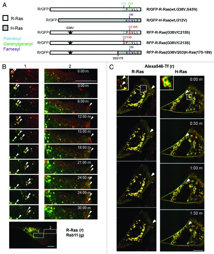
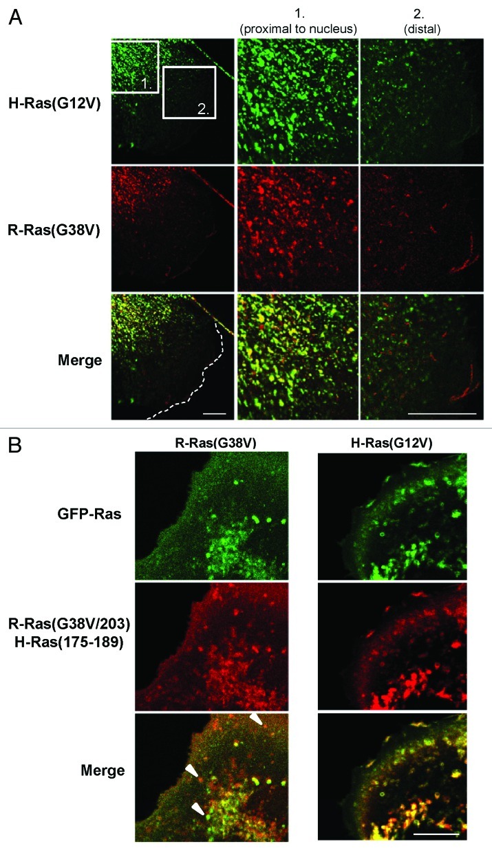
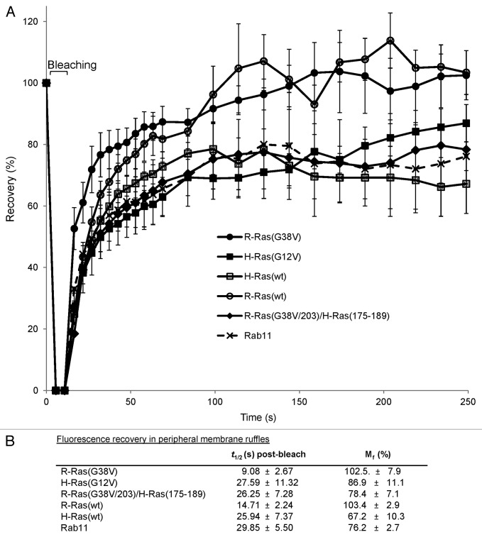
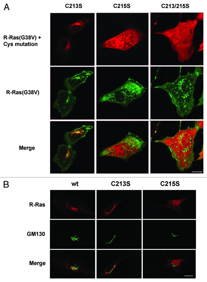
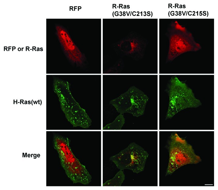
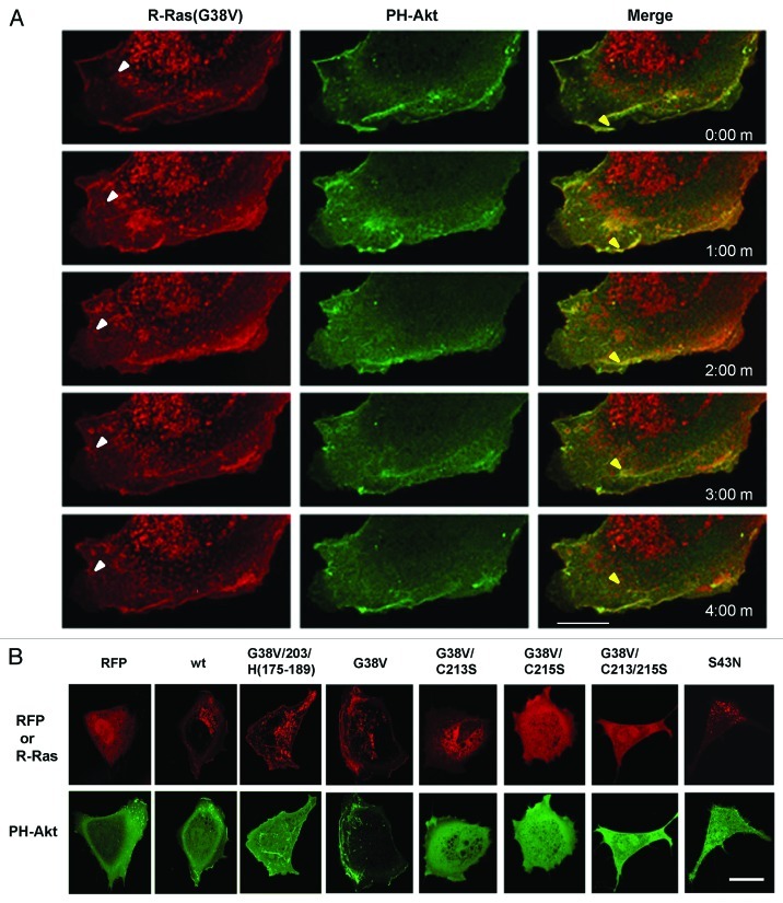
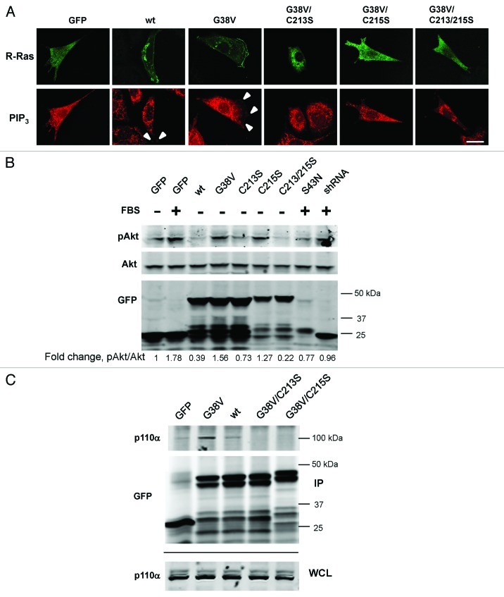
In this study we investigated the dynamics of R-Ras intracellular trafficking and its contributions to the unique roles of R-Ras in membrane ruffling and cell spreading. Wild type and constitutively active R-Ras localized to membranes of both Rab11- and transferrin-positive and -negative vesicles, which trafficked anterograde to the leading edge in migrating cells. H-Ras also co-localized with R-Ras in many of these vesicles in the vicinity of the Golgi, but R-Ras and H-Ras vesicles segregated proximal to the leading edge, in a manner dictated by the C-terminal membrane-targeting sequences. These segregated vesicle trafficking patterns corresponded to distinct modes of targeting to membrane ruffles at the leading edge. Geranylgeranylation was required for membrane anchorage of R-Ras, whereas palmitoylation was required for exit from the Golgi in post-Golgi vesicle membranes and trafficking to the plasma membrane. R-Ras vesicle membranes did not contain phosphatidylinositol (3,4,5)-trisphosphate (PtdIns(3,4,5)P(3)), whereas R-Ras co-localized with PtdIns(3,4,5)P(3) in membrane ruffles. Finally, palmitoylation-deficient R-Ras blocked membrane ruffling, R-Ras/PI3-kinase interaction, enrichment of PtdIns(3,4,5)P(3) at the plasma membrane, and R-Ras-dependent cell spreading. Thus, lipid modification of R-Ras dictates its vesicle trafficking, targeting to membrane ruffles, and its unique roles in localizing PtdIns(3,4,5)P(3) to ruffles and promoting cell spreading.


Figures

None
Figure 1. R-Ras traffics to membrane ruffles in recycling vesicles. (A) R-Ras and H-Ras constructs used in this study, expressed as red or green fluorescent protein (RFP or GFP) fusions. G38V and G12V (starred) are constitutively active mutants of R-Ras and H-Ras, respectively. HVR C-terminal sequences are shown with post-translational modification sites indicated: palmitoyl, blue; geranylgeranyl, green; farnesyl, purple; mutations are shown in red. R-Ras(G38V/203)H-Ras(175–189) is activated R-Ras, residues 1–203, deleted in the HVR and replaced with the farnesyl-specific HVR of H-Ras. R-Ras(S43N) is constitutively inactive. (B) R-Ras and Rab11 trafficking in live, spread cells. RFP-R-Ras(G38V) (red) and GFP-Rab11 (green) were tracked in live NIH 3T3 cells by confocal microscopy. Images were acquired every 30 sec to facilitate vesicle tracking; representative images are shown. Left panels (1) show R-Ras anterograde transport with recycling endosomes. R-Ras transport vesicles included Rab11-containing RE and Rab11-negative puncta. Right panels (2) show retrograde R-Ras transport from membrane ruffles that do not contain Rab11. Arrowheads point to individual R-Ras/Rab11 vesicles tracked across images. The lower panel shows a representative image of the whole cell used for imaging, at lower magnification, with zones 1 and 2 indicated by white boxes. (C) R-Ras and H-Ras (wt) partially traffic in transferrin (Tf)-containing RE. Cells expressing GFP-R-Ras or -H-Ras (green) were labeled with Alexa546-Tf (red) by a pulse-chase scheme (see Materials and Methods) and viewed by live confocal microscopy with images acquired every 30 sec. Areas of co-localization are seen as yellow. Inset areas in white boxes are shown at 2x. Arrowheads point to Tf-negative R-Ras or H-Ras vesicles. Bars, 10 μm.
None
Figure 2. R-Ras and H-Ras segregate into distinct vesicles near the leading edge in a HVR-dependent manner. (A) Cells co-transfected with GFP-H-Ras(G12V) and RFP-R-Ras(G38V) were seeded at low density to allow for random migration, then fixed and imaged by confocal microscopy; merged images are shown at the bottom. The two boxed zones are shown at high magnification. H-Ras and R-Ras co-localized in vesicles proximal to the nucleus (1), but occupied distinct compartments (2) closer to the leading edge of the cell (dotted white line). (B) GFP-R-Ras(G38V) (green) and RFP-R-Ras(G38V/203)H-Ras(175–189) were co-expressed and imaged in live, randomly migrating cells. Merged images are shown at the bottom. White arrowheads point to selected vesicles containing either R-Ras(G38V) or R-Ras(G38V/203)/H-Ras(175–189). Bars, 7.5 μm.
None
Figure 3. Fast R-Ras trafficking to ruffles requires the R-Ras HVR, and is distinct from slow H-Ras trafficking. (A) Fluorescence recovery after photobleaching (FRAP) was assessed in a ~100 μm2 boxed region at the leading edge of migrating cells expressing GFP-R-Ras, -H-Ras or -Rab11 fusions as indicated. GFP fluorescence intensities were measured for the selected area before (set to a normalized value of 100%) and after bleaching at 5 sec for the first minute, followed by 10 sec intervals for 3 min. Intensities were normalized with an adjacent, non-bleached zone, and are shown as percent recovery ± SD. At least 15 cells per type were measured; representative of three independent experiments. (B) Time of recovery (t1/2) and mobile fraction (Mf) for fluorescence in leading edge membrane ruffles are shown ± S.D, calculated from exponential curve fits with a minimum R2 of 0.95.
None
Figure 4. R-Ras palmitoylation is required for localization in vesicles and at the leading edge. (A) Cells co-expressing GFP-R-Ras(G38V) (green) with RFP-R-Ras(G38V) (red) harboring C-terminal site mutations were seeded in chamber slides and imaged live by confocal microscopy. Merged images are shown at the bottom. (B) RFP-R-Ras variants (red) co-expressed with Golgi marker GFP-GM130 (green). R-Ras(wt) partially localized to the Golgi (overlap seen as yellow). The C213S mutant was sequestered in the Golgi, whereas Golgi association was blocked by C215S mutation. Bars, 10 μm.
None
Figure 5. H-Ras vesicular targeting does not require R-Ras trafficking. RFP alone or fused to R-Ras variants as indicated (red) was co-expressed with GFP-H-Ras(wt) (green) and live cells were imaged by confocal microscopy. R-Ras Cysteine mutations altered R-Ras localization and blocked membrane ruffling but did not prevent H-Ras vesicular targeting. Bar, 10 μm.
None
Figure 6. Cell spreading and ruffling regulated by R-Ras lipid modification. Cells were transiently transfected with RFP vector alone (RFP), RFP-R-Ras fusions or RFP plus R-Ras shRNA as indicated. After 24 h, cells were seeded onto fibronectin-coated surfaces and membrane ruffling (A) and cell spreading (B) in RFP-positive cells were determined as described in Materials and Methods. (A) Percentages of transfected cells displaying edge ruffles for at least 100 cells per sample are shown + SEM *, p < 0.001. (B) Average cell areas for at least 100 cells per sample are shown + SEM *, p < 0.05; **, p < 0.004; ***, p < 0.003. Data are from three independent experiments each.
None
Figure 7. R-Ras vesicles are PI(3,4,5)P3-negative, whereas R-Ras co-localizes with PI(3,4,5)P3 in membrane ruffles from which R-Ras is recycled. RFP-R-Ras (red) was co-transfected with GFP-PH-Akt (green), as a marker for PI(3,4,5)P3. (A) Dynamics of R-Ras(G38V) and PH-Akt monitored in a migrating cell. Images were acquired every 30 sec; 1 min intervals are shown. R-Ras was in perinuclear vesicles which trafficked toward the plasma membrane (e.g., white arrowheads), and in retrograde membrane ruffles. In contrast, PH-Akt was restricted to the retrograde ruffles, where it co-localized with R-Ras. Both proteins moved in retrograde fashion in the ruffles. In some cases R-Ras recycled from ruffles through vesicular structures (e.g., yellow arrowheads) that lacked PH-Akt. (B) RFP or RFP-R-Ras fusions as indicated, co-expressed with GFP-PH-Akt and imaged in live cells by confocal microscopy. Bars, 7.5 μm.
None
Figure 8. PtdIns(3,4,5)P3 in membrane ruffles requires R-Ras lipid modification. (A) GFP-R-Ras fusions as indicated were imaged along with PtdIns(3,4,5)P3 antibody staining in fixed cells. White arrowheads point to PtdIns(3,4,5)P3 in ruffles, co-localized with R-Ras. Bar, 7.5 μm. (B) Cells transfected with the indicated GFP-R-Ras fusions were serum-starved (0.5%) for 2 d and either fed with serum (10%) for 30 min (+ FBS) or kept in starvation medium (-) before being lysed. Lysates were subjected to western blotting with antibodies to GFP, total Akt (Akt) or Akt phosphorylated at Ser473 (pAkt). Densitometric ratios of pAkt:Akt are shown as fold change relative to the starved cell ratio. Representative of three independent experiments. (C) GFP or GFP-R-Ras fusions as indicated were transfected, then immunoprecipitated from cell extracts using GFP antibodies. GFP and R-Ras fusions, and endogenous p110α subunit of PI3K were detected by immunoblotting the immunoprecipitate (IP) or whole cell lysate (WCL) fractions using GFP and p110α-specific antibodies. Representative of four independent experiments.

Similar articles

Cited by

References

    1. Conklin MW, Ada-Nguema A, Parsons M, Riching KM, Keely PJ. R-Ras regulates beta1-integrin trafficking via effects on membrane ruffling and endocytosis. BMC Cell Biol. 2010;11:14. doi: 10.1186/1471-2121-11-14. - DOI - PMC - PubMed
    1. Sethi T, Ginsberg MH, Downward J, Hughes PE. The small GTP-binding protein R-Ras can influence integrin activation by antagonizing a Ras/Raf-initiated integrin suppression pathway. Mol Biol Cell. 1999;10:1799–809. - PMC - PubMed
    1. Kwong L, Wozniak MA, Collins AS, Wilson SD, Keely PJ. R-Ras promotes focal adhesion formation through focal adhesion kinase and p130(Cas) by a novel mechanism that differs from integrins. Mol Cell Biol. 2003;23:933–49. doi: 10.1128/MCB.23.3.933-949.2003. - DOI - PMC - PubMed
    1. Cole AL, Subbanagounder G, Mukhopadhyay S, Berliner JA, Vora DK. Oxidized phospholipid-induced endothelial cell/monocyte interaction is mediated by a cAMP-dependent R-Ras/PI3-kinase pathway. Arterioscler Thromb Vasc Biol. 2003;23:1384–90. doi: 10.1161/01.ATV.0000081215.45714.71. - DOI - PubMed
    1. Holly SP, Larson MK, Parise LV. The unique N-terminus of R-ras is required for Rac activation and precise regulation of cell migration. Mol Biol Cell. 2005;16:2458–69. doi: 10.1091/mbc.E03-12-0917. - DOI - PMC - PubMed

Publication types

Substances