. 2013 Feb;27(2):315-24.
doi: 10.1038/leu.2012.180. Epub 2012 Jul 3.

Downregulation of Mcl-1 through GSK-3β activation contributes to arsenic trioxide-induced apoptosis in acute myeloid leukemia cells

Affiliations

Downregulation of Mcl-1 through GSK-3β activation contributes to arsenic trioxide-induced apoptosis in acute myeloid leukemia cells

R Wang et al. Leukemia. 2013 Feb.

Abstract

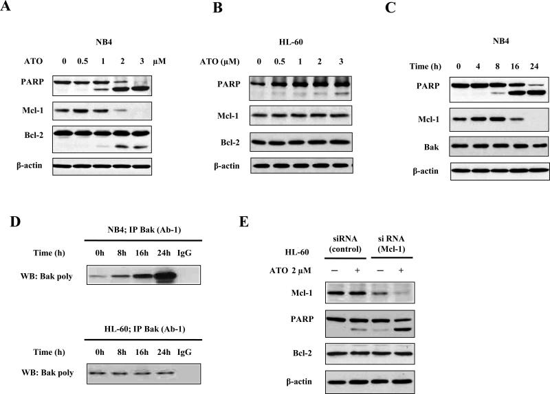
Arsenic trioxide (ATO) induces disease remission in acute promyelocytic leukemia (APL) patients, but not in non-APL acute myeloid leukemia (AML) patients. ATO at therapeutic concentrations (1-2 μM) induces APL NB4, but not non-APL HL-60, cells to undergo apoptosis through the mitochondrial pathway. The role of antiapoptotic protein Mcl-1 in ATO-induced apoptosis was determined. The levels of Mcl-1 were decreased in NB4, but not in HL-60, cells after ATO treatment through proteasomal degradation. Both glycogen synthase kinase-3β (GSK-3β) inhibitor SB216763 and siRNA blocked ATO-induced Mcl-1 reduction as well as attenuated ATO-induced apoptosis in NB4 cells. Silencing Mcl-1 sensitized HL-60 cells to ATO-induced apoptosis. Both ERK and AKT inhibitors decreased Mcl-1 levels and enhanced ATO-induced apoptosis in HL-60 cells. Sorafenib, an Raf inhibitor, activated GSK-3β by inhibiting its phosphorylation, decreased Mcl-1 levels and decreased intracellular glutathione levels in HL-60 cells. Sorafenib plus ATO augmented reactive oxygen species production and apoptosis induction in HL-60 cells and in primary AML cells. These results indicate that ATO induces Mcl-1 degradation through activation of GSK-3β in APL cells and provide a rationale for utilizing ATO in combination with sorafenib for the treatment of non-APL AML patients.


Figures

Fig. 1
Fig. 1. Down-regulation of Mcl-1 contributes to ATO-induced apoptosis in leukemia cells
(A) Down-regulation of Mcl-1 levels by ATO treatment in NB4 cells. (B) Down-regulation of Mcl-1 levels by ATO treatment in HL-60 cells. (C) Time-dependent down-regulation of Mcl-1 by ATO treatment in NB4 cells. NB4 and HL-60 cells were treated with ATO at the indicated concentrations for 24 h or with 2 μM ATO for the indicated times. The relative levels of PARP, Mcl-1, Bcl-2, and β-actin were determined using specific antibodies with Western blot analysis. β-actin was used as a loading control. (D) Bak activation by ATO treatment in both NB4 and HL-60 cells. NB4 and HL-60 cells were treated with ATO at 2 μM for the indicated times and lysed in CHAPS lysis buffer. Total Bak protein was immunoprecipitated with anti-Bak (AB-1) antibody and conformationally changed Bak was probed using poly anti-Bak antibody. (E) Silencing Mcl-1 enhances ATO-induced apoptosis in HL-60 cells. HL-60 cells transfected with Mcl-1 siRNA or control siRNA were treated with 2 μM ATO for 24 h. The relative levels of PARP, Mcl-1, Bcl-2, and β-actin were determined using specific antibodies with Western blot analysis.
Fig. 2
Fig. 2. ATO reduces Mcl-1 and phosphorylated ERK levels in NB4 cells
(A, B) Reduction of phosphorylated ERK and MEK following ATO treatment in NB4 cells. NB4 cells were treated with ATO at the indicated concentrations for 24 h or treated with 2 μM ATO for the indicated times. The levels of Mcl-1, p-MER, ERK, p-ERK, p-Mcl-1(Thr163), and β-actin were determined using specific antibodies with Western blot analysis. (C, D, E) The combined effects of ATO with MEK/ERK inhibitors on Mcl-1 levels. NB4 cells were pretreated with 5 μM U0126 (C), 1 μM PD184352 (C), or 5 μM sorafenib (D) for 2 h and then treated with 1 μM ATO for another 24 h. The levels of PARP, Mcl-1, p-Mcl-1(Thr163), p-MEK, p-ERK, and β-actin were determined using specific antibodies with Western blot analysis. (E) The effect of ATO plus sorafenib on inducing apoptosis. NB4 were treated with 5 μM sorafenib, 1 μM ATO, or their combination and apoptotic cells were analyzed with annexin V-FITC using flow cytometry.
Fig. 3
Fig. 3. Inhibition of the AKT/mTOR pathway does not enhance ATO-induced apoptosis
(A, B) Inhibition of the AKT/mTOR signaling pathway by ATO in a dose- and time-dependent manner. NB4 cells were treated with ATO at the indicated concentrations for 24 h or treated with 2 μM ATO for the indicated times. The levels of Mcl-1, AKT, p-mTOR, p-p70S6K(Thr389), p-4E-BP1, p-S6(Ser235/236), and β-actin were determined using specific antibodies with Western blot analysis. (C, D) The combined effects of rapamycin plus ATO on both Mcl-1 levels and apoptosis. NB4 cells were pretreated with 40 nM rapamycin for 2 h and then treated with 1 μM ATO for another 24 h. The levels of PARP, Mcl-1, p-ERK, p-p70S6K (Thr421/Ser424), p-p70S6K (Thr389), p-S6 (Ser235/236), and β-actin were determined using specific antibodies with Western blot analysis (C). Apoptotic cells were detected with annexin V-FITC using flow cytometry (D).
Fig. 4
Fig. 4. Inhibition of GSK-3β phosphorylation contributes to reduction in Mcl-1 levels in NB4 cells
(A) ATO treatment decreases p-GSK-3β(Ser9) levels. NB4 cells were treated with ATO at the indicated concentrations for 24 h. (B) The GSK-3β inhibitor, SB216763, blocks ATO-induced decreases in Mcl-1 levels. NB4 cells were pretreated with 5 μM SB216763 for 2 h and then treated with 2 μM ATO for another 24 h. (C) Silencing GSK-3β blocks ATO-induced reduction of Mcl-1 levels. NB4 cells transfected with GSK-3β siRNA or control siRNA were treated with 2 μM ATO for 24 h. (D) Proteasome inhibitor MG132 blocks ATO-induced reduction of Mcl-1 levels. NB4 cells were pretreated with 0.5 μM MG132 for 2 h and then treated with 2 μM ATO for another 24 h. The relative levels of Mcl-1, PARP, GSK-3β, p-ERK, AKT and β-actin were determined with Western blot analyses using specific antibodies. (E) The potential mechanisms of ATO-induced reduction of Mcl-1 levels in NB4 cells.
Fig. 5
Fig. 5. ERK and AKT inhibitors enhance ATO-induced apoptosis in HL-60 cells
(A) The regulation by ATO treatment on the levels of AKT, p-ERK, and p-GSK-3β in HL-60 cells. HL-60 cells were treated with ATO at the indicated concentrations for 24 h and the levels of Mcl-1, p-ERK, AKT, GSK-3β, p-GSK-3β(Ser9), and β-actin were determined with Western blot analysis using specific antibodies. (B) The combined effects of an ERK inhibitor or an AKT inhibitor with ATO on apoptosis induction. HL-60 cells were pretreated with 20 μM LY294002, 1 μM PD184352, or 5 μM sorafenib for 2 h and then treated with or without 2 μM ATO for another 24 h. Apoptotic cells were detected with annexin V-FITC using flow cytometry. (C, D, E) The combined effects of either ERK inhibitors or an AKT inhibitor with ATO on the regulation of Mcl-1 levels and PARP cleavage. HL-60 cells were pretreated with 20 μM LY294002 (C), 1 μM PD184352 (D), or 5 μM sorafenib (E) for 2 h and then treated with or without 2 μM ATO for another 24 h. The levels of PARP, Mcl-1, GSK-3β, GSK-3β(Ser9), and β-actin were determined with Western blot analysis using specific antibodies.
Fig. 6
Fig. 6. Sorafenib decreases intracellular GSH levels and enhances ROS production due to ATO treatment
(A) Sorafenib decreases GSH levels in HL-60 cells. HL-60 cells were pretreated with 5 μM sorafenib for 2 h and then treated with or without 2 μM ATO for 12 h. Then the intracellular GSH levels were determined as described in Material and Methods. (B) ROS levels of HL-60 cells treated with ATO plus sorafenib. HL-60 cells were pretreated with 5 μM sorafenib for 2 h and then treated with or without 2 μM ATO for another 16 h. The intracellular H2O2 content was determined by DCFH-DA staining with FACS. (C) The combined effects of sorafenib plus ATO on apoptosis induction. HL-60 and HP100-1 cells were treated with 5 μM sorafenib with or without 2 μM ATO for 24 h. Apoptotic cells were detected with annexin V-FITC using flow cytometry. (D) The combined effects of sorafenib plus ATO on Mcl-1 levels. HL-60 and HP100-1 cells were treated by 5 μM sorafenib with or without 2 μM ATO for 24 h. Then the levels of PARP, Mcl-1, p-GSK-3β(Ser9), and β-actin were determined with Western blot analysis using specific antibodies.
Fig. 7
Fig. 7. Sorafenib plus ATO augment apoptosis induction in primary AML cells
Primary AML cells were isolated as described in Material and Methods from four AML patients and treated with 5 μM sorafenib, 2 μM ATO and their combination for 24 h. Apoptotic cells were detected with annexin V-FITC using flow cytometry (A). The number listed are average and ** p < 0.01 compared to cells treated with either ATO or sorafenib alone (A). The levels of PARP, Mcl-1, p-GSK-3β(Ser9), and β-actin were determined with Western blot analysis using specific antibodies in cells isolated from AML#2 patient (B).
Fig. 8
Fig. 8. The mechanisms of augmented apoptosis induction by ATO in combination with inhibitors of ERK or AKT in AML cells
Inhibitors of ERK and AKT activate GSK3β by inhibiting phosphorylation due to ERK/AKT. Inhibitors of ERK and AKT also decrease intracellular GSH levels which enhances ATO production of ROS as well as ATO inhibition of ERK/AKT. Activated GSK3β phosphorylates Mcl-1 and leads to its proteasomal degradation. Enhanced ROS production plus decreased Mcl-1 levels by these combination treatment result in synergistic induction of apoptosis via activation of Bak.

References

    1. Park JH, Tallman MS. Treatment of acute promyelocytic leukemia without cytotoxic chemotherapy. Oncology (Williston Park) 2011;25:733–741. - PubMed
    1. Soignet SL, Maslak P, Wang ZG, Jhanwar S, Calleja E, Dardashti LJ, et al. Complete remission after treatment of acute promyelocytic leukemia with arsenic trioxide. N Engl J Med. 1998;339:1341–1348. - PubMed
    1. Jing Y, Wang L, Xia L, Chen GQ, Chen Z, Miller WH, et al. Combined effect of all-trans retinoic acid and arsenic trioxide in acute promyelocytic leukemia cells in vitro and in vivo. Blood. 2001;97:264–269. - PubMed
    1. Chen GQ, Shi XG, Tang W, Xiong SM, Zhu J, Cai X, et al. Use of arsenic trioxide (As2O3) in the treatment of acute promyelocytic leukemia (APL): I. As2O3 exerts dose-dependent dual effects on APL cells. Blood. 1997;89:3345–3353. - PubMed
    1. Dai J, Weinberg RS, Waxman S, Jing Y. Malignant cells can be sensitized to undergo growth inhibition and apoptosis by arsenic trioxide through modulation of the glutathione redox system. Blood. 1999;93:268–277. - PubMed

Publication types

MeSH terms