Review
. 2012 Jul 2;10(8):563-74.
doi: 10.1038/nrmicro2820.

Viroporins: structure and biological functions

Affiliations
Review

Viroporins: structure and biological functions

José Luis Nieva et al. Nat Rev Microbiol. .

Abstract

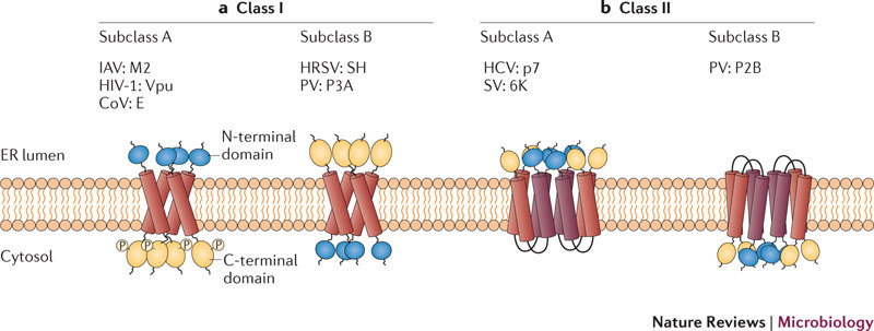
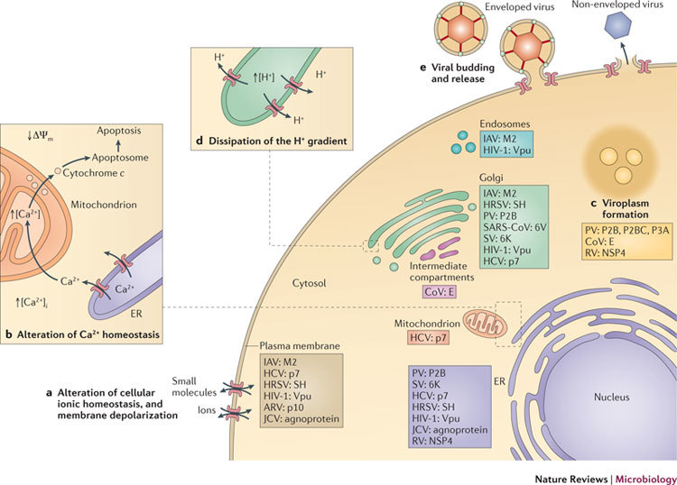
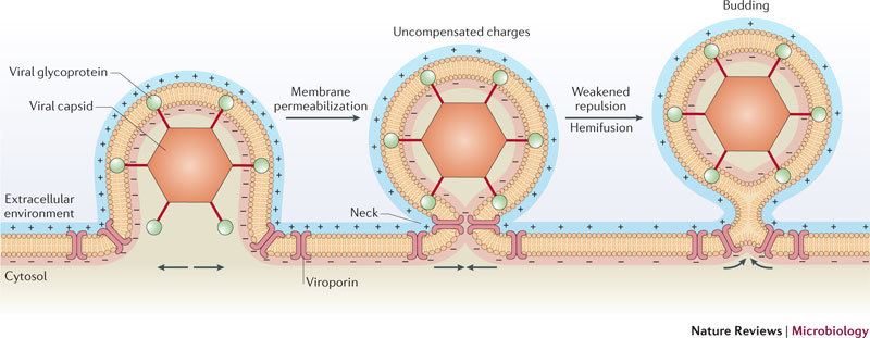
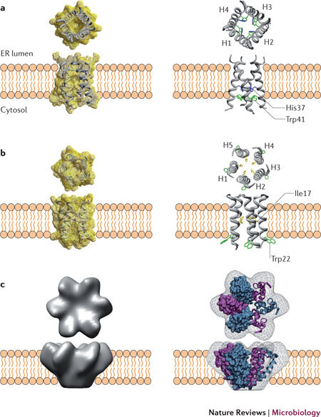
Viroporins are small, hydrophobic proteins that are encoded by a wide range of clinically relevant animal viruses. When these proteins oligomerize in host cell membranes, they form hydrophilic pores that disrupt a number of physiological properties of the cell. Viroporins are crucial for viral pathogenicity owing to their involvement in several diverse steps of the viral life cycle. Thus, these viral proteins, which include influenza A virus matrix protein 2 (M2), HIV-1 viral protein U (Vpu) and hepatitis C virus p7, represent ideal targets for therapeutic intervention, and several compounds that block their pore-forming activity have been identified. Here, we review recent studies in the field that have advanced our knowledge of the structure and function of this expanding family of viral proteins.


Conflict of interest statement

The authors declare no competing financial interests.

Figures

Figure 1
Figure 1. Classification of viroporins according to the number of transmembrane domains and the membrane topology of the constituent monomers.
a | Class I viroporins have a single membrane-spanning domain. The A and B subclasses contain proteins that are inserted into the membrane with either a lumenal amino terminus and cytosolic carboxyl terminus (class IA) or a cytosolic amino terminus and lumenal carboxyl terminus (class IB). In addition, class IA members are usually phosphorylated at the C terminus. Known viroporins of each subclass are shown. b | Class II viroporins form helix–turn–helix hairpin motifs that span the membrane. Subclass A members have lumenal N and C termini, whereas members of subclass B have cytosolic N and C termini. Known viroporins of each subclass are shown. CoV, coronavirus; E, envelope small membrane protein; HCV, hepatitis C virus; HRSV, human respiratory syncytial virus; IAV, influenza A virus; M2, matrix protein 2; PV, poliovirus; SH, small hydrophobic protein; SV, Sindbis virus; Vpu, viral protein U.
Figure 2
Figure 2. Cytopathic effects of viroporins and their functions during the viral life cycle.
The main host cell organelles that are targeted by viroporins, and the cytopathic effects that are induced by viroporins and their functions during the viral life cycle are represented. a | Alteration of plasma membrane potential. Viroporins that are located at the plasma membrane can dissipate the ionic gradient across the membrane, leading to depolarization. b | Alteration of cellular Ca2+ homeostasis. The poliovirus (PV) viroporin protein 2B (P2B) assembles pores in the ER membrane and induces the release of Ca2+ from the ER lumen into the cytosol. Uptake of Ca2+ by the mitochondria can lead to dissipation of the inner-mitochondrial-membrane potential (ΔΨm), permeabilization of the outer mitochondrial membrane and, finally, the release of cytochrome c. In the cytosol, cytochrome c promotes the formation of the so-called apoptosome, a molecular platform that is involved in apoptosis. c | Certain viroporins, such as P2B, polyprotein P2BC and P3A from PV or envelope small membrane protein (E) from coronavirus (CoV), induce intracellular membrane remodelling to generate new membrane vesicles (called the viroplasm) that serve as viral replication sites. d | Dissipation of the proton gradient in the Golgi and the trans-Golgi network. The viroporins influenza A virus (IAV) matrix protein 2 (M2) and hepatitis C virus (HCV) p7 reduce the acidification of vesicular acidic compartments by equilibrating the proton concentration with the cytosol. Alteration of the intracellular ionic gradient in the vesicular system impairs glycoprotein trafficking. e | During the viral replication cycle, viroporins play an essential part in assembly, budding and release of the viral progeny. ARV, avian reovirus; [Ca2+]i, intracellular Ca2+ concentration; HRSV, human respiratory syncytial virus; JCV, JC polyomavirus; NSP4, non-structural protein 4; RV, rotavirus; SARS-CoV, severe acute respiratory syndrome CoV; SH, small hydrophobic protein; SV, Sindbis virus; Vpu, viral protein U.
Figure 3
Figure 3. Model of a viroporin promoting viral budding at the plasma membrane.
Viroporins localize at the plasma membrane in specific sites surrounding the neck of the budding virus particle, as described for the influenza A virus matrix protein 2 (M2) viroporin. Viroporins alter membrane permeability by conducting the flux of different ions (for example, Na+ and K+) across the membrane in favour of their concentration gradients and so reducing the transmembrane potential, which is essentially determined by three factors: the concentration of ions inside and outside the cell; the permeability of the cell membrane to those ions (that is, the ion conductance) through specific ion channels; and the activity of electrogenic pumps (for example, the (Na++K+)ATPase and Ca2+ transport pumps) that require energy to maintain the ion gradients across the membrane. Depolarization of the membrane (that is, decreasing the imbalance of charges across the membrane) leads to a reduction in the charge density on the membrane surface. This phenomenon would result in a decrease in the electrical or contact repulsion between opposing monolayers of the membrane at the neck of the budding site (middle) and could provide the stimulus and even the energy to locally promote budding and release,, as occurs in depolarization-dependent exocytosis.
Figure 4
Figure 4. Three-dimensional structures of selected viroporins.
Each viroporin structure is shown both oriented in the membrane bilayer and from a down-top view. a | High-resolution (1.65 Å) X-ray structure of the oligomers formed by a peptide representing the transmembrane region (residues 25–46) of the class IA viroporin matrix protein 2 (M2) from influenza A virus. Crystals were obtained at pH 6.5 in the presence of N-octylglucoside (Protein Data Bank (PDB) accession 3LBW). In the ribbon representation (right), constituent helices (H1–H4) are indicated, and side chains of His37 and Trp41 are depicted in blue and green, respectively. Structures were generated using Swiss-PdbViewer. b | Solid-state NMR structure of the transmembrane region (residues 2–30) from the class IA viroporin viral protein U (Vpu) from HIV-1, in lipid bilayers (PDB accession 1PI7). The oligomeric form was calculated using energy minimization protocols, and side chains were added to a backbone structure that was generated from solid-state NMR spectroscopy data. Ribbon representations (right) display the constituent helices (H1–H5) and side chains of Ile17 and Trp22 in yellow and green, respectively. Structures were generated using Swiss-PdbViewer. c | Density map contours of oligomers of p7, a class IIA viroporin from hepatitis C virus, solubilized in detergent. Simulated p7 monomers were fitted with their amino and carboxyl termini oriented towards the petal tips (right). Structures in part c are modified, with permission, from Ref. © (2009) US National Academy of Sciences.

References

    1. Carrasco L. Modification of membrane permeability by animal viruses. Adv. Virus Res. 1995;45:61–112. doi: 10.1016/S0065-3527(08)60058-5. - DOI - PMC - PubMed
    1. Gonzalez ME, Carrasco L. Viroporins. FEBS Lett. 2003;552:28–34. doi: 10.1016/S0014-5793(03)00780-4. - DOI - PubMed
    1. Hout DR, et al. Vpu: A multifunctional protein that enhances the pathogenesis of human immunodeficiency virus type 1. Curr. HIV Res. 2004;2:255–270. doi: 10.2174/1570162043351246. - DOI - PubMed
    1. Griffin SD. Plugging the holes in hepatitis C virus antiviral therapy. Proc. Natl Acad. Sci. USA. 2009;106:12567–12568. doi: 10.1073/pnas.0906760106. - DOI - PMC - PubMed
    1. Wang J, Qiu JX, Soto C, DeGrado WF. Structural and dynamic mechanisms for the function and inhibition of the M2 proton channel from influenza A virus. Curr. Opin. Struct. Biol. 2011;21:68–80. doi: 10.1016/j.sbi.2010.12.002. - DOI - PMC - PubMed

Publication types

MeSH terms

Substances